S H O R T G E N O M E R E P O R T
Open Access
Non-contiguous finished genome sequence and
description of the gliding bacterium
Flavobacterium seoulense sp. nov.
Su-Kyoung Shin
1, Heemoon Goo
2, Yong-Joon Cho
3, Soonsung Kwon
4, Dongeun Yong
4and Hana Yi
1,2,5*Abstract
Flavobacterium seoulense strain EM1321
Tis the type strain of Flavobacterium seoulense sp. nov., a proposed novel
species within the genus Flavobacterium. This strain is a Gram-reaction-negative, aerobic, rod-shaped bacterium
isolated from stream water in Bukhansan National Park, Seoul. This organism is motile by gliding. Here, we describe
the features of Flavobacterium seoulense EM1321
T, together with its genome sequence and annotation. The genome
comprised 3,792,640 bp, with 3,230 protein-coding genes and 52 RNA genes.
Keywords: Flavobacterium, Gliding motility, Aerobic, Flavobacteriaceae
Introduction
Flavobacterium
is the type genus of the family
Flavobac-teriaceae
in the phylum Bacteroidetes. Flavobacterium
was proposed by Bergey et al. [1,2] and the description
was emended by Bernardet et al. [3]. Flavobacterium
species have been isolated from various environments,
including seawater, freshwater, river sediments, and soil
[4-8]. Members of the genus Flavobacterium are
Gram-negative, rod-shaped, yellow-pigmented, aerobic
bac-teria. At the time of writing, about 118 Flavobacterium
species with validly published names have been
de-scribed [9]; however, the genomes of only 14 type
strains in this genus have been sequenced.
Flavobacterium seoulense
sp. nov. strain EM1321
T(=
KACC 18114
T= JCM 30145
T) was isolated from stream
water in Bukhansan National Park, Seoul, Korea. Here, we
present a summary classification and the features of
Flavo-bacterium seoulense
EM1321
Tas well as its genome
se-quence and annotation.
Classification and features
Based on its 16S rRNA gene phylogeny and phenotypic
characteristics, strain EM1321
Twas classified as a
mem-ber of the genus Flavobacterium (Table 1). Preliminary
sequence-based identification using the 16S RNA gene
se-quences in the EzTaxon database [10] indicated that strain
EM1321
Twas most closely related to F. granuli Kw05
T(GenBank accession no. AB180738) with a sequence
simi-larity of 96.54%. This value was lower than the 98.7% 16S
rRNA gene sequence similarity as a threshold
recom-mended by Stackebrandtia and Ebers [11] to delineate a
new species without carrying out DNA-DNA hybridization.
Subsequent phylogenetic analysis was performed using the
16S rRNA gene sequences of strain EM1321
Tand related
species. Sequences were aligned according to the bacterial
rRNA secondary structure model using the jPHYDIT [12].
Phylogenic trees were constructed using neighbor-joining
(NJ) and maximum-likelihood (ML) methods implemented
in MEGA version 5 [13]. The resultant tree topologies were
evaluated by bootstrap analyses with 1,000 random
sam-plings. Strain EM1321
Tformed a monophyletic clade
to-gether with Flavobacterium soli [5] in both the NJ and ML
trees; however, the clustering was not supported by the
bootstrap analysis (Figure 1). Flavobacterium
nitratiredu-cens
[8] was further recovered as a sister group of the
monophyletic clade in the ML tree only. Based on these
phylogenetic trees, F. soli KACC 17417
Tand F.
nitratiredu-* Correspondence:[email protected]
1Department of Public Health Sciences, BK21PLUS Program in Embodiment:
Health-Society Interaction, Graduate School, Korea University, Seoul, Republic of Korea
2
School of Biosystem and Biomedical Science, Korea University, Seoul, Republic of Korea
Full list of author information is available at the end of the article
© 2014 Shin et al.; licensee BioMed Central. This is an Open Access article distributed under the terms of the Creative Commons Attribution License (http://creativecommons.org/licenses/by/4.0), which permits unrestricted use, distribution, and reproduction in any medium, provided the original work is properly credited. The Creative Commons Public Domain Dedication waiver (http://creativecommons.org/publicdomain/zero/1.0/) applies to the data made available in this article, unless otherwise stated.
cens
JCM 17678
Twere selected as reference strains and
were obtained from the corresponding culture collections
for comparative study.
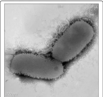
Strain EM1321
Twas Gram-reaction negative. Cells of
strain EM1321
Twere rod shaped with rounded ends and
motile by gliding. The cells were 1.0–1.5 μm × 0.3–
0.5
μm in size (Figure 2). No flagellum was observed.
The colonies were yellow in color and translucent on
R2A agar medium. Growth occurred aerobically at 4–35°C,
and optimal growth was observed at 30°C. The cells
grew in 0–4% (w/v) NaCl. Strain EM1321
Texhibited
catalase and oxidase activities. Physiological and
bio-chemical properties were tested by using the API 20NE,
API 50CH, and API ZYM systems (BioMérieux). In the
API ZYM system, enzyme activity was detected for
alka-line phosphatase, esterase (C4), esterase lipase (C8),
leucine arylamidase, acid phosphatase,
naphthol-AS-BI-phosphohydrolase,
β-galactosidase, and valine
arylami-dase (Table 2). No activity was detected for lipase,
trypsin,
α-chymotrypsin, α-galactosidase,
β-glucuroni-dase,
α-glucosidase, N-acetyl-β-glucosaminidase, cystine
arylamidase,
α-mannosidase, and α-fucosidase. In the
API 20NE system, positive reactions were observed for
nitrate reduction and negative reactions were observed
for indole production, glucose fermentation, arginine
dihydrolase, urease activity, and aesculin and gelatin
hy-drolysis. The strain assimilated d-glucose and
l-arabinose, but not d-mannitol, d-mannose, d-maltose,
potassium gluconate, N-acetylglucosamine, capric acid,
adipic acid, malic acid, trisodium citrate, or phenylacetic
acid. Acid was produced from l-arabinose, xylose,
galactose, glucose, fructose, mannose, and
d-lactose (API 50CH).
Matrix-assisted
laser-desorption/ionization
time-of-flight (MALDI-TOF) MS protein analysis was carried
out as previously described [24]. Deposits were done
from 12 isolated colonies for each strain (strain
EM1321
Tand reference strains). Measurements were
Table 1 Classification and general features of
Flavobacterium seoulense EM1321
Taccording to the MICG
recommendations [14]
MIGS ID Property Term Evidence code Current classification Domain Bacteria TAS [15]
Phylum Bacteroidetes TAS [16,17] Order Flavobacteriales TAS [17,18] Family Flavobacteriaceae TAS [3,19-21] Genus Flavobacterium TAS [1-3,22] Species F. seoulense IDA Strain EM1321T IDA
Gram stain Negative IDA Cell shape Rod-shaped IDA
Motility Gliding IDA
Sporulation Non-sporulating IDA Temperature range 4–35°C IDA Optimum temperature 30°C IDA MIGS-6 Habitat Freshwater IDA
MIGS-6.3 Salinity 0–4% IDA
MIGS-22 Oxygen requirement Aerobic IDA Carbon source d-glucose, l-arabinose IDA MIGS-15 Biotic relationship Free-living IDA MIGS-14 Pathogenicity Non-pathogenic NAS MIGS-4 Geographic location Seoul, South Korea IDA MIGS-5 Sample collection time September 2013 IDA MIGS-4.1 Latitude 37°36′52′′N IDA MIGS-4.2 Longitude 126°59′19′′E IDA Isolation Stream water IDA
Evidence codes - IDA: Inferred from Direct Assay; TAS: Traceable Author Statement (i.e., a direct report exists in the literature); NAS: Non-traceable Author Statement (i.e., not directly observed for the living, isolated sample, but based on a generally accepted property for the species, or anecdotal evidence). These evidence codes are from the Gene Ontology project [23]. If the evidence is IDA, the property was directly observed by one of the authors.
Shin et al. Standards in Genomic Sciences 2014, 9:34 Page 2 of 8
made with a Microflex spectrometer (Bruker Daltonics,
Leipzig, Germany). Spectra were recorded in the positive
linear mode for the mass range of 2,000 to 20,000 Da
(parameter settings: ion source 1 (IS1), 20 kV; IS2,
18.5 kV; lens, 7 kV). The time of acquisition was
be-tween 30 seconds and 1 minute per spot. The twelve
EM1321
Tspectra were imported into the MALDI
BioTyper software (version 2.0; Bruker) and analyzed
by standard pattern matching (with default parameter
settings) against 4,613 bacterial spectra including
eight Flavobacterium species, used as reference data,
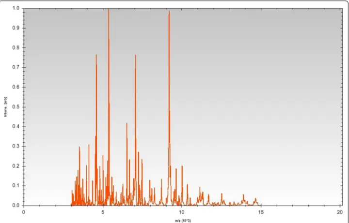
in the BioTyper database. For strain EM1321
Tspectrum (Figure 3), no significant score was obtained,
suggesting that our isolate was not a member of the
eight known species in the database. Spectrum
differ-ences with the two closely related Flavobacterium
spe-cies are shown in Figure 4.
Genome sequencing information
Genome project history
Flavobacterium seoulense
EM1321
Twas selected for
genome sequencing based on its phylogenetic position and
its 16S rRNA similarity to other members of the genus
Fla-vobacterium. The genome sequence was deposited in
Gen-Bank under accession number JNCA00000000.1. A
summary of the project and the Minimum Information
about a Genome Sequence (MIGS) [14] are shown in
Table 3.
Growth conditions and DNA isolation
Flavobacterium seoulense
EM1321
Twas cultured
aerob-ically on R2A agar medium at 30°C. Genomic DNA was
extracted using the QIAamp DNA mini kit (Qiagen).
Genome sequencing and assembly
The genome of strain EM1321
Twas sequenced at
Chun-Lab, Inc. by using an Illumina Miseq_PE_300 system
Figure 1 Phylogenetic tree highlighting the position of Flavobacterium seoulense EM 1321Trelative to the type strains of other species
within the genus Flavobacterium. The strains and their corresponding GenBank accession numbers of 16S rRNA genes are indicated in parentheses. The sequences were aligned using jPHYDIT and the phylogenetic inferences were obtained using neighbour-joining method with MEGA version 5 [13]. The numbers at nodes are the percentage of bootstrap values obtained by 1,000 replicates. Solid circles indicate that the corresponding nodes were also recovered in maximum-likelihood tree. Bar, 0.01 substitutions per nucleotide position.
Figure 2 Transmission electron micrograph of Flavobacterium seoulense EM1321T. Scale bar, 200 nm.
with 2 × 300 paired-end reads. The Illumina platform
provided 166× coverage (for a total of 3,792,640
sequen-cing reads) of the genome. CLC Genomics Workbench
(ver. 6.5.1) was used for sequence assembly and
qual-ity assessment. The final draft assembly contained 56
contigs.
Genome annotation
The genes in the assembled genome were predicted with
Rapid Annotation using Subsystem Technology (RAST)
server databases [25] and the gene-caller GLIMMER 3.02
[26]. The predicted ORFs were annotated by searching
clusters of orthologous groups (COGs) [11] using the
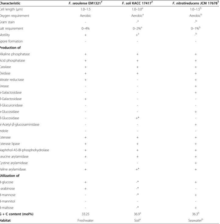
Table 2 Phenotypic characteristics of
Flavobacterium seoulense EM1321
Tand phylogenetically related
Flavobacterium
species
Characteristic F. seoulense EM1321T
F. soli KACC 17417T
F. nitratireducens JCM 17678T
Cell length (μm) 1.0–1.5 1.0–3.0a 1.0–1.5b
Oxygen requirement Aerobic Aerobica Aerobicb
Gram stain - -a -b Salt requirement 0–4% 0–2%a 0–1%b Motility + +a -b Spore formation - - -Production of Alkaline phosphatase + + + Acid phosphatase + + + Catalase + + + Oxidase + + + Nitrate reductase + - + Urease - - + α-Galactosidase - - + β-Galactosidase + - -β-Glucuronidase - - -α-Glucosidase - - + β-Glucosidase - +* -N-Acetyl-β-glucosaminidase - - + Indole - - -Esterase + + + Esterase lipase + + + Naphthol-AS-BI-phosphohydrolase + + + Leucine arylamidase + + + Cystine arylamidase - - + Valine arylamidase + +* + Utilization of d-glucose + -* + l-arabinose + -* -d-mannose - -* + d-mannitol - - -d-maltose - -* + G + C content (mol%) 33.25 36.9a 36.3b
Habitat Freshwater Soila Seawaterb
+: positive result,−: negative result.
a
Data from Yoon et al. [5].
b
Data from Nupur et al. [8].
*Data incongruent with a previous study [5].
Shin et al. Standards in Genomic Sciences 2014, 9:34 Page 4 of 8
Figure 3 Reference mass spectrum from Flavobacterium seoulense EM1321T. Spectra from 12 individual colonies were compared and a
reference spectrum was generated.
Figure 4 Gel view comparing the Flavobacterium seoulense EM1321Tspectrum with those of other members in the genus
Flavobacterium. The gel view displays the raw spectra of all loaded spectrum files arranged in a pseudo-gel-like look. The x-axis records the m/z value. Peak intensity is shown as a gray-scale scheme code. The color bar and the right y-axis indicate the relation between the color of a peak and peak intensity in arbitrary units.
SEED database [27]. RNAmmer 1.2 [28] and
tRNAscan-SE 1.23 [29] were used to identify rRNA genes and tRNA
genes, respectively. CRISPR repeats were examined using
CRISPR recognition tool (CRT) [30]. CLgenomics™ 1.06
(ChunLab) was used to visualize the genomic features.
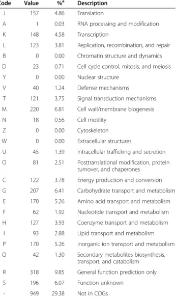
Genome properties
The genome comprised a circular chromosome with a
length of 3,792,640 bp and 33.25% G + C content (Figure 5
and Table 4). It is composed of 56 contigs. Of the 3,282
predicted genes, 3,230 were protein-coding genes and 52
were RNA genes (2 rRNA genes and 50 tRNA genes). The
sequencing coverage of rRNA operon (673×) indicated
that 4 copies of rRNA operons are exist in this genome.
The majority of the protein-coding genes (2,054 genes,
62.58%) were assigned putative functions, while the
remaining genes were annotated as hypothetical proteins
(1,176 genes, 35.83%). The properties of and statistics for
the genome are summarized in Table 4. The distribution
of genes into COG functional categories is presented in
Table 5 and Figure 5.
Conclusions
Based on the results from phylogenetic and phenotypic
analyses, we formally propose the creation of the new
spe-cies Flavobacterium seoulense sp. nov. for strain EM1321
T.
The non-contiguous genome sequence of the type strain
was determined and described here.
Description of Flavobacterium seoulense sp. nov.
Flavobacterium seoulense
(seo.ul.en’se. N.L. neut. adj.,
named after Seoul, Korea, the geographical origin of the
type strain).
Aerobic, Gram-reaction negative. Cells are rod shaped
and motile by gliding. Does not have a flagellum. The
colonies are yellow in color and translucent on R2A agar
medium. Grows at 4–35°C, with optimum growth at 30°C
and in 0–4% (w/v) NaCl. Catalase- and oxidase-positive.
Positive for alkaline phosphatase, esterase (C4), esterase
lipase
(C8),
leucine
arylamidase, acid
phosphatase,
naphthol-AS-BI-phosphohydrolase,
β-galactosidase, and
valine arylamidase. Positive for nitrate reduction, but
negative for indole production, glucose fermentation,
ar-ginine dihydrolase, urease activity, and aesculin and gelatin
Table 3 Genome sequencing project information
MIGS ID Property Term
MIGS-31 Finishing quality High-quality draft
MIGS-28 Libraries used One paired-end Illumina library MIGS-29 Sequencing platforms Illumina MiSeq
MIGS-31.2 Fold coverage 166×
MIGS-30 Assemblers CLCbio CLC Genomics Workbench, version 6.5.1 MIGS-32 Gene calling method Glimmer 3.0
Genbank ID JNCA00000000.1 Genbank Date of Release 2014/05/27 BIOPROJECT PRJNA248341
Project relevance Environmental, Biotechnological MIGS-13 Source Material Identifier KACC 18114, JCM 30145
Figure 5 Graphical circular map of the genome. Starting from the outmost circle and moving inwards, each ring of the circle contains information on a genome: rRNA/tRNA, genes on the reverse strand (colored according to the COG categories), genes on the forward strand (colored according to the COG categories), GC skew, and GC ratio.
Table 4 Genome statistics
Attribute Value % of totala Genome size (bp) 3,792,640 100 DNA coding region (bp) 3,386,688 89.30 G + C content (bp) 1,261,070 33.25 Total genes 3,282 100 RNA genes 52 1.58 rRNA operons 4 -Protein-coding genes 3,230 98.42 Pseudo genes 45 1.37 Genes with function prediction 2,054 62.58 Genes assigned to COGs 2,281 69.50 Genes assigned Pfam domains 1,997 60.85 Genes with signal peptides 119 3.63 Genes with transmembrane helices 682 20.78 CRISPR repeats 0
-a
The total is based on either the size of the genome in base pairs or the total number of protein-coding genes in the annotated genome.
Shin et al. Standards in Genomic Sciences 2014, 9:34 Page 6 of 8
hydrolysis. Negative for lipase, trypsin,
chymotrypsin,
α-galactosidase,
β-glucuronidase, α-glucosidase,
β-glucosi-dase, N-acetyl-β-glucosaminiβ-glucosi-dase, or cystine arylamidase
activity. This strain assimilated d-glucose and l-arabinose,
but not d-mannitol, d-mannose, d-maltose,
N-acetylgluco-samine, potassium gluconate, capric acid, adipic acid,
malic acid, trisodium citrate, or phenylacetic acid.
Pro-duces acid from l-arabinose, xylose, galactose,
d-glucose, d-fructose, d-mannose, and d-lactose.
The G + C content of the genome is 33.25%. The 16S
rRNA and genome sequences are deposited in GenBank
under accession numbers KJ461685 and JNCA00000000.1,
respectively. The type strain EM1321
T(= KACC 18114
T=
JCM 30145
T) was isolated from stream water in Bukhansan
National Park, Seoul, Korea.
Competing interests
The authors declare that they have no competing interests.
Authors’ contributions
SS drafted the manuscript, performed laboratory experiments, and analyzed the data. HG cultured samples and performed the electron micrograph and phylogenetic analysis. YC, SK and DY sequenced, assembled, and annotated the genome. HY organized the study and drafted the manuscript. All authors read and approved the final manuscript.
Acknowledgements
This work was supported by the Basic Science Research Programs through the National Research Foundation of Korea (NRF) funded by the Ministry of Science, ICT, and Future Planning (NRF-2013R1A1A3010041) and supported by a Korea University Grant.
Author details
1
Department of Public Health Sciences, BK21PLUS Program in Embodiment: Health-Society Interaction, Graduate School, Korea University, Seoul, Republic of Korea.2School of Biosystem and Biomedical Science, Korea University, Seoul, Republic of Korea.3Chunlab, Inc., Seoul, Republic of Korea.4Department of
Laboratory Medicine and Research Institute of Bacterial Resistance, Yonsei University College of Medicine, Seoul, Republic of Korea.5Korea University Guro
Hospital, Korea University, Seoul, Republic of Korea.
Received: 8 July 2014 Accepted: 24 November 2014 Published: 29 December 2014
References
1. Bergey DH, Harrison FC, Breed RS WHB, Huntoon FM. Bergey’s Manual of Determinative Bacteriology, Volume 4. 1st ed. Baltimore: The Williams and Wilkins Co; 1923: p. 1–442.
2. Skerman VBD, Mcgowan V, Sneath PHA. Approved lists of bacterial names. Int J Syst Bacteriol. 1980; 30:225–420.
3. Bernardet JF, Segers P, Vancanneyt M, Berthe F, Kersters K, Vandamme P. Cutting a gordian knot: Emended classification and description of the genus Flavobacterium, emended description of the family
Flavobacteriaceae, and proposal of Flavobacterium hydatis nom nov (basonym, Cytophaga aquatilis Strohl and Tait 1978). Int J Syst Bacteriol. 1996; 46:128–48.
4. Kim BY, Weon HY, Cousin S, Yoo SH, Kwon SW, Go SJ, Stackebrandt E. Flavobacterium daejeonense sp. nov. and Flavobacterium suncheonense sp. nov., isolated from greenhouse soils in Korea. Int J Syst Evol Microbiol. 2006; 56:1645–49.
5. Yoon JH, Kang SJ, Oh TK. Flavobacterium soli sp nov., isolated from soil. Int J Syst Evol Microbiol. 2006; 56:997–1000.
6. Tamaki H, Hanada S, Kamagata Y, Nakamura K, Nomura N, Nakano K, Matsumura M. Flavobacterium limicola sp. nov., a psychrophilic, organic-polymer-degrading bacterium isolated from freshwater sediments. Int J Syst Evol Microbiol. 2003; 53:519–26.
7. Kim JH, Kim KY, Cha CJ. Flavobacterium chungangense sp. nov., isolated from a freshwater lake. Int J Syst Evol Microbiol. 2009; 59:1754–58. 8. Nupur Bhumika V, Srinivas TN, Kumar PA. Flavobacterium nitratireducens
sp. nov., an amylolytic bacterium of the family Flavobacteriaceae isolated from coastal surface seawater. Int J Syst Evol Microbiol. 2013; 63:2490–96. 9. NamesforLife, LLC. http://doi.namesforlife.com/10.1601/tx.8071. 10. Kim OS, Cho YJ, Lee K, Yoon SH, Kim M, Na H, Park SC, Jeon YS, Lee JH,
Yi H, Won S, Chun J. Introducing EzTaxon-e: a prokaryotic 16S rRNA gene sequence database with phylotypes that represent uncultured species. Int J Syst Evol Microbiol. 2012; 62:716–21.
11. Stackebrandt E, Ebers J. Taxonomic parameters revisited: tarnished gold standards. Microbiol Today. 2006; 33:152–55.
12. Jeon YS, Chung H, Park S, Hur I, Lee JH, Chun J. jPHYDIT: a JAVA-based integrated environment for molecular phylogeny of ribosomal RNA sequences. Bioinformatics. 2005; 21:3171–713.
13. Tamura K, Peterson D, Peterson N, Stecher G, Nei M, Kumar S. MEGA5: molecular evolutionary genetics analysis using maximum likelihood, evolutionary distance, and maximum parsimony methods. Mol Biol Evol. 2011; 28:2731–39.
14. Field D, Garrity G, Gray T, Morrison N, Selengut J, Sterk P, Tatusova T, Thomson N, Allen MJ, Angiuoli SV, Ashburner M, Axelrod N, Baldauf S,
Table 5 Number of genes associated with the 25 general
COG functional categories
Code Value %a Description
J 157 4.86 Translation
A 1 0.03 RNA processing and modification K 148 4.58 Transcription
L 123 3.81 Replication, recombination, and repair B 0 0.00 Chromatin structure and dynamics D 23 0.71 Cell cycle control, mitosis, and meiosis Y 0 0.00 Nuclear structure
V 40 1.24 Defense mechanisms
T 121 3.75 Signal transduction mechanisms M 220 6.81 Cell wall/membrane biogenesis N 18 0.56 Cell motility
Z 0 0.00 Cytoskeleton W 0 0.00 Extracellular structures
U 45 1.39 Intracellular trafficking and secretion O 81 2.51 Posttranslational modification, protein
turnover, and chaperones C 122 3.78 Energy production and conversion G 207 6.41 Carbohydrate transport and metabolism E 170 5.26 Amino acid transport and metabolism F 62 1.92 Nucleotide transport and metabolism H 127 3.93 Coenzyme transport and metabolism
I 93 2.88 Lipid transport and metabolism P 170 5.26 Inorganic ion transport and metabolism Q 42 1.30 Secondary metabolites biosynthesis,
transport, and catabolism R 318 9.85 General function prediction only S 196 6.07 Function unknown
- 949 29.38 Not in COGs
a
The total is based on the total number of protein coding genes in the annotated genome.
Ballard S, Boore J, Cochrane G, Cole J, Dawyndt P, De Vos P, dePamphilis C, Edwards R, Faruque N, Feldman R, Gilbert J, Gilna P, Glockner FO, Goldstein P, Guralnick R, Haft D, Hancock D, et al. The minimum information about a genome sequence (MIGS) specification. Nat Biotechnol. 2008; 26:541–47. 15. Woese CR, Kandler O, Wheelis ML. Towards a natural system of organisms:
proposal for the domains Archaea, Bacteria, and Eucarya. Proc Natl Acad Sci USA. 1990; 87:4576–79.
16. Krieg NR, Ludwig W, Euzéby J, Whitman WB, Phylum XIV. Bacteroidetes phyl. nov. In: Krieg NR, Staley JT, Brown DR, Hedlund BP, Paster BJ, Ward NL, Ludwig W, Whitman WB, editors. Bergey’s Manual of Systematic Bacteriology, Volume 4. 2nd ed. New York: Springer; 2011: p. 25.
17. Euzéby JP. Validation List No. 143. List of new names and new combinations previously effectively, but not validly, published. Int J Syst Evol Microbiol. 2012; 62:1–4.
18. Bernardet JF, Order I. Flavobacteriales ord. nov. In: Krieg NR, Staley JT, Brown DR, Hedlund BP, Paste RBJ, Ward NL, Ludwig W, Whitman WB, editors. Bergey’s Manual of Systematic Bacteriology, Volume 4. 2nd ed. New York: Springer; 2011: p. 105.
19. Bernardet JF, Family I. Flavobacteriaceae. In: Krieg NR, Staley JT, Brown DR, Hedlund BP, Paste RBJ, Ward NL, Ludwig W, Whitman WB, editors. Bergey’s Manual of Systematic Bacteriology, Volume 4. 2nd ed. New York: Springer; 2011: p. 106–11.
20. Bernardet JF, Nakagawa Y, Holmes B. Proposed minimal standards for describing new taxa of the family Flavobacteriaceae and emended description of the family. Int J Syst Evol Microbiol. 2002; 52:1049–70. 21. List Editor. Validation List No. 41. Validation of publication of new names
and new combinations previously effectively published outside the IJSEM. Int J Syst Bacteriol. 1992; 42:327–28.
22. Holmes B, Owen RJ. Proposal that Flavobacterium breve be substituted as the type species of the genus in place of Flavobacterium aquatile and emended description of the genus Flavobacterium: status of the named species of Flavobacterium. Request for an Opinion. Int J Syst Bacteriol. 1979; 29:416–26.
23. Ashburner M, Ball CA, Blake JA, Botstein D, Butler H, Cherry JM, Davis AP, Dolinski K, Dwight SS, Eppig JT, Harris MA, Hill DP, Issel-Tarver L, Kasarskis A, Lewis S, Matese JC, Richardson JE, Ringwald M, Rubin GM, Sherlock G. Gene ontology: tool for the unification of biology. The Gene Ontology Consortium. Nat Genet. 2000; 25:25–9.
24. Seng P, Drancourt M, Gouriet F, La Scola B, Fournier PE, Rolain JM, Raoult D. Ongoing revolution in bacteriology: routine identification of bacteria by matrix-assisted laser desorption ionization time-of-flight mass spectrometry. Clin Infect Dis. 2009; 49:543–51.
25. Aziz RK, Bartels D, Best AA, DeJongh M, Disz T, Edwards RA, Formsma K, Gerdes S, Glass EM, Kubal M, Meyer F, Olsen GJ, Olson R, Osterman AL, Overbeek RA, McNeil LK, Paarmann D, Paczian T, Parrello B, Pusch GD, Reich C, Stevens R, Vassieva O, Vonstein V, Wilke A, Zagnitko O. The RAST Server: rapid annotations using subsystems technology. BMC Genomics. 2008; 9:75. 26. Delcher AL, Bratke KA, Powers EC, Salzberg SL. Identifying bacterial genes
and endosymbiont DNA with Glimmer. Bioinformatics. 2007; 23:673–79. 27. Overbeek R, Begley T, Butler RM, Choudhuri JV, Chuang HY, Cohoon M, de
Crecy-Lagard V, Diaz N, Disz T, Edwards R, Fonstein M, Frank ED, Gerdes S, Glass EM, Goesmann A, Hanson A, Iwata-Reuyl D, Jensen R, Jamshidi N, Krause L, Kubal M, Larsen N, Linke B, McHardy AC, Meyer F, Neuweger H, Olsen G, Olson R, Osterman A, Portnoy V, et al. The subsystems approach to genome annotation and its use in the project to annotate 1000 genomes. Nucleic Acids Res. 2005; 33:5691–702.
28. Lagesen K, Hallin P, Rodland EA, Staerfeldt HH, Rognes T, Ussery DW. RNAmmer: consistent and rapid annotation of ribosomal RNA genes. Nucleic Acids Res. 2007; 35:3100–08.
29. Lowe TM, Eddy SR. tRNAscan-SE: a program for improved detection of transfer RNA genes in genomic sequence. Nucleic Acids Res. 1997; 25:955–64. 30. Bland C, Ramsey TL, Sabree F, Lowe M, Brown K, Kyrpides NC, Hugenholtz P.
CRISPR recognition tool (CRT): a tool for automatic detection of clustered regularly interspaced palindromic repeats. BMC Bioinformatics. 2007; 8:209.
doi:10.1186/1944-3277-9-34
Cite this article as: Shin et al.: Non-contiguous finished genome sequence and description of the gliding bacterium Flavobacterium seoulense sp. nov.. Standards in Genomic Sciences 2014 9:34.
Submit your next manuscript to BioMed Central
and take full advantage of:
• Convenient online submission • Thorough peer review
• No space constraints or color figure charges • Immediate publication on acceptance
• Inclusion in PubMed, CAS, Scopus and Google Scholar • Research which is freely available for redistribution
Submit your manuscript at www.biomedcentral.com/submit
Shin et al. Standards in Genomic Sciences 2014, 9:34 Page 8 of 8
![Table 1 Classification and general features of Flavobacterium seoulense EM1321 T according to the MICG recommendations [14]](https://thumb-ap.123doks.com/thumbv2/123dokinfo/5096693.78108/2.892.89.808.163.691/table-classification-general-features-flavobacterium-seoulense-according-recommendations.webp)