혈액암
세포주(HL-60)에서 제주조릿대
(Sasa quelpaertensis Nakai) 잎
추출물의
세포사멸 유도 활성
지도교수
김 세 재
장
미 경
이
논문을 이학 석사학위 논문으로 제출함
2006년 12월
장미경의
이학 석사학위 논문을 인준함
심사위원장
_____________________
○
印
위
원 _____________________
○
印
위
원 _____________________
○
印
제주대학교
대학원
2006년 12월
Apototic activity of the leaf extract of
Sasa quelpaertensis Nakai in acute leukemia
Mi-Gyeong Jang
(Supervised by Professor Se-Jae Kim)
A thesis submitted in partial fulfillment of the requirement for the degree of
Master of Science
2006 . 12 .
This thesis has been examined and approved.
________________________________________________
Thesis director, Sun-Ryung Lee of life science
________________________________________________
________________________________________________
(Name and signature)
DEPARTMENT OF LIFE SCIENCE
GRADUATE SCHOOL
i
Abstract
Sasa quelpaertensis Nakai is a native plant, which is distributed widely
throughout Halla mountain of JeJu Island. In order to evaluate the utilizing possibility as bioresource in human health, the antioxidant activities and cell growth inhibitory activity in HL-60 cells were investigated using methanol extract and its fractions prepared from S. quelpaertensis leaf. The ethylacetate
(EtOAc) fractions showed the strongest antioxidant activities (DPPH scavenging activity, IC50 = 288 ug/ml; nitric oxide scavenging activity, IC50 = 259 ug/ml; and
superoxide scavenging activity and xanthine oxidase inhibitory activities, IC50 =
21.9, 32.4 ug/ml) comparing with those of other fractions. Also, EtOAc fraction showed high inhibitory effect (IC50 = 57 ug/ml) on the growth of HL-60 cells.
The characteristic apoptotic symptoms, such as the nuclear condensation, DNA fragmentation and the accumulated Sub-G1 DNA content, an exposure of phosphatidylserine from the inner cell membrane to the outer cell membrane and changes in the levels of apoptosis-related proteins expression were confimed in EtOAc fraction-treated cells. These results strongly demonstrate that EtOAc fraction of S. quelpaertensis leaf induce the apoptosis in human
leukemia cells.
ii
목 차
목 차 ··· ABSTRACT ··· List of Tables ··· List of Figures ··· Ⅰ. 서 론 ··· Ⅱ. 재료 및 방법 ··· 1. 시료의 준비 ··· 2. 시료의 추출 ··· 3. 항산화 활성 측정 ··· 3-1. DPPH 자유유리기 소거 활성 ··· 3-2. Nitric oxide 소거 활성 ··· 3-3. Xanthine oxidase 억제 및 superoxide 소거 활성 ··· 4. 세포 배양 ··· 5. 세포증식 억제활성 측정 ··· 6. 핵의 형태학적 변화 관찰 ··· 7. DNA 분절화 관찰 ··· 8. Flow cytometry 분석 ··· 9. Western blot ··· 10. 통계처리 ··· Ⅲ. 결 과 ··· ⅰ ⅲ ⅳ ⅴ 1 4 4 4 4 5 5 6 6 6 7 7 7 8 9 10iii
1. 제주조릿대 잎 추출물의 항산화 활성 ··· 1) DPPH 자유 유리기 소거 활성 ··· 2) NO 소거 활성 ··· 3) Xanthine oxidase 억제 및 superoxide 소거 활성 ··· 2. HL-60 세포에서 제주조릿대 잎 추출물(EtOAc 분획물)의 세포사멸 유도 활성 ··· 1) 세포 증식억제 활성 ··· 2) 핵의 형태학적 변화 및 DNA 절편화 효과 ··· 3) Flow cytometry 분석 ··· 4) 세포사멸 관련 단백질 발현 변화 ··· Ⅳ. 고 찰 ··· Ⅴ. 요 약 ··· Ⅵ. 참고문헌 ··· 10 10 10 10 18 18 18 18 19 27 32 33
iv
List of Tables
Table 1. Antioxidant activities of the methanol extract and its various fractions from Sasa quelplaertensis leaf ··· 17
v
List of figures
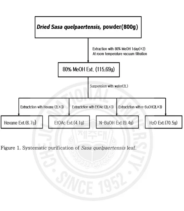
Figure 1. Systematic purification of Sasa quelpaertensis leaf ···
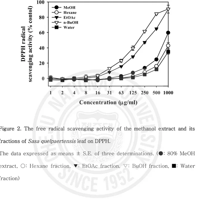
Figure 2. The free radical scavenging activity of the methanol extract and its fractions of Sasa quelpaertensis leaf on DPPH ···
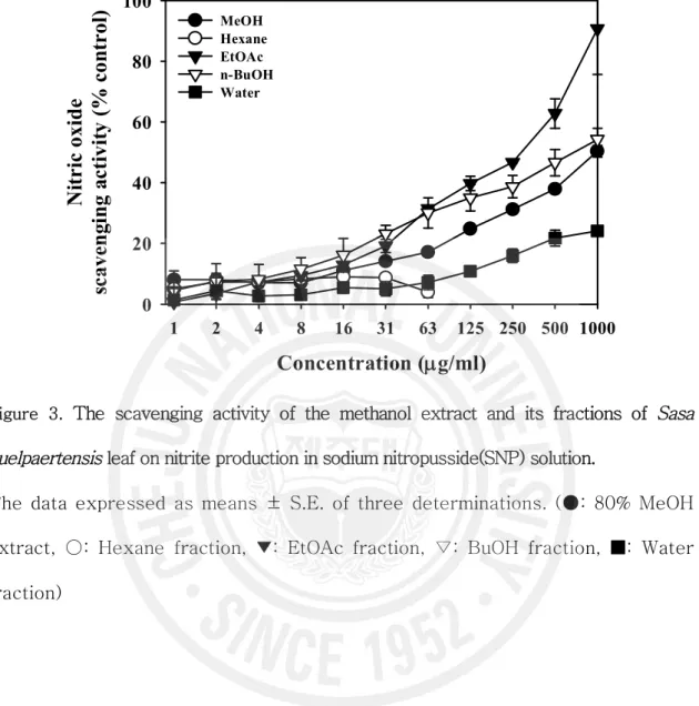
Figure 3. The scavenging activity of the methanol extract and its fractions of Sasa
quelpaertensis leaf on nitrite production in sodium nitropusside(SNP)
solution ···
Figure 4. Xanthine oxidase inhibitory activities of the methanol extract its various fractions of Sasa quelpaertensis leaf ···
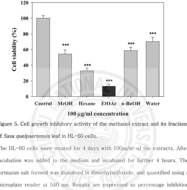
Figure 5. Cell growth inhibitory activity of the methanol extract and its fractions of Sasa quelpaertensis leaf in HL-60 cells ···
Figure 6. Cell growth inhibitory activity of the EtOAc fraction of Sasa quelpaertensis
leaf in HL-60 and Hs-68 cells ···
Figure 7. Morphological features of HL-60 cells after a treatment with the EtOAc fraction of Sasa quelpaertensis leaf ···
12 13 14 16 20 21 22
vi
Figure 8. DNA fragment by the methanol extract and the EtOAc fraction of Sasa
quelpaertensis leaf in HL-60 cells ···
Figure 9. Cell cycle analysis in EtOAc fraction – treated cells ···
Figure 10. Apoptosis was also studied by annexin-V-FITC and PI double staning ···
Figure 11. The expression patterns of apoptosis-related proteins in Hl-60 cells ··· 23
24
25
1
Ⅰ. 서 론
암으로 인한 한국인의 사망률은 전체사망자의 25.6%로 사망원인 1 위로 나타나고 있다 (통계청, 2003). 암은 식생활 습관의 변화와 평균수명의 연장으로 증가추세에 있으며, 이러한 암의 치료제를 개발하기 위한 많은 보고들이 발표되고 있다(Baguley 등, 1981; Boyd 와 Nash, 1989; Geran 등, 1975; Thayer 등, 1971). 현재 고도의 경제성장에 따른 국민소득의 수준 향상이 삶의 질 향상이라는 웰빙 욕구로 이어지면서 각종 질병에 대한 치료제 및 예방제 또는 건강보조제로서 천연식물자원에 대한 국민들이 관심도는 급증하고 있다. 또한, 천연물에 대한 항암 및 여러 생리활성 기능들이 밝혀지고, 이러한 천연물에서 추출된 생리활성물질은 인공 합성물질에 비해 부작용이 적고, 인체 내에서 안전성이 높다고 보고되면서 천연물을 이용한 생리활성 물질을 탐색하는 연구는 더욱 더 활발히 진행되고 있다 (남, 1995). 현재 사용되고 있는 대부분의 항암제들은 궁극적으로 세포의 DNA 에 직접적인 손상을 주거나 혹은 암세포의 성장시 DNA 의 정상적 형성에 이상을 초래함 으로써 암세포의 사멸을 유도한다. 그러나 이러한 기전을 바탕으로 개발된 항암제는 빠른 시간 내에 내성을 유발한다는 심각한 문제점을 가지고 있었다. 따라서 최근에는 세계적으로 기존의 항암제에서 나타난 문제점을 보완하여 암세포의 세포주기 조절 억제와 세포 사멸 유도를 통해 암세포를 괴사 시킬 수 있는 새로운 항암 물질을 찾는데 초점이 맞춰지고 있다 (채와 최, 2006).
세포사멸(apoptosis)은 세포예정사(programmed cell death)로서, 다세포 생물의 발생과 분화 과정에서 세포내부의 프로그램화된 신호를 따라 여러 유전자들이 발현 됨으로써 일어나는 조절이 가능한 능동적 세포의 죽음 과정이며, 불필요해진 세포 또는 원하지 않는 세포를 제거하는 정상적인 생리과정이다. 또한, 생체의 발생 및
2 항상성 유지에 매우 중요하게 작용 한다(Thompson, 1995; Wyllie 등, 1980). 그리 고, 형태적으로 세포의 비중감소, 세포막의 변형, 염색체의 응축, 활성화된 세포내 부의 물질들이 사멸체(apoptotic body)라는 포낭을 형성하면서 식세포작용을 거치 는 작용과 함께 진행되며, 생화학적으로는 DNA 분절이 일어나고, 원형질막의 phosphatidylserine의 외부유출, 세포내 Ca2+ 농도의 증가, caspase로 알려진 시스 테인 프로테아제의 활성등으로 특징 지워진다(Custodilo 등, 2001; Duvall와 Wyllie, 1985; Shrivastava 등, 2000). 조릿대류는 식물분류학적으로 벼과(Gramineae), 대나무아과(Bambusoideae)의 조릿대속(Sasa)에 속하는 식물군으로서, 현재 전 세계적으로 대나무아과에 6속 250종이 알려져 있다 (이, 1980). 조릿대는 키 작은 대나무 식물로 산죽, 지죽, 조 죽, 입죽, 죽엽 또는 담죽엽, 임하죽, 토맥이라고도 불리우며, 우리나라 중부 이남 지방과 일본, 중국이 속하는 극동아시아 지역에서 서식한다(이, 1980). 동의보감, 본 초강목, 신농본초경에 따르면 대나무는 중풍, 고혈압을 치료하는데 탁월한 효과가 있고, 특히 대나무 잎은 해열, 거담, 청량 등의 목적으로 폐렴, 기관지염 등에 사용 되었다고 기록되어 있다. 또한, 대나무아과에 속하는 조릿대는 황달, 암, 고열, 위 및 십이지장염, 고혈압, 당뇨병, 습진, 해열 등의 증세를 완화시키는 효과가 있는 것으로 알려져 민간 약재로 사용되고 있다(Lee 와 Moon, 2003; 윤 등, 2000). 현 재 대나무에는 orientin, homoorientin, vitexin, isovitexin, naringin-7-rhamnoglucoside, quercetin, leteolin, rutin, tricin, caffeic acid, chlorogenic acid, p-hydroxy coumaric acid와 같은 flavone 배당체, cinnamic acid 유도체 그리고, coumaric lactone 등의 화합물들이 존재한다고 알려져 있다(Zhang, 1995; Zhang, 2002). 대나무류의 생리활성에 관한 연구들(Baek 등, 2002; Kim, 1996; Kim 등, 2001; Kim 등, 2001; Kweon 등, 2001; Michiko, 1990; Zang 과 Wu. 2002)은 성 분분석과 항균활성에 관한 연구가 대부분을 차지하고 있다. 그리고, 조릿대에 관한
3
연구는 조릿대(Sasa borealis Makino)의 약리학적 연구(임, 1984)와 조릿대(Sasa
borealis) 잎의 Flavone 성분분리(윤, 2000) 등이 보고된 바 있지만, 제주조릿대에 관한 연구는 생태학적 연구(Kim, 1996)를 제외하고는 거의 전무한 실정이다. 현재 제주조릿대는 한라산 중산간 지역을 중심으로 기하급수적으로 늘어나고 있으며 더 욱이 한라산의 자연생태계 흐름을 파괴하는 주범으로도 인식되고 있다. 따라서, 본 연구에서는 제주도에만 서식한다는 지역특성과 항산화 활성 및 여러 약리작용에 효 과를 가지고 있는 것으로 보고되어 있는 제주조릿대를 이용하여 항암활성을 규명하 여 제주도의 자원활용화 및 항암 질병치료제로서의 가능성을 모색하는 데 목적을 두고 수행하였다.
4
Ⅱ. 재료 및 방법
1. 시료의 준비 제주조릿대는 2005 년 4 월, 한라산 해발 717m 인 물찻오름 지역에 분포한 조릿대 중 일년생 잎만을 분별하여 채취하였다. 채집된 시료는 동결건조기를 사용하여 완전히 건조한 후 파쇄기로 미세하게 파쇄시킨 후 냉동보관 (-20℃)하여 시료로 사용 하였 다. 2. 시료의 추출 제주조릿대 잎 건조물 800g을 80% 메탄올(MeOH) 20L를 가하여 상온에서 24시 간 동안 추출하여 여과 (90 mm, ADVENTEC)한 후, 상층액을 회수하였다. 이 과정 을 2회 반복하여 얻어진 상층액을 모아 감압 농축하여 조릿대 메탄올 추출물 115.69g을 수득하였다. 그리고 조릿대 메탄올 추출물 115.69g을 증류수 2L에 현 탁시킨 후에, 각각 핵산(Hexane, 2L × 3), 에틸아세테이트(EtOAc, 2L × 3), 부탄 올(n-BuOH, 2L × 3), 물(H2O) 순으로 극성이 낮은 용매부터 극성이 높은 용매 순 으로 순차적으로 용매분획하여 4개의 분획물을 얻었고, 각각 진공 건조하여 시료로 사용하였다(Figure 1). 시료는 PBS (phosphate buffered saline)와 에탄올(EtOH)용 매에 녹여 실험에 사용하였고, 에틸아세테이트 분획물은 100% 에탄올에 녹여 실험 에 사용하였다.3. 항산화 활성 측정
5
전자공여능 측정은 Blosis 방법(Blois, 1958)에 의한 DPPH 자유 라디칼 소거법 에 따라 측정하였다. 즉, 메탄올에 녹인 시료를 96 well plate에 100㎕씩 분주하고 0.4mM DPPH (1.1-diphenyl-2-picrylhydrazyl)용액을 동량 첨가하여 실온에서 10 분간 방치한 후 517 ㎚에서 흡광도를 측정하였고, 대조군으로는 butylated hydroxy anisole (BHA)과 trolox를 사용하였다. DPPH 라디칼 소거활성은 아래의 식으로부터 산출하였고 DPPH의 흡광도가 50% 감소할 때 나타나는 시료의 농도 (IC50)로 표시하였으며, 각 시료는 3회 반복 실험을 하여 평균값을 구하였다.
DPPH radical 소거활성(%) = {(Acontrol - Asampl)/Acontrol}× 100
Asample = 시료를 첨가한 반응액의 흡광도
Acontrol = 시료대신 메탄올을 첨가한 반응액의 흡광도
3-2. Nitric oxide 소거 활성
자연적으로 nitric oxide를 생성하는 물질인 sodium nitroprusside (SNP)를 사용 하여 nitric oxide 소거 활성을 분석하였다(Green 등, 1982; Marcocci L 등, 1994). 10 mM SNP 용액 200 ㎕에 시료를 농도별로 첨가하고 25℃에서 3시간 동안 반응 시켰다. 반응이 끝난 후 반응액과 동일한 양의 Griess시약 [1% (w/v) sulfanilamide, 0.1% (w/v) naphylethylenediamine를 포함하는 2.5% (v/v) phosphoric acid]을 첨 가하였다. 실온에서 10분 동안 반응시킨 후 540 nm에서 흡광도를 측정하여 잔존하 는 아질산염(nitrite)의 양으로 nitric oxide 소거활성을 산출하였다. Nitric oxide 소 거활성은 흡광도가 50% 감소할 때 나타나는 시료의 농도(IC50)로 표시하였다.
6
Xanthine/xanthine oxidase에 의한 uric acid생성은 290 nm에서 증가된 흡광도 에 의해 측정하였고(Noro등, 1983), superoxide의 양은 nitroblue tetrazolium (NBT) 환원방법에 의해 측정하였다(Cheng 등, 1998). 반응액은 각 시료와 0.5 mM xanthine과 1 mM EDTA를 200 mM phosphate buffer (pH 7.5) 100 ㎕에서 준비하 였고, 50 mU/ml xanthine oxidase를 첨가하여 uric acid의 생성을 유도하였다. Superoxide 소거활성은 위 반응액에 0.5 mM NBT를 첨가하여 반응하였다. Xanthine oxidase 억제 및 superoxide 소거 활성은 각각 생성된 uric acid와 superoxide의 흡광도가 50% 감소할 때 나타나는 시료의 농도(IC50)로 표시하였다.
4. 세포 배양
급성 전골수성 백혈병 세포주인HL-60 (human promyelocytic leukemia) 세포주와 피부 섬유아세포주인 Hs-68 (skin fibroblast) 세포주는 한국 세포주은행(KCLB)으 로부터 분양 받았다. 세포는 100 units/ml의 penicillin 과 100 ug/ml streptomycin (GIBCO Inc, USA), 10 %의 fetal bovine serum (FBS, GIBCO Inc, USA)가 함유된 RPMI 1640 배지와 DMEM (GIBCO Inc, USA)배지를 사용하여 37℃, 5% CO2 항온 기에서 배양하였다.
5. 세포증식 억제 활성 측정
제주조릿대 추출물 처리에 의한 세포증식 억제는 MTT assay를 이용하여 측정하 였다(Carmichael 등, 1987). HL-60 세포 (2.5×10⁵ cells/ml) 와 Hs-68 세포 (1.0
×105 cells/ml)를 96 well plate또는 48 well plate 의 각 well에 분주하여 24시간 배양 후 HL-60 세포에는 제주조릿대 추출물과 분획물을 100 ug/ml의 농도로, Hs-68 세포에는 에틸아세테이트 분획물만 농도별로 처리하였다. 이를 4 일간 배양한 다음, MTT용액(20 ug/ml)을 첨가하고 4시간 동안 반응시켰다. 1000 rpm에서 10분
7
간 원심분리하고 조심스럽게 배지를 제거한 다음, dimethylsulfoxide (DMSO, Sigma) 150 ㎕를 가하여 MTT의 환원에 의해 생성된 formazan 침전물을 용해시 킨 후 microplate reader(BIO-TEK INSTRUMENIS. INC)를 사용하여 540 nm에서 흡광도를 측정하였고, 대조군과 비교하여 성장 억제정도와 세포독성을 조사하였다.
6. 핵의 형태학적 변화 (Morphological changes) 관찰
HL-60 세포 (2.5×10⁵ cells/ml)를 48 시간 동안 배양한 후, DNA 에 특이적으로 결합하는 형광색소인 H33342 (Hoechest 33342, Sigma, USA)용액을 가하여 37℃에서 30 분간 염색한 후 형광현미경 (IX-71, Olympus, Japan)하에서 관찰하였다.
7. DNA 분절화 (fragmentation) 관찰
배양된 세포를 수집한 후 PBS (Sigma, USA)용액으로 2 회 세척한 후, Promega Wizard ®Genomic DNA Purification Kit (Promega, USA)를 사용하여 DNA 를 분리 하 였다. 분리한 DNA 를 1.5 % agarose gel 에서 30 분(100 V)동안 전기영동 한 다음 ethidium bromide 로 염색하고 UV transilluminator (SLB MylmaggerTM) 하에서 DNA 분절화 현상을 관찰 하였다(Purohit 등, 2000).
8. Flow cytometry 분석
DNA 함량을 측정하는 세포주기 분석을 위해 HL-60 세포 (2.5×10⁵ cells/ml)를 48시간 동안 배양하였다. 배양이 끝난 세포는 수확하여 PBS로 세척하였고, -20℃ 에서 70% 에탄올로 30분 동안 고정시킨 후, PBS로 세척하였다. 그 후에 RNase A(1mg/ml)을 30분간 처리한 다음에 propidium iodide (PI: Sigma, USA)로 염색하 여, FACScalibur Flow Cytometer (BD Bioscience)로 분석하였다(Nicoletti 등, 1991; Sherwood 등, 1994). 세포막 성분인 Phosphatidylserine의 분포를 측정하기
8
위해서는Annexin V-binding buffer(10 mM HEPES/NaOH, pH 7.4, 140mM NaCl, 5mM CaCl2 )를 가하여 세포를 현탁한 후, Annexin V-FITC (BD Bioscience)와 propidium iodide (PI)를 5 ug/ml 농도로 첨가하였다. 상온 암실에서 15분간 반응한 후 binding buffer를 400 ㎕ 넣어서 희석하여 FACScalibur Flow Cytometerm (BD Bioscience)로 분석하였다.
9. Western Blot
HL-60 세포 (2.5×10⁵ cells/ml)에 제주조릿대의 에틸아세테이트 분획물을
농도별로 각각 처리 한 후 24 시간 동안 배양하여 세포를 수집하였다. 1 ml 의 RIPA buffer (0.1M PMSF, 0.1M Na3O4V, 0.5M NaF, 5mg/ml aporotinin, 5mg/ml leupeptin)로 lysis 시킨 후 12,000 rpm 에서 20 분간 원심분리 하여 얻어진 상층액을 사용 하였다. 단백질정량은 Bradford 방법을 이용하여 Bio-Rad Protein Assay Kit 를 사용하여 정량 하였다(Bradford 등 1976). 30~50 ㎍의 단백질을 8~15% gel SDS-PAGE (Poly Acrylamide Gel Electrophoresis)로 변성 분리하여, 이를 PVDF membrane (Milipore, Massachusetts, USA)에 200mA 로 2 시간 동안 transfer 하였다. membrane 의 blocking 은 5% skim milk 가 함유된 TTBS (TBS + 0.1% Tween 20)용액을 가하여 상온에서 2 시간 동안 실시하였다. 단백질 발현 분석에 사용된 항체로는 anti-mouse Bcl-2(1: 1000, Santa Cruz Biotech), anti-mouse Bax(1: 1000, Santa Cruz Biotech), anti-mouse procaspase-3(1: 1000, Santa Cruz Biotech)와 anti-rabbit cleave caspase-3(1: 1000, Cell signaling), anti-mouse PARP(1: 1000, Santa Cruz Biotech)를 TTBS 용액에서 희석하여 4℃에서 overnight 한 후 TTBS 로 4 회 세정하였다. 2 차 항체로는 HRP (Horse Radish Peroxidase)가 결합된 anti-mouse 또는 anti-rabbit IgG (Jackson ImmunoResearch Inc. USA )를 1: 5000 으로 희석하여 상온에서 1 시간 반응시킨
9
후, TTBS 로 5 회 세정하여 ECL 기질(Amersham Co.)과 1~3 분간 반응 후 X-ray 필름에 감광하였다.
10. 통계처리
표시된 결과는 3번 이상의 독립적인 실험결과이며, 이들의 평균(mean)과 표준오 차(standard error, SE)를 산출하여 통계적 유의성을 검증하였다.
10
Ⅲ. 결 과
1. 제주조릿대 잎 추출물의 항산화 활성
1) DPPH 자유유리기 소거 활성
DPPH는 안정한 유리기로 cysteine, glutathione과 같은 아미노산과 ascorbic acid, aromatic amine (ρ-phenylenediamine, ρ-aminophenol) 등에 의해 환원되어 탈색된다. 제주조릿대 잎 메탄올 추출물 및 용매 분획물을 시료로 사용하여 DPPH 의 자유유리기 소거 활성을 측정한 결과, IC50 값은 메탄올 추출물과 에틸아세테이
드 분획물, 부탄올 분획물에서 각각 862.5, 288.9 과 166.4 mg/ml을 나타내었다 (Table 1, Figure 2). 이들의 활성은 대조군인 BHA 와 Trolox에 비교할 때 낮은 IC50 값이지만 이 중 부탄올 분획물에서 비교적 높은 자유유리기 소거 활성을 보이 는 것으로 조사되었다. 2) NO 소거 활성 제주조릿대 메탄올 추출물과 각 분획물의 nitric oxide 소거 활성을 산출하였다. Nitric oxide 소거 활성의 IC50 값은 에틸아세테이트 분획물에서만 259.4 mg/ml을 나타내었고, 다른 용매 분획물에서의 NO 소거 활성은 거의 없는 것으로 나타났다 (Figure 3, Table 1).
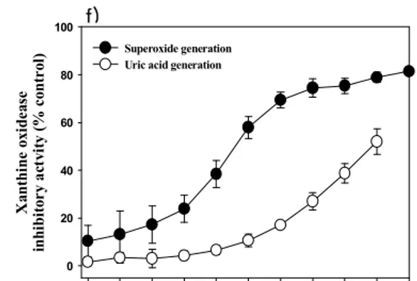
3) Xanthine oxidase 억제 및 superoxide 소거 활성
제주조릿대 메탄올 추출물과 각 분획물의 xanthine oxidase 억제활성은 xanthine oxidase 억제제로 알려진 allopurinol과 superoxide 소거능을 가진
11
superoxide dismutase (SOD)을 양성 대조군으로 사용하여 비교하였으며, 그 결과 는 Figure 4에 나타내었다. Allopurinol은 농도의존적으로 superoxide와 xanthine oxidase 억제활성을 보였다. Allopurinol의 그래프를 기준으로 하여 비교해보면 메 탄올 추출물과 각 분획물 모두에서 superoxide 소거 활성이 나타남을 확인 할수 있었다. 또한 메탄올 추출물과 헥산, 에틸아세테이트, 부탄올 분획물에서 xanthine oxidase 억제활성을 보였고, 특이적으로 물 분획물에서는 superoxide 억제 활성만 을 나타내었다. Table 1에서 보여주는 바와 같이메탄올 추출물과 용매 분획물에 대 한 uric aicd 생성저해와 superoxdie 생성저해 활성은 에틸아세테이트 분획물에서 가장 양호하였다.
12
13
Concentration (
mg/ml)
1 2 4 8 16 31 63 125 250 500 1000D
P
P
H
r
ad
ic
al
sc
av
en
gi
n
g
ac
ti
vi
ty
(
%
c
on
to
l)
0 20 40 60 80 100 MeOH Hexane EtOAc n-BuOH WaterFigure 2. The free radical scavenging activity of the methanol extract and its fractions of Sasa quelpaertensis leaf on DPPH.
The data expressed as means ± S.E. of three determinations. (●: 80% MeOH extract, ○: Hexane fraction, ▼: EtOAc fraction, ▽: BuOH fraction, ■: Water fraction)
14
Concentration (
mg/ml)
1 2 4 8 16 31 63 125 250 500 1000N
it
ri
c
ox
id
e
sc
av
en
gi
n
g
ac
ti
vi
ty
(
%
c
on
tr
ol
)
0 20 40 60 80 100 MeOH Hexane EtOAc n-BuOH WaterFigure 3. The scavenging activity of the methanol extract and its fractions of Sasa
quelpaertensis leaf on nitrite production in sodium nitropusside(SNP) solution.
The data expressed as means ± S.E. of three determinations. (●: 80% MeOH extract, ○: Hexane fraction, ▼: EtOAc fraction, ▽: BuOH fraction, ■: Water fraction)
15 Concentration (mg/ml) 1.0 2.0 3.9 7.8 15.6 31.3 62.5 125.0 250.0 500.0 1000.0 X an th in e ox id ea se in h ib it or y ac tv it y (% c on tr ol ) 0 20 40 60 80 100 Superoxide generation Uric acid generation
d) Concentration (mg/ml) 1.0 2.0 3.9 7.8 15.6 31.3 62.5 125.0 250.0 500.0 1000.0 X an th in e ox id ea se in h ib it or y ac tv it y (% c on tr ol ) 0 20 40 60 80 100 Superoxide generation Uric acid generation
(mg/ml) Concentration (mg/ml) 1.0 2.0 3.9 7.8 15.6 31.3 62.5 125.0 250.0 X an th in e ox id ea se in h ib it or y ac tv it y (% c on tr ol ) 0 20 40 60 80
100 Superoxide generationUric acid generation
Concentration (mg/ml) 1.0 2.0 3.9 7.8 15.6 31.3 62.5 125.0 250.0 500.01000.0 X an th in e ox id ea se in h ib it or y ac tv it y (% c on tr ol ) 0 20 40 60 80 100 Superoxide generation Uric acid generation
a) b)
(mg/ml)
c)
(mg/ml)
16
Figure 4. Xanthine oxidase inhibitory activities of the methanol extract and its various fractions of Sasa quelpaertensis leaf.
The data represent the mean ± S.E. of three determinations. (●: Superoxide generation, ○: Uric acid generation, a) Allopurinol, b) SOD, c) MeOH extract, d) Hexane fraction, e) EtOAc fraction, f) BuOH fraction, g) Water fraction)
Concentration (mg/ml) 1.0 2.0 3.9 7.8 15.6 31.3 62.5 125.0 250.0 500.0 X an th in e ox id ea se in hi bi to ry a ct vi ty ( % c on tr ol ) 0 20 40 60 80 100 120 Superoxide generation Uric acid generation
Concentration (mg/ml) 1.0 2.0 3.9 7.8 15.6 31.3 62.5 125.0 250.0 500.01000.0 X an th in e ox id ea se in hi bi to ry a ct vi ty ( % c on tr ol ) 0 20 40 60 80 100 Superoxide generation Uric acid generation
f) (mg/ml) e) Concentration (mg/ml) 1.0 2.0 3.9 7.8 15.6 31.3 62.5 125.0 250.0 500.01000.0 X an th in e ox id ea se in hi bi to ry a ct vi ty ( % c on tr ol ) 0 20 40 60 80 100 Superoxide generation Uric acid generation
g)
(mg/ml)
17
Table 1. Antioxidant activities of the methanol extract and its various fractions from Sasa quelplaertensis leaf.
IC50 (mg/ml) a) Tretment DPPH radical scavenging activity Nitric oxide scavenging activity Uric acid generation activity Superoxide generation activity Methanol extract 862.5 ± 6.4 * 352.9 ± 16.0 113.5 ± 13.4 Hexane fraction * * 238.4 ± 5.6 62.8 ± 4.3 EtOAc fraction 288.9 ± 12.7 259.4 ± 1.6 32.4 ± 1.6 21.9 ± 5.4 BuOH fraction 166.4 ± 9.4 * 473.5 ± 15.4 23.4 ± 5.9 Water fraction * * - 305.2 ± 6.9 Trolox 3.49 ± 0.3 * 220.3 ± 12.3 32.5 ± 1.9 BHA b) 7.6 ± 0.2 * 801.7 ± 11.7 *
Allopurinol N/A N/A 1.33 ± 0.1 5.1 ± 0.1
SOD c) N/A N/A * 4.9 ± 1.9
a) IC
50 values were calculated from regression lines using five different
concentrations in triplicate experiments. b) Butylated hydroxyl anisole
c)
Superoxide dismutase
N/A: Not assay
* : Can’t calculate the value of IC50
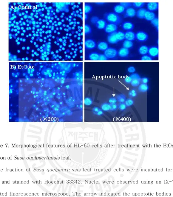
18 2. HL-60 세포에서 제주조릿대 잎 추출물(EtOAc 분획물)의 세포사멸 유도 활성 1) 세포 증식억제 활성 제주조릿대 잎 메탄올 추출물 및 용매 분획물의 세포증식 억제 활성은 MTT의 환원에 의해 생성되는 formazan의 흡광도를 측정하여 분석하였다. HL-60 세포주 에 메탄올 추출물과 용매 분획물을 100 mg/ml 농도로 처리한 결과 메탄올 추출물 은 54.2%, 에틸아세테이트 분획물은 13.2%의 세포증식 억제 활성을 보였다(Figure 5). 가장 높은 세포증식억제 활성을 갖는 에틸아세테이트 분획물의 IC50 값은 57.6 ㎍/㎖을 나타내었다. 그리고 에틸아세테이트 분획물을 혈액암 세포주와 정상세포주 에 처리하여 확인한 결과 혈액암 세포주에서만 농도 의존적으로 세포증식 억제활성 을 보였다(Figure 6). 2) 핵의 형태학적 변화 및 DNA 절편화 효과 HL-60 세포 증식억제 작용이 제주조릿대 잎 용매 분획물에 의한 세포사멸 유도 에 의한 것인지 알아보기 위하여, 세포사멸시 나타나는 여러 현상들을 관찰하였다. 핵의 형태학적 변화를 조사한 결과 대조군에 비해 에틸아세테이트 분획물 처리군에 서 축소된 세포, 불규칙적인 핵의 응집, 사멸체(apoptotic body)들을 관찰할 수 있 었고, 이는 DNA 절편화 현상과 일치하였다(Figure 7, 8). HL-60 세포에 제주조릿 대 추출물과 용매 분획물을 처리하여 48시간 동안 배양한 후 분석한 결과, 대조군 과 제주조릿대 메탄올 추출물에 비해 제주조릿대 에틸아세테이트 분획물에서 DNA 절편화 현상을 뚜렷하게 관찰할 수 있었다(Figure 8). 3) Flow cytometry 분석 HL-60 세포주에 에틸아세테이트 분획물을 농도별로 처리하여 48시간 동안 배양
19
한 후, DNA 함량에 의한 세포주기의 변화를 관찰하였다(Figure 9). 대조군은 apoptotic peak (Sub-G1)가 5.67%로 나타났고, 에틸아세테이드 분획물은 200 ug/ml의 농도에서는 sub-G1이 78%로 증가된 것을 관찰 할 수 있었다. 그러므로, 분획물의 농도 증가에 따라 세포사멸의 지표인 G1 arrest가 증가됨을 확인할 수 있 었다(Figure 9F). 그리고 원형질막의 내층에서 외층으로의 Phosphatidylserine(PS)의 분포를 FITC-Annexin V binding assay로 측정하였다. 에틸아세테이트 분획물을 처리한 HL-60 세포에서 PS의 이동이 관찰된 세포(Annexin V에 양성이고 propidium iodide에 음성)의 비율은 대조군보다 증가 되는 것을 확인하였다(Figure 10).
4) 세포사멸 관련 단백질 발현 변화
미토콘드리아로부터 세포사멸을 조절하는 Bcl-2 와 Bax의 변화를 확인하기 위해 HL-60 세포에 에틸아세테이트 분획물을 농도별로 처리하여 세포사멸을 억제하는 Bcl-2와 세포사멸을 촉진하는 Bax의 발현을 Western blot으로 확인 하였다. 그 결 과 처리한 에틸아세테이드 분획물의 농도가 증가할수록 Bcl-2 변화는 감소하였고, Bax의 변화는 증가되는 경향을 나타내었다(Figure 11). 세포사멸의 실행자인 caspase-3는 불활성화 상태인 32KDa의 procaspase-3 형태로 있다가 17KDa으로 cleavage되면서 활성형으로 전환하게 된다. 제주조릿대 잎의 에틸아세테이트 분획 물의 처리는 procaspase-3의 발현양을 감소시켰고, 이와 반대로 활성화된 caspase-3 발현양은 증가시킴으로 인해 세포사멸이 유도되는 것을 확인하였다. PARP (poly(ADP-ribose) polymerase)는 핵 내에 존재하여 DNA의 손상을 치료하 는 것으로, caspase-3의 활성으로 인해 116kDa 에서 85kDa으로 cleavage가 일어 나게 된다. PARP의 cleavage 변화 또한 에틸아세테이트 분획물의 농도가 증가함에 따라 PARP의 양은 감소한 반면, cleavage 산물은 증가되었다.
20
100
mg/ml concentration
Control MeOH Hexane EtOAc n-BuOH Water
C
el
l v
ia
bi
li
ty
(
%
)
0 20 40 60 80 100 120***
***
***
***
***
Figure 5. Cell growth inhibitory activity of the methanol extract and its fractions of Sasa quelpaertensis leaf in HL-60 cells.
The HL-60 cells were treated for 4 days with 100㎍/㎖ of the extracts. After incubation was added to the medium and incubated for further 4 hours. The formazan salt formed was dissolved in dimethylsulfoxide, and quantified using a microplate reader at 540 nm. Results are expressed as percentage inhibition that produced a reduction in the absorbance in extract-treated cells when compared to the untreated controls. Each value represents mean± S.E. * P < 0.05; **, P < 0.01; ***, P < 0.001 Student’s t- test from three independent experiments.
21
Figure 6. Cell growth inhibitory activity of the EtOAc fraction of Sasa quelpaertensis
leaf in HL-60 and Hs-68 cells.
HL-60 and Hs-68 cells were treated with EtOAc fraction of S. quelpaertensis
leaf at the indicated concetration for 4 days. Cell viability in each treatment was expressed as a percentage of the control. Each value represents mean± S.E. * P < 0.05; **, P < 0.01; ***, P < 0.001 Student’s t- test from three independent experiments. 0 63 33 65 125 250 C el l v ia vi li ty ( % ) 0 20 40 60 80 100 120 HL-60 ** ** *** *** ** control 25 50 100 200 C el l v ia bi lit y (% ) 0 20 40 60 80 100 120 140 Hs-68 * ** ** **
22
Figure 7. Morphological features of HL-60 cells after treatment with the EtOAc fraction of Sasa quelpaertensis leaf.
EtOAc fraction of Sasa quelpaertensis leaf treated cells were incubated for 2
days and stained with Hoechst 33342. Nuclei were observed using an IX-71 inverted fluorescence microscope. The arrow indicated the apoptotic bodies of HL-60 cells that undergoing cell death.
23
Figure 8. DNA fragment by the methanol extract and the EtOAc fraction of Sasa
quelpaertensis leaf in HL-60 cells.
HL-60 cells were treated with methanol extract and its EtOAc fraction at a final concentration of 100㎍/㎖ for 2 days. The DNA was isolated and subjected to 1.5% agarose gel electrophoresis for staining with ethidium bromide. Lane M: 100 bp DNA ladder size maker, Lane 1: control, Lane 2: 80% MeOH extracts, Lane 3: EtOAc fraction.
24
Figure 9. Cell cycle analysis in EtOAc fraction – treated cells.
HL-60 cells were treated with various concentrations with EtOAc fraction of
sasa quelpaerteasis leaf for 2 days. The cells collected and stained with PI, and
then the degree of apoptosis represented as the DNA contents measured by the flow cytometric analysis.
25
Figure 10. Apoptosis was also studied by annexin-V-FITC and PI double staining. Apoptotic cell population with FITC-Annexin V-positive but PI-negative cells increased gradually from less than 2% at control and to more then 42% at final concentration. The EtOAc fraction-treated cells were co-stained with PI and FITC conjugated annexin-V and analyzed wing a FACSCalibur flow cytometer Fluorescence intensities are shown as log scale.
26
Figure 11. The expression patterns of apoptosis- related proteins in HL-60 cellsCells were cultured with EtOAc fraction of for 1 day and whole cell lysates were sybjcted to SDS-PAGE then anylyzed by western blotting.
27
Ⅳ. 고 찰
본 연구는 예전부터 제주지역에서 민간요법 및 구황작물로 이용되어 왔던 제주조 릿대의 생리활성을 조사하기 위한 기초 연구로써 제주조릿대 잎의 메탄올추출물 및 용매 분획물에 대한 항산화 활성과 암세포주 성장억제 활성을 분석하고자 수행되었 다. 또한 암세포주 성장억제 활성이 세포사멸(apoptosis) 유도에 기인한 것인지를 분석하였다. 최근 보고된 대나무과에 속하는 식물의 항산화 활성에 대한 연구에 의하면 에탄 올 추출물 및 그 분획물의 DPPH 자유유리기 소거 활성 및 NO 소거 활성이 비교 적 높은 것으로 보고 되었다(Ju, 2005; Lee, 2003). Lee (2003)는 왕대 에탄올 추출 물이 DPPH 자유유리기 소거 및 NO 소거 활성이 100 mg/ml 에서 50%가 넘는 활 성을 나타내며, 용매별 추출물의 NO 소거 활성은 100 mg/ml 에서 디클로로메탄> 에틸아세테이트> 부탄올> 물> 핵산 순으로 높게 나타난다고 보고하였다. 또한 Ju 등 (2005)의 대나무과 식물의 비교 연구에서 DPPH 자유유리기 소거 활성은 왕대 > 조릿대> 솜대> 맹족죽> 오죽의 순으로 활성을 갖는다고 보고되었다. 그러나 현 재까지 제주조릿대의 항산화활성 탐색에 대한 연구는 보고된 바 없으며, 기존 대나 무과 식물의 항산화 활성 비교도 DPPH 소거활성에 대한 연구 보고가 대부분이다. 이에 본 연구는 제주조릿대의 항산화 활성을 측정하고자 DPPH 자유유리기 활성, NO 소거 활성, xanthine oxidase 억제 및 superoxide 소거 활성을 용매별로 비교 분석하였다. 제주조릿대의 항산화 활성은 메탄올추출물인 경우 왕대추출물보다 낮 은 활성을 갖는 것으로 나타났지만, EtOAc 분획물은 항산화 활성 모두에서 100 mg/ml 농도에서 높은 소거활성을 나타내었다(Figure 2,3,4). 이런 결과는 비록 DPPH 자유유리기 소거 및 NO 소거 활성에서 다른 대나무과 추출물의 활성보다28
유사하거나 낮은 경향을 보이지만, 보고된 문헌의 분석방법이 화학적인 소거활성능 에 의해 분석된 것이므로 세포내에서 역시 높은 항산화 활성을 보인다고는 생각할 수 없다. 따라서 제주조릿대의 잎 추출물은 물론 다른 대나무류 식물의 in vitro 세 포연구, in vivo 동물실험에서의 항산화 활성 효능을 검증할 필요가 있다고 사료된 다. 또한 본 연구에서 수행한 superoxide 소거 및 xanthine oxidase 억제 활성능 에 연구는 다른 대나무류에서의 보고가 없기 때문에 대나무류 식물들과의 직접적인 활성 비교는 할 수 없었다. 나아가 기존 DPPH 및 NO 소거능과의 관련성에 대한 연구도 앞으로 제주조릿대와 다른 대나무과의 비교 분석을 통해 진행되어야 할 것 으로 사료된다. 제주조릿대 잎 에틸아세테이트 추출물이 높은 항산화 활성을 보이기 때문에 혈액 암 세포주인 60 세포에서 항암활성인 세포증식 억제 활성을 측정하였다. HL-60 세포주에 제주조릿대 잎의 메탄올 추출물과 용매 분획물을 100 mg/ml 농도로 처리하였을 때 에틸아세테이트 분획물의 약 13.2%로 가장 높은 세포증식억제 활성 을 나타내었다(Figure 5). 또한, 에틸아세테이트 분획물만을 혈액암 세포주와 정상 세포주에 처리하여 세포증식억제 활성을 확인 한 결과 혈액암세포에서만 농도의존 적으로 세포증식 억제활성을 나타내었고, 정상세포주에서는 세포독성이 없는 것으 로 나타났다(Figure 6). 제주조릿대 잎의 메탄올추출물과 용매 분획물에 의한 세포 증식억제 효과가 세포사멸(apoptosis)의 유도에 의한 것인지 확인하기 위해 세포사 멸에서 가장 특징적인 DNA 분절과 핵의 형태적 변화를 관찰 하였다. 그 결과 제주 조릿대 잎의 메탄올 추출물보다는 에틸아세테이트 분획물에서 DNA 분절 현상을 뚜렷하게 관찰 할 수 있었다. 그리고 핵의 형태적 변화 관찰에서도 정상대조군의 핵의 형태는 정상적이고 둥근 형태이나 에틸아세테이트 분획물을 처리한 세포에서 는 세포의 크기가 축소되고, 핵의 모양이 불규칙하고 부분적으로 핵의 염색질이 응 축된 현상을 관찰 할 수 있었다(Figure 7, 8). 또한, flow cytometry를 이용한 DNA
29 함량과 FITC-Annexin V에 의한 원형질막 내외에서의 PS 변화를 측정 한 결과, 에 틸아세테이트 분획물을 처리한 세포에서 Sub-G1이 농도 의존적으로 증가하였으며, PS 이동에 의해 관찰되는 apopotic 세포도 농도 의존적으로 증가하는 것을 확인하 였다(Figure 9, 10). 이런 결과로서 에틸아세테이트 분획물에 의한 세포증식 억제현 상은 세포괴사(necrosis)에 의한 것 보다는 세포사멸 유도에 의한 것임을 확인하였 다. 그리고 이 결과를 바탕으로 에틸아세테이트 분획물이 세포사멸을 일으키는 기 전을 알아보기 위하여 apoptosis 유도에 관련된 단백질 발현을 분석하였다. 세포사 멸의 주요한 경로는 내재성인 미토콘드리아의 변화에 의한 경로와 외재성인 death receptor에 의한 Fas 리간드와 TNFR family 경로가 있다고 알려져 있으며, 외재 성과 내재성 경로 모두 세포사멸의 최종산물인 caspase-3를 활성화 시키고 cell death를 일으킨다고 알려져 있다(Duane 등 2003). 내재성 세포사멸 경로는 자극에 의해 미토콘드리아 외부에 있던 Bcl-2 family 단백질을 활성화시키고, 미토콘드리 아에서 cytochrome c가 방출된다(Susin 등, 1996). 또한, 세포사멸에서 내재적 경 로는 Bcl-2 의 overexpression에 의해 차단된다. cytochrome c는 Apaf-1 (apoptotic protease activating factor), procaspase-9와 복합체를 형성하여 cytochrome c를 방출하게 되며, Apaf-1는 adaptor 단백질로서 procaspase-9과 procaspase-3의 활성화를 유도한다. 따라서 cytochrome c와 ATP 또는 dATP의 존재하에서 caspase-9이 활성화가 된다(Li 등 1997). 이들 caspase family들의 활 성화를 통해 세포사멸이 유도된다고 알려져 있다. 이때 Bcl-2 는 세포사멸 억제 인 자로 작용하며 cell death의 촉진자인 Bax 나 Bid 와 관련이 있다. 이러한 Bcl-2 family 들은 세포사멸 경로의 다른 수준에서의 death 와 survival 신호, 사이토카인 에 의해 조절이 되며, cytochrome c, AIF (apoptosis inducing factor), ATP 또는 dATP, caspase와 함께 cell death를 촉진하거나 억제 한다고 알려져 있다(Duane 등 2003). 본 연구에서 제주조릿대 에틸아세테이트 분획물을 처리한 HL-60 세포
30 는 농도 의존적인 Bcl-2 단백질 감소와 Bax 단백질의 증가되는 현상을 관찰할 수 있었다. 그러나, 이는 미토콘드리아 경로 중 가장 중요한 Bcl-2 와 Bax의 변화만 을 확인하고 미토콘드리아 경로라고 확신하기는 어렵다. 본 연구에서 결과로 제시 하는 않았지만 caspase-9의 변화 역시 관찰할 수 있었으므로 미토콘드리아 경로를 통해 세포사멸이 유도된다는 것으로 사료되며, 미토콘드리아 경로를 통해 이루어짐 을 확실하게 밝히기 위해서는 추후에 cytchrom-c, Procaspase-9 등의 변화를 검 증할 필요가 있을 것으로 생각된다. 또한, procaspase-3는 단백질 가수분해 효소에 의해 전환되어 caspase-3를 활성화시켜 세포사멸을 유도하고, caspase-3 의 활성 은 caspase-3 기질 중의 하나인 PARP가 단백질 분해 효소에 의해 분절되어 cleavage 되는 것으로 알려져 있다(Ischiropoulos, 1998). 따라서 제주조릿대 에틸 아세테이트 분획물을 처리한 HL-60 세포에서 농도 의존적으로 procaspse-3가 감 소하였고, caspase-3는 분절되며 증가하였다(Figure 11). DNA 손상복구와 세포괴 사의 중개, 면역반응을 조절하는 PARP도 분절되며 증가하는 결과로 볼 때 에틸아 세테이트 분획물에 의해서 세포사멸이 유도되었다고 생각된다. 최근 보고에 의하면
대나무에 많이 함유된 성분에는 flavone glycosides, phenolic acids, coumarin
lactones, anthraquinones 등이 보고된 바 있다(Chen 등, 2002; Li 등 2003; Lu and Liao, 2003; Luo and Chen, 2003;Meng 등, 2002; Zhang and Ding, 1996a, b; Zhou, 1992). 또한, 윤 등(2000)에 의해 조릿대(Sasa borealis (Hackel)Makino)잎
메탄올 추출물을 CH2Cl2 와 EtOAc 분획으로 나눈 후 EtOAc 분획에서 tricin,
tricin 7-o-ß-D-glucopyranoside, luteolin 배당체를 동정한 바 있으며, 이 중 tricin 7-o-ß-D-glucopyranoside, luteolin 배당체는 조릿대속에서 처음 분리된 물 질로 알려져 있다. 따라서 제주조릿대에도 이와 유사한 물질들이 존재할 것으로 생 각되며, 이러한 화합물 중 하나의 화합물이나 혹은 여러 화합물의 조합에 의한 시 너지(synergy)효과에 의해 apoptosis를 유도한다고 사료된다. 따라서, 좀 더 명확
31
한 기전을 밝히기 위해서는 제주조릿대 추출물에서 지표물질을 분리하여 구조를 규 명하고 차후에 이러한 화합물이 apoptosis와 어떠한 상관관계를 유지하며 작용하는 지 확인해 봐야 할 필요성이 있다.
32
Ⅴ. 요 약
제주조릿대는 제주도 한라산 일대에서만 자생하는 지방 고유의 식물이다. 본 연구는 제주조릿대가 유용 생물자원으로 활용가능성을 평가하기 위하여 제주조릿대 잎 메탄올 추출물과 용매 분획물의 항산화 활성과 HL-60 세포의 증식억제 활성을 분석하였다. 제주조릿대 용매 분획물 중에서 에틸아세테이트 분획물이 가장 높은 항산화 활성(DPPH 소거활성 IC50 값, 288 ug/ml; NO 소거활성 IC50값, 259 ug/ml;superoxide 소거활성 및 xanthine oxidase 억제 활성 IC50 값 21.9, 31.4 ug/ml)을
보여 주었다. 또한 에틸아세트 분획물은 HL-60 세포에서 가장 높은 세포증식억제 효과(IC50 값, 57 ug/ml)도 나타내었다. 에틸아세테이트 분획물을 처리한 HL-60 세포에서 세포사멸의 특징인 핵의 응축과 DNA 절편화, DNA 함량 변화, 그리고 phosphatidylserine 이 원형질막 내층에서 외층으로 이동하는 것을 관찰할 수 있었다. 또한, 에틸아세테이트 분획물을 처리한 HL-60 세포에서 anti-apoptotic 단백질 발현은 농도 의존적으로 감소되는 반면에 apoptotic 신호와 관련 되는 단백질 발현은 농도 의존적으로 증가됨을 확인하였다. 본 연구 결과는 제주조릿대 잎 에틸아세테이트 분획물이 혈액암세포주에서 apoptosis 를 유도한다는 강력한 증거를 제시하였다.
33
Ⅵ
. 참 고 문 헌
2002 년 사망원인통계결과, (2003) 통계청 남상해 (1995) 산국으로부터 항암활성 성분의 분리, 한국 농화학학회지, 38(3): 273-277 임미송 (1985) 조릿대의 약리학적 연구, 숙명여자대학교, 1-2 이창복 (1980) 원색대한식물도감, 향문사, p430-432 윤기동, 김철영, 허훈 (2000) 조릿대 잎의 flavone 배당체 성분, 한국 생약학회, 31(2): 224-227 채진아, 최부영 (2006) 항암활성 신물질 MCS-C2 의 난소암세포에서의 세포주기 조절 및 세포사멸 유도 효과, 대한 암학회, p 5Baek JW, Jung SH, Moon GS (2002) Antimicrobial activities of ethanol extracts from Korean bamboo culms and leaves. Korean J Food Sci Technol 34: 1073-1078
Baguley BC, and Nash R (1981) Antitumor activity of substituted 9-anillinoacridines comparison of in vivo and in vitro testing system. Eur J
34
Blois MS (1958) Antioxidant determination by the use a stable free radical. Nature 26: 1199-1200
Body MR (1989) Status of the NCI preclinical antitumor drug discovery screen. Prinicples and practice of Oncology 3: 1-12
Bradford MM (1976) A rapid and sensitive method for the quantitation of microgram quantities of protein utilizing the principle of ptotein-dye binding. Anal Biochem 72: 248-254
Carmichael J, DeGraff WG, Gazdar AF, Minna JD and Mitchell JB (1987) Evaluation of a tetrazolium-based semiautonated colorimetric assay: assessment of chemisensitivity testing. Cancer Research 47(4): 936-942
Chen Q, Wu LJ, Ruan LJ (2002) Chemical studies on the constituents of lophatherum gracile brongn. Journal of shenyang pharmaceutical university 19(4): 257-259
Cheng ZJ, Kuo SC, Chan SC, Ko FN and Teng CM (1998) Antioxidant properties of butein isolated from Dalbergia odorifera. Biochim Biophys Acta 1392:
35
Custodilo JB, Cardoso CM, Madeira VM, Almeida LM (2001) Mitochondrial permeability transition induced by the anticancer drug etoposide. Toxicology in Vitro: an international journal Published in Association with BIBRA 15: 265-270
Duane RS and William J, Harrington Jr (2003) Apoptosis: programmed cell death at a molecular level. Semin Arthritis Rheum 32: 345-369
Duvall E, Wyllie AH, Morris RG (1985) Macrophage recognition of cells undergoing programmed cell death (apoptosis). Immunology 56(2): 351-358
Geran RI, Greeberg NH, Macdonald MM and Abbott BJ (1975) Modified protocols the testing of new synthetics in the L1210lymphoid leukemic murine model in the R & D program. Cancer chemother Rep 3(5): 15-19
Green LC, Wagner DA, Glogowski J, Skipper PL, Wishnok JS, Tannenbaum SR, (1994) Analysis of nitrate, nitrite, and (15N) nitrate in biological. fluidsAnal Biochem 126: 131-136
Ischiropoulos H (1998) Living and dying with reactive species focus on “peroxynitrite induces apoptosis of HL-60 cells by activation of a caspase-3 family protease”. Am J Physol cell physiol 274: 853-854
36
Ju IO, Jung GT, Ryu J, Choi JS, Choi YG (2005) Chemical componets and physiological activities of boomboo (Phyllostachys bambusoides starf)
extracts prepared with different methods. Korean J Food SCI Technol 37(4): 542-548
Kim CH (1996) Ecotypic Variation of Sasa quelpaertensis Nakai According to
the Environmental Gradient of Habitats. Journal of Natural Science of Pusan Women's University (2): 21~36
Kim MJ, Kim BK and Jang MS (1996) Effect of bamboo (pseudasasa japonica
Makino) leaves on the qulity and sensory characteristics of dongchimi. J.
Food Sci. Nut 1: 159-167
Kim NK, Cho SH, Lee SD, Ryu JS, Shim KH (2001a) Chemical properties of hot water extracts from bamboos (Phyllostachys sp.). Korean J Postharvest Sci
Technol 8: 469-474
Kim NK, Cho SH, Lee SD, Ryu JS, Shim KH (2001b) Functional properties and antimicrobial activity of bamboo (Phyllostachys sp.). Korean J Postharvest Sci
Technol 8: 175-480
Kweon MH, Hwang HJ and Sung HC (2001) Identification and antioxidant activity of novel chlorogenic acid derivatives from bamboo (phyllostachys
37
Lee MJ, Moon GS (2003) Antioxidative Effexts of Korean Bomboo Trees,
Wang-dae, Som-dae, Maengjong-juk, Jolit-dae and O-juk. Korean J Food Sci
Technol 35(6): 1226-1232
Li HT, Sun JW, Dai SW (2003) Study on chemical components of bamboo leaf. Chinese medical material 26(8): 562-563
Li P, Nijhawan D, Budihardjo I, Srinivasula SM, Ahmad M, Alnemri ES (1997) Cytochrome c and dATP-dependent formation of Apaf-1/caspase-9 complex initiates an apoptotic protease cascade. Cell 91:479-489
Lu ZK, Liao W (2003) Prelininary determination of chemical components for leaves of Phllostachys pubescens. Journal of shanxi university 26(1): 46-48
Luo jY, Chen WY (2003) Study on extracting tea polyphenols from leaf of
Indocalamus. Chemistry and Industry of Forest products 37: 15-19
Marcocci L, Maguire JJ, Droy-Lefaix MT, Packer L (1994) The nitric oxide-scavenging properties of Ginkgo biloba extract EGb 761. Biochem.Biophys Res Commun 201: 748-755
Meng DL, Li X, Xiong YH, Wang JH (2002) Study on the chemical constituents of
38
19(1): 23-25
Michiko F (1990) Difference between bamboo shoots and vegetables in thermal disintegration of tissues and polysaccharides fractionated by successive extraction. J Food Sci 55: 739-745
Nicoletti I, Migliorati G, Pagliacci MC, Grignani F and Riccardi C (1991) A rapid and simple method for measuring thymocyte apoptosis by propodium iodide stanining and flow cytometry. J Immunol Methods 139(2): 271-279
Noro T, Oda Y, Miyase T, Ueno A, Fukushima S (1983) Inhibitories of xanthine oxidase from the flowers and buds of Dafne genkwa. Chemical Pharmaceutical
Bulletin 31: 3984–3987
Purohit A, Hejaz HA, Walden L, MacCarthy-Morogh L, Packham G, Potter BV and Reed MJ (2000) The effect of 2-methoxyoestrone-3-O-sulphamate on the growth of breast cancer cells and induced mammary tumors. Int J Cancer 85(4); 584-589
Sherwood SW, Sheridan JP, Schimke RT (1994) Induction of apoptosis by the anti-tubulin drug colcemid: relationship of mitotic checkpoint control to the induction of apoptosis in HeLa S3 cells. Exp. Cell. Res 215; 373-379
Shrivastava P, Sodhi A, Ranjan P (2000) Anticancer drug-induced apoptosis in human monocytic leukemic cell line U937 requires activation of endonuclease.
39
Anticancer Drugs Jan 11(1); 39-48
Susin SA, Zamzami N, Cawtedo M, Hirsch T, Marchetti P, Macho A (1996) Bcl-2 inhitis the mitochondrial release of an appoptogenic protease. J Exp Med 1884: 1331-1334
Thayer PS, Himmelfarb P and Watte GL (1971) Cytotoxicity assay with L1210 in vivo and KB cell in vitro. Cancer chemother Rep 2(2): 1-25
Thompson (1995) Apoptosis in the pathogenesis and treatment of disease. Science 267: 1456-1462
Wyllie AH, Kerr JF and Currie AR (1980) Cell death: the significance of apoptosis. Int Rew Cytol 68: 251-603
Zhang Y (1995) Studies of the functional factors in bamboo lesves. Ph D Thesis of Wuxi University of light Industry
Zhang Y (2002) Natural functional extract of Bamboo leaves-bamboo leaf anthoxanthin. China Food Additives 3: 54-58
Zhang Y, Ding XL (1996a) Studies on anti-oxidative fraction in bamboo leaves and its capacity to scavenge active oxygen radicals. Journal of bamboo research 15(3): 17-24
40
Zhang Y, Ding XL (1996b) Studies on antioxidative fraction in bamboo leaves and capacity to scavenge active oxygen radicals. Journal of bamboo research 15(3): 17-24
Zhang Y, Wu XQ and Yu ZY (2002) Comparison study on total flavonoid content and anti-free radical activity of the leaves of bamboo, phyllostachys nigra, and Ginko bilabo. China J Chinese Mat Medica27: 254-257
Zhou AX (1992) The studies on the chemical constituents of bamboo leaves. Research and Development of Natural Product 4(1): 44-51