저작자표시-비영리-변경금지 2.0 대한민국 이용자는 아래의 조건을 따르는 경우에 한하여 자유롭게 l 이 저작물을 복제, 배포, 전송, 전시, 공연 및 방송할 수 있습니다. 다음과 같은 조건을 따라야 합니다: l 귀하는, 이 저작물의 재이용이나 배포의 경우, 이 저작물에 적용된 이용허락조건 을 명확하게 나타내어야 합니다. l 저작권자로부터 별도의 허가를 받으면 이러한 조건들은 적용되지 않습니다. 저작권법에 따른 이용자의 권리는 위의 내용에 의하여 영향을 받지 않습니다. 이것은 이용허락규약(Legal Code)을 이해하기 쉽게 요약한 것입니다. Disclaimer 저작자표시. 귀하는 원저작자를 표시하여야 합니다. 비영리. 귀하는 이 저작물을 영리 목적으로 이용할 수 없습니다. 변경금지. 귀하는 이 저작물을 개작, 변형 또는 가공할 수 없습니다.
Beta-blocker therapy in the era of
primary percutaneous intervention
for ST elevation myocardial infarction
by
You Hong Lee
Major in Medicine
Department of Medical Science
The Graduate School, Ajou University
Beta-blocker therapy in the era of primary percutaneous
intervention for ST elevation myocardial infarction
by
You hong Lee
A Dissertation Submitted to The Graduate School of Ajou
University in Partial fulfillment of The Requirements for
The Degree of Master of Medicine
Supervised by
Seung Jea Tahk, M.D., Ph.D.
Major in Medicine
Department of Medical Science
The Graduate School, Ajou University
This certifies that the dissertation
of You Hong Lee is approved
.
SUPERVISORY COMMITTEE
Seung Jea Tahk
Seung Jea Tahk
Joon Han Shin
Joon Han Shin
Myeong Ho Yoon
Myeong Ho Yoon
The Graduate School, Ajou University
December 13th, 2013
i
- ABSTRACT -
Beta-blocker therapy in the era of primary percutaneous
intervention for ST elevation myocardial infarction
Background: The potential benefit of beta-blocker for ST-elevation myocardialinfarction (STEMI) was believed as therapeutic effects of left ventricular (LV) dysfunction. With present therapeutic advance, numbers of STEMI patients with LV dysfunction have been decreased. We studied the long term clinical outcomes of beta-blocker therapy after successful primary percutaneous coronary intervention (PCI) on STEMI.
Methods: We analyzed the data and clinical outcomes of 901 STEMI patients who
underwent primary PCI . We classified patients into blocker (N=598) and non beta-blocker groups (N=303) according to its use at discharge. Mean follow up month was 54±30 months. Primary end point was all-cause death and secondary end point was major adverse cardiac event (MACE; all-cause death, recurrent MI, target vessel
revascularization).
Results: The beta-blocker group had lower Killip class, higher ejection fraction,
younger ages, more male gender, statin use, hypertension, obesity, use of drug eluting stent and left anterior descending artery infarct. Cumulative incidence of all-cause death was 10.0% (60 patients) in beta-blocker group during mean follow up months of 56±28 and 25.4% (77 patients) in non beta-blocker group during mean follow up months of 49±32 (p<0.001) (Table 3). Incidence of MACE was 22.1% (132 patients) in beta-blocker group and 34.3% (104 patients) in non beta-beta-blocker group (p<0.001). The relative hazard ratios (HR) for all-cause death and MACE, in beta-blocker group with abnormal LVEF (left ventricle ejection fraction, EF<50%), were 0.54 (95% confidence interval (CI) 0.34-0.85, P=0.008) and 0.72 (95% CI 0.50-1.04, P=0.082). In beta-blocker
ii
group with normal LVEF (EF≥50%), the relative HR for death and MACE were 0.50 (95% CI 0.29-0.86, P=0.012) and 0.80 (95% CI 0.54-1.19, P=0.275). After propensity score matching to adjust difference of baseline characteristics, Kaplan-Meier survival curve of beta-blocker group in abnormal LVEF and normal LVEF demonstrated significant lower mortality (P=0.02, P=0.001, respectively).
Conclusions: Beta-blocker has benefit on clinical outcomes in patients with STEMI
after primary PCI regardless of LVEF.
Keyword: beta-blocker, ST-elevation myocardial infarction (STEMI), primary
iii
TABLE OF CONTENTS
ABSTRACT ··· i
TABLE OF CONTENTS ··· iii
LIST OF FIGURES ··· iv
LIST OF TABLES ··· v
ABBREVIATION ··· vi
I. INTRODUCTION ··· 1
II. MATERIAL AND METHODS ··· 2
A. STUDY DESIGN AND POPULATION ··· 2
B. STUDY END POINTS ··· 2
C. STATISTICAL ANALYSIS ··· 2 III. RESULTS ··· 4 A. BETA-BLOCKER USE ··· 4 B. BASELINE CHARACTERISTICS ··· 4 C. CLINICAL OUTCOMES ···7 IV. DISCUSSION ··· 22 V. CONCLUSION ··· 25 REFERENCES ··· 26 국문요약 ··· 30
iv
LIST OF FIGURES
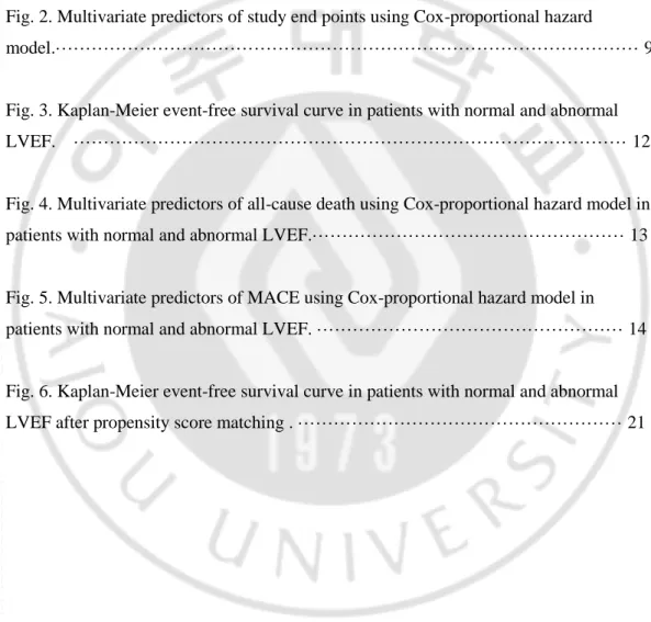
Fig 1. Kaplan-Meier event-free survival curve in all patients ··· 8 Fig. 2. Multivariate predictors of study end points using Cox-proportional hazard
model.··· 9 Fig. 3. Kaplan-Meier event-free survival curve in patients with normal and abnormal LVEF. ··· 12 Fig. 4. Multivariate predictors of all-cause death using Cox-proportional hazard model in patients with normal and abnormal LVEF.··· 13 Fig. 5. Multivariate predictors of MACE using Cox-proportional hazard model in patients with normal and abnormal LVEF. ··· 14 Fig. 6. Kaplan-Meier event-free survival curve in patients with normal and abnormal LVEF after propensity score matching . ··· 21
v
LIST OF TABLES
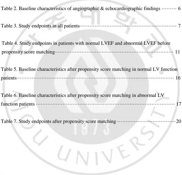
Table 1. Baseline characteristics of clinical findings ··· 4 Table 2. Baseline characteristics of angiographic & echocardiographic findings ··· 6 Table 3. Study endpoints in all patients··· 7
Table 4. Study endpoints in patients with normal LVEF and abnormal LVEF before propensity score matching··· 11 Table 5. Baseline characteristics after propensity score matching in normal LV function patients··· 16 Table 6. Baseline characteristics after propensity score matching in abnormal LV
function patients ··· 17 Table 7. Study endpoints after propensity score matching ···20
vi
ABBREVIATION
STEMI : ST-elevation myocardial infarction PCI : Percutaneous coronary intervention LV : Left ventricle
LVEF : Left ventricular ejection fraction MACE : Major adverse cardiac events MI : Myocardial infarction
1
I. INTRODUCTION
Beta-blocker had been well known for its beneficial effect on ST elevation myocardial infarction (STEMI) as primary medical treatment and for secondary prevention. Many randomized controlled studies and meta analysis have showed the benefits of beta-blockers on clinical outcomes in STEMI patients (1981; Hjalmarson et al., 1981; Hawkins et al., 1983; Olsson et al., 1985; Gottlieb et al., 1998; Freemantle et al., 1999). But firm confidence about the benefit of beta-blocker has gotten weak in low risk patients of STEMI, as the clinical outcomes of STEMI have been improved due to generalized primary percutaneous coronary intervention (PCI) and advance of medical treatment such as anti-platelet agent, angiotensin converting enzyme inhibitor and statin (Kezerashvili et al., 2012). In Asian people especially, there have been concerns of beta blocker about more frequent coronary artery vasospasm and more sensitive response of heart rate and blood pressure with lower dose of beta-blocker (Pristipino et al., 2000; Hori et al., 2004; Konishi et al., 2010). While some retrospective studies which were published recently demonstrated that there were no clinical benefits of beta-blocker in patients of normal or near normal left ventricular function, last updated ACC/AHA guideline still recommended beta blocker use in all patients with STEMI regardless of presence of left ventricular (LV) dysfunction (class I, level of evidence B) (Kernis et al., 2004; Ozasa et al., 2010; Bao et al., 2013; O'Gara et al., 2013).
To provide additive evidence to this controversy, we designed present study about the long term clinical benefits of beta-blocker in patients of STEMI with normal and abnormal LVEF (left ventricle ejection fraction).
2
II. Material and Method
A. Study deign and population
We analyzed data collected from single center (Department of cardiology, Ajou University school of Medicine, Suwon, Korea) retrospectively which included STEMI patients who underwent successful primary PCI without mortality after 30 days from 2003 to 2009. All clinical data sources of in hospital or follow-up information was based on hospital charts and the information about death of follow-up loss patients was collected from national health insurance service. In our data, 901 patients (716 males, 58±13 year-old) who were diagnosed as STEMI and underwent successful primary PCI without mortality after 30 days were recruited. These 901 patients were classified into beta-blocker (N=598, 491 males, 56±12 year-old) and non beta-blocker groups (N=303, 225 males, 61±13 year-old) according to beta-blocker use at discharge. After subdivided of each group into normal LVEF (left ventricular ejection fraction) group and abnormal LVEF group, long-term clinical outcomes were evaluated and compared as use of beta-blocker or not. Normal LVEF was defined as ≥50%
B. Study end points
Primary end point was all cause death and secondary end point was major adverse cardiac event (MACE) composed of death, recurrent myocardial infarction (MI), target vessel revascularization. MI was defined according to the universal definition of myocardial infarction (Thygesen et al., 2007).
C. Statistical analysis
PASW statistics (version 18) was used for statistical analysis. Continuous variables were expressed as mean ± standard deviation and were compared by independent t test or Mann-Whitney U test. A 2-sided p value of <0.05 was regarded as statistical significance.
Categorical variables were expressed as number and percentages and were compared with chi-square test or Fisher’s exact test. Cumulative incidences of primary and secondary end
3
point in each group were estimated by the Kaplan-Meier curves and differences were evaluated with log-rank test. To assess adjusted relative hazard ratio of beta-blocker to the study end point, we used Cox proportional hazard model with potential variables associated with clinical outcomes. Adjusted covariates for Cox proportional hazard model were old age (>65 year-old), Diabetes mellitus, hypertension, smoking, family history of cardiovascular disease, history of cerebrovascular disease, use of statin, use of angiotensin blockade agent (angiotensin converting enzyme inhibitor or angiotensin receptor blocker), multi-vessel disease, Killip stage >1 and end-stage renal disease. Results of multivariable analysis are expressed as adjusted hazard ratios and their 95% confidence intervals of PCI for clinical outcomes. To adjust difference of baseline characteristics, we used propensity score matching about two groups (beta blocker group and non beta blocker group) in normal LVEF and abnormal LVEF patients. Covariate used for matching were age, sex, LVEF, Killip stage, hypertension, infarct related vessel of left anterior descending artery and use of drug eluting stent which showed significant difference of two groups. After propensity score matching, cumulative incidences of primary and secondary end point were also estimated by the Kaplan-Meier curves with log-rank test.
4
III. Results
A. Beta-blocker use
Among enrolled 901 patients, 598 (66.4%) patients discharged with beta-blocker after successful primary PCI and in-hospital management. Carvedilol was used most frequently in 277 patients (46.3% in beta blocker group) and metoprolol in 268 patients (44.8%). These two beta-blockers were majority (91.1%) in beta-blocker group. The other beta-blockers (8.9%) used in this study were bisoprolol, bevantolol, atenolol and propranolol.
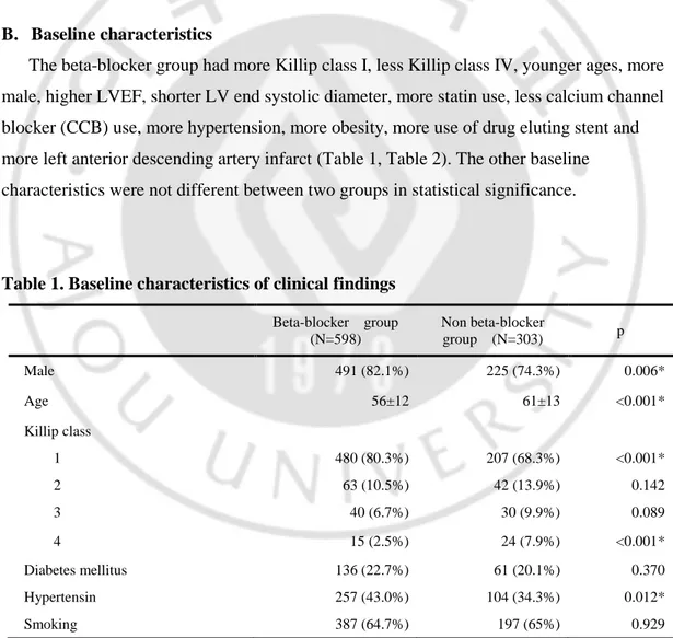
B. Baseline characteristics
The beta-blocker group had more Killip class I, less Killip class IV, younger ages, more male, higher LVEF, shorter LV end systolic diameter, more statin use, less calcium channel blocker (CCB) use, more hypertension, more obesity, more use of drug eluting stent and more left anterior descending artery infarct (Table 1, Table 2). The other baseline characteristics were not different between two groups in statistical significance.
Table 1. Baseline characteristics of clinical findings Beta-blocker group (N=598) Non beta-blocker group (N=303) p Male 491 (82.1%) 225 (74.3%) 0.006* Age 56±12 61±13 <0.001* Killip class 1 480 (80.3%) 207 (68.3%) <0.001* 2 63 (10.5%) 42 (13.9%) 0.142 3 40 (6.7%) 30 (9.9%) 0.089 4 15 (2.5%) 24 (7.9%) <0.001* Diabetes mellitus 136 (22.7%) 61 (20.1%) 0.370 Hypertensin 257 (43.0%) 104 (34.3%) 0.012* Smoking 387 (64.7%) 197 (65%) 0.929
5
Dyslipidemia 48 (8%) 19 (6.3%) 0.343 Family history of CAD 34 (5.7%) 12 (4.0%) 0.266 Old cerebrovascular attack 16 (2.7%) 13 (4.3%) 0.194 End stage renal disease 3 (0.3%) 0 (0%) 0.218 Obecity (BMI>25, kg/m2) 218 (45.1%) 68 (33.8%) 0.006* Prior MI 19 (3.2%) 10 (3.3%) 0.921 Prior CABG 1 (0.2%) 1 (0.3%) 0.624 Prior PCI 40 (6.7%) 25 (8.3%) 0.392 Discharge medication Aspirin 593 (99.2%) 301 (99.3%) 0.564 clopidogrel 552 (92.3%) 268 (88.4%) 0.056 Other antiplatelet agent 146 (24.4%) 75 (24.8%) 0.911 ACE inhibitor or ARB 567 (94.8%) 273 (90.1%) 0.008 Calcium channel blocker 72 (12%) 87 (28.7%) <0.001* Statin 421 (70.4%) 176 (58.1%) <0.001* Nitrate & other anti-anginal drugs 475 (79.4%) 234 (77.2%) 0.445 Values are mean ± SD or n (%), p<0.05 were expressed as ‘*’
CAD, coronary artery disease; BMI, body mass index; MI, myocardial infarction; CABG, coronary artery bypass surgery; PCI, percutaneous coronary intervention; ACE, angiotensin converting enzyme; ARB, angiotensin receptor blocker
6
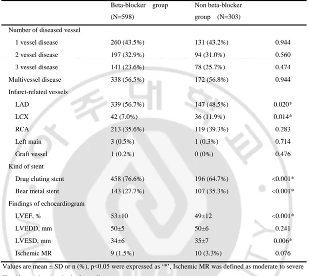
Table 2. Baseline characteristics of angiographic & echocardiographic findings Beta-blocker group
(N=598)
Non beta-blocker group (N=303) Number of diseased vessel
1 vessel disease 260 (43.5%) 131 (43.2%) 0.944 2 vessel disease 197 (32.9%) 94 (31.0%) 0.560 3 vessel disease 141 (23.6%) 78 (25.7%) 0.474 Multivessel disease 338 (56.5%) 172 (56.8%) 0.944 Infarct-related vessels LAD 339 (56.7%) 147 (48.5%) 0.020* LCX 42 (7.0%) 36 (11.9%) 0.014* RCA 213 (35.6%) 119 (39.3%) 0.283 Left main 3 (0.5%) 1 (0.3%) 0.714 Graft vessel 1 (0.2%) 0 (0%) 0.476 Kind of stent
Drug eluting stent 458 (76.6%) 196 (64.7%) <0.001* Bear metal stent 143 (27.7%) 107 (35.3%) <0.001* Findings of echocardiogram
LVEF, % 53±10 49±12 <0.001* LVEDD, mm 50±5 50±6 0.241 LVESD, mm 34±6 35±7 0.006* Ischemic MR 9 (1.5%) 10 (3.3%) 0.076 Values are mean ± SD or n (%), p<0.05 were expressed as ‘*’, Ischemic MR was defined as moderate to severe MR without primary valvular heart disease.
LAD, Left anterior descending artery; LCX, Left circumflex artery; RCA, Right coronary artery; LVEF, left ventricular ejection fraction; LVEDD, left ventricular diastolic dimension; LVESD, left ventricular end-sistolic dimension; MR, mitral regurgitation
7 C. Clinical outcomes
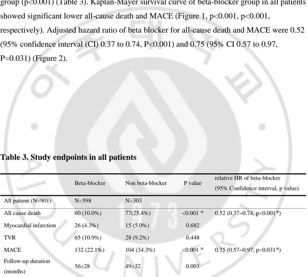
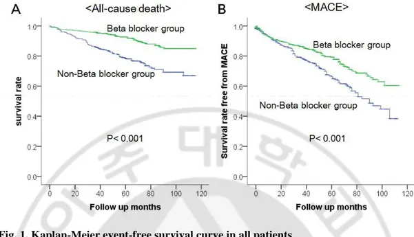
Cumulative incidence of all-cause death was 10.0% (60 patients) in beta-blocker group during mean follow up months of 56±28 and 25.4% (77 patients) in non beta-blocker group during mean follow up months of 49±32 (p<0.001) (Table 3). Incidence of MACE was 22.1% (132 patients) in beta-blocker group and 34.3% (104 patients) in non beta-blocker group (p<0.001) (Table 3). Kaplan-Mayer survival curve of beta-blocker group in all patients showed significant lower all-cause death and MACE (Figure 1, p<0.001, p<0.001,
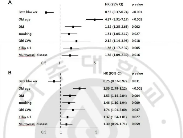
respectively). Adjusted hazard ratio of beta blocker for all-cause death and MACE were 0.52 (95% confidence interval (CI) 0.37 to 0.74, P<0.001) and 0.75 (95% CI 0.57 to 0.97,
P=0.031) (Figure 2).
Table 3. Study endpoints in all patients
Values are mean ± SD or n (%), p value was analyzed by Chi-Square test and p<0.05 were expressed as ‘*’, relative HR of beta-blocker was derived from Cox-proportional hazard model with multivariate analysis before propensity score matching
TVR, target vessel revascularization; MACE, major adverse cardiac events; HR, hazard ratio
Beta-blocker Non beta-blocker P value relative HR of beta-blocker (95% Confidence interval, p value) All patient (N=901) N=598 N=303
All cause death 60 (10.0%) 77(25.4%) <0.001 * 0.52 (0.37~0.74, p<0.001*) Myocardial infarction 26 (4.3%) 15 (5.0%) 0.682
TVR 65 (10.9%) 28 (9.2%) 0.448
MACE 132 (22.1%) 104 (34.3%) <0.001 * 0.75 (0.57~0.97, p=0.031*) Follow-up duration
8
Fig. 1. Kaplan-Meier event-free survival curve in all patients
A. Beta-blocker group in all patients showed significant higher survival rate free from all-cause death. B. Beta-blocker group also showed significant higher survival rate free from MACE.
9
Fig. 2. Multivariate predictors of study end points using Cox-proportional hazard model.
A. All-cause death. B. Major adverse cardiac events.
HR, hazard ratio; CI, confidence interval; DM, diabetes mellitus; CVA, cerebrovascular attack
10
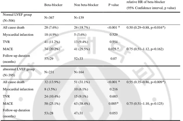
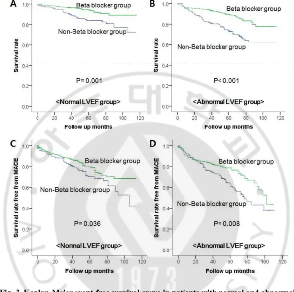
Table 4 showed study end points in patients with normal and abnormal LVEF. In patients with normal LVEF (N=506), the cumulative incidence of all-cause death was 7.6% (28 patients) in beta-blocker group during mean follow up months of 57±29 and 18.7% (26 patients) in non beta-blocker group during mean follow up months of 52±33 (p<0.001). And with abnormal LVEF, the cumulative incidence of all-cause death was 13.9% (32 patients) in beta-blocker group during mean follow up months of 53±28 and 31.1% (51 patients) in non beta-blocker group during mean follow up months of 47±31 (p<0.001). The cumulative incidence of MACE was 20.2% (74 patients) in beta-blocker group and 29.5% (41 patients) in non beta-blocker group with normal LVEF (p=0.025). And the incidence of MACE was 25.1% (58 patients) in beta-blocker group and 38.4% (63 patients) in non beta-blocker group with abnormal LVEF (p=0.005). Kaplan-Mayer survival curve of beta-blocker group in patients with both of normal and abnormal LVEF demonstrated significant higher survival rate of all-cause death (p=0.001, p<0.001, respectively) (Figure 3 A,B). And beta-blocker group also showed significant higher survival rate free from MACE in normal LVEF and abnormal LVEF patients (p=0.036, p=0.008, respectively) (Figure 3 C,D).
Adjusted hazard ratio of beta-blocker for all-cause death in patients with normal and abnormal LVEF were 0.50 (95% CI 0.29 to 0.88, P=0.016) and 0.55 (95% CI 0.35 to 0.86, P=0.009) (Figure 4). Adjusted hazard ratio of beta-blocker for MACE in patients with normal and abnormal LVEF were 0.075(95% CI 0.51 to 1.12, P=0.162) and 0.75 (95% CI 0.51 to 1.09, P=0.125) (Figure 5).
11
Table 4. Study endpoints in patients with normal LVEF and abnormal LVEF before propensity score matching
Values are mean ± SD or n (%), p value was analyzed by Chi-Square test and p<0.05 were expressed as ‘*’, relative HR of beta-blocker was derived from Cox-proportional hazard model with multivariate analysis before propensity score matching
TVR, target vessel revascularization; MACE, major adverse cardiac events; HR, hazard ratio
Beta-blocker Non beta-blocker P value relative HR of beta-blocker (95% Confidence interval, p value) Normal LVEF group
(N=506) N=367 N=139
All cause death 28 (7.6%) 26 (18.7%) <0.001 * 0.50 (0.29~0.88, p=0.016*) Myocardial infarction 18 (4.9%) 5 (3.6%) 0.529
TVR 41 (11.2%) 13 (9.4%) 0.554
MACE 74 (20.2%) 41 (29.5%) 0.025 * 0.75 (0.51~1.12, p=0.162) Follow-up duration
(months) 57±29 52±33 0.07
abnormal LVEF group
(N=395) N=231 N=164
All cause death 32 (13.9%) 51 (31.1%) <0.001 * 0.55 (0.35~0.86, p=0.009*) Myocardial infarction 8 (3.5%) 10 (6.1%) 0.216
TVR 24 (10.4%) 15 (9.1%) 0.683
MACE 58 (25.1%) 63 (38.4%) 0.005* 0.75 (0.51~1.10, p=0.125) Follow-up duration
12
Fig. 3. Kaplan-Meier event-free survival curve in patients with normal and abnormal LVEF.
A. Survival rate free from all-cause death in patients with normal LVEF. B. Survival rate free from all-cause death in patients with abnormal LVEF. C. Survival rate free from MACE in patients with normal LVEF. D. Survival rate free from MACE in patients with abnormal LVEF.
P value was derived from the Log-rank test; LVEF, left ventricular ejection fraction; MACE, major adverse cardiac events
13
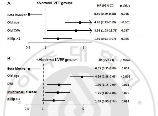
Fig. 4. Multivariate predictors of all-cause death using Cox-proportional hazard model in patients with normal and abnormal LVEF.
A. Predictors of all-cause death in patients with normal LVEF. B. In patients with abnormal LVEF
HR, hazard ratio; CI, confidence interval; DM, diabetes mellitus; CVA, cerebrovascular attack
14
Fig. 5. Multivariate predictors of MACE using Cox-proportional hazard model in patients with normal and abnormal LVEF.
A. Predictors of MACE in patients with normal LVEF. B. In patients with abnormal LVEF
HR, hazard ratio; CI, confidence interval; DM, diabetes mellitus; CVA, cerebrovascular attack
15
After propensity score matching (1:1) to adjust difference of baseline characteristics, 140 patients of each group (beta-blocker versus non beta-blocker) in patients with abnormal LVEF were selected and 137 patients of each group in patients with normal LVEF were selected. Baseline characteristics of two groups in normal LVEF and abnormal LVEF patients showed no significant difference in all variables except the use of CCB (Table 5, Table 6). We allowed the difference in the use of CCB intentionally through not including it in matching variables because it was reasonable to give more CCB as anti-anginal drug to patients who did not take beta-blocker and CCB had little effect to clinical outcomes in STEMI patients.
16
Table 5. Baseline characteristics after propensity score matching in normal LV function patients Beta-blocker group (N=137) Non beta-blocker group (N=137) p Male 97 (70.8%) 100 (73.0%) 0.687 Age 58±13 59±13 0.496 Killip class 0.98 1 114 (83.2%) 112 (81.8%) 0.751 2 15 (10.9%) 15 (10.9%) 1 3 5 (3.6%) 6 (4.4%) 0.758 4 3 (2.2%) 4 (2.9%) 0.702 Diabetes mellitus 28 (20.4%) 28 (20.4%) 1 Hypertensin 55 (40.1%) 52 (38.0%) 0.71 Smoking 78 (56.9%) 88 (64.2%) 0.216 Dyslipidemia 13 (9.5%) 8 (5.8%) 0.256 Family history 8 (5.8%) 6 (4.4%) 0.583 Creatine level 0.97±0.69 1.02±0.43 0.363 Obecity (BMI>25) 45 (42.9%) 32 (35.6%) 0.298 Discharge medication Aspirin 137 (100%) 136 (99.3%) 1 Plavix 122 (89.1%) 117 (85.4%) 0.366 Cilostazole 20 (14.6%) 25 (18.2%) 0.415 ACEi 90 (65.7%) 90 (65.7%) 1 ARB 37 (27.0%) 36 (26.3%) 0.891 Calcium channel blocker 21 (15.%) 50 (36.5%) <0.001* Statin 91 (66.4%) 87 (63.5%) 0.612 Nitrate 103 (75.2%) 99 (72.3%) 0.583 Multivessel disease 83 (60.6%) 74 (54.0) 0.272 Culprit vessel LAD 43 (31.4%) 36 (26.3%) 0.351 LCX 16 (11.7%) 19 (13.9%) 0.587 RCA 78 (56.9%) 82 (59.9%) 0.624 Kind of stent
17
Drug eluting stent 94 (68.6%) 84 (61.3%) 0.205 Bear metal stent 46 (33.6%) 50 (36.5%) 0.612 Findings of echocardiogram
LVEF, % 59±7 59±8 0.744 LVEDD, mm 48±5 49±5 0.179 LVESD, mm 31±5 31±5 0.541 Ischemic MR 3 (2.2%) 2 (1.5%) 1 Values are mean ± SD or n (%), p<0.05 were expressed as ‘*’. Ischemic MR was defined as moderate to severe MR without primary valvular heart disease.
BMI, body mass index; ACE, angiotensin converting enzyme; ARB, angiotensin receptor blocker; LAD, Left anterior descending artery; LCX, Left circumflex artery; RCA, Right coronary artery; LVEF, left ventricular ejection fraction; LVEDD, left ventricular end-diastolic dimension; LVESD, left ventricular end-sistolic dimension; MR, mitral regurgitation
Table 6. Baseline characteristics after propensity score matching in abnormal LV function patients Beta-blocker group (N=140) Non beta-blocker group (N=140) p Male 112 (80.0%) 108 (77.1%) 0.560 Age 58±13 60±12 0.147 Killip class 0.557 1 92 (65.7%) 88 (62.9%) 0.618 2 20 (14.3%) 26 (18.6%) 0.333 3 21 (15%) 16 (11.4%) 0.378 4 7 (5%) 10 (7.1%) 0.453 Diabetes mellitus 31 (22.1%) 29 (20.7%) 0.771 Hypertensin 50 (35.7%) 46 (32.9%) 0.615 Smoking 95 (67.9%) 96 (68.6%) 0.898 Dyslipidemia 8 (5.7%) 11 (7.9%) 0.476 Family history 10 (7.1%) 5 (3.6%) 0.184 Creatine level 1.02±0.32 1.01±0.44 0.829 Obecity (BMI>25, kg/m2) 45 (40.5%) 32 (34.0%) 0.338 Discharge medication
18 Aspirin 137 (97.9%) 139 (99.3%) 0.622 Plavix 128 (91.4%) 128 (91.4%) 1 Cilostazole 25 (17.9%) 20 (14.3%) 0.416 ACEi 83 (59.3%) 79 (56.4%) 0.628 ARB 54 (38.6%) 47 (33.6%) 0.384 Calcium channel blocker 14 (10%) 35 (25%) 0.001* Statin 90 (64.3%) 81 (57.9%) 0.27 Nitrate 103 (73.6%) 101 (72.1%) 0.788 Multivessel disease 76 (54.3%) 81 (57.9%) 0.547 Culprit vessel LAD 101 (72.1%) 99 (70.7%) 0.791 LCX 5 (3.6%) 12 (8.6%) 0.08 RCA 31 (22.1%) 29 (20.7%) 0.771 Left main 3 (2.1%) 0 (0%) 0.247 Kind of stent
Drug eluting stent 101 (72.1%) 96 (68.6%) 0.513 Bear metal stent 40 (28.6%) 46 (32.9%) 0.437 Findings of echocardiogram
LVEF, % 42±6 41±6 0.41 LVEDD, mm 51±6 52±6 0.326 LVESD, mm 37±7 38±7 0.424 Ischemic MR 2 (1.4%) 5 (3.6%) 0.447 Values are mean ± SD or n (%), p<0.05 were expressed as ‘*’. Ischemic MR was defined as moderate to severe MR without primary valvular heart disease.
BMI, body mass index; ACE, angiotensin converting enzyme; ARB, angiotensin receptor blocker; LAD, Left anterior descending artery; LCX, Left circumflex artery; RCA, Right coronary artery; LVEF, left ventricular ejection fraction; LVEDD, left ventricular end-diastolic dimension; LVESD, left ventricular end-sistolic dimension; MR, mitral regurgitation
19
In matched group, cumulative incidence of death was 5.8% (8 patients) in beta-blocker group during mean follow up months of 58±32 and 19% (26 patients) in non beta-blocker group during mean follow up months of 51±33 with normal LVEF (p=0.001). And in patients with abnormal LVEF, cumulative incidence of death was 15.7% (22 patients) in beta-blocker group during mean follow up months of 53±29 and 27.9% (39 patients) in non beta-blocker group during mean follow up months of 49±31 (p=0.014) (Table 7). Incidence of MACE defined as a composite of all-cause death, recurrent MI, TVR was 19% (26 patients) in beta-blocker group and 29.2% (40 patients) in non beta-blocker group with normal LVEF (p=0.048). With abnormal LVEF, the incidence of MACE was 27.9% (39 patients) in beta-blocker group and 36.4% (51 patients) in non beta-blocker group (p=0.125) (Table 7).
Kaplan-Mayer survival curve of beta-blocker group also showed significantly higher survival rate free from all-cause death in patients with normal LVEF and abnormal LVEF after propensity score matching (p=0.001, p=0.020, respectively) (Figure 6 A,B). The survival rate free from MACE was significantly higher in beta-blocker group with normal LVEF, but not significantly different in both group with abnormal LVEF (p=0.046, p=0.184, respectively) (Figure 6 C,D).
20
Table 7. Study endpoints after propensity score matching
Values are mean ± SD or n (%), p value was analyzed by Chi-Square test and p<0.05 were expressed as ‘*’, relative HR of beta-blocker was derived from Cox-proportional hazard model with multivariate analysis before propensity score matching
TVR, target vessel revascularization; MACE, major adverse cardiac events; HR, hazard ratio
Beta-blocker Non beta-blocker P value
normal LVEF group N=137 N=137 274
Death 8 (5.8%) 26 (19%) 0.001*
Myocardial infarction 6 (4.4%) 5 (3.6%) 0.758
TVR 18 (13.1%) 12 (8.8%) 0.246
MACE 26 (19%) 40 (29.2%) 0.048*
Follow-up duration (months) 58±32 51±33 0.068
abnormal LVEF group N=140 N=140 280
Death 22 (15.7%) 39 (27.9%) 0.014* MI 6 (4.3%) 9 (6.4%) 0.426 TVR 15 (10.7%) 14 (10.0%) 0.845 MACE 39 (27.9%) 51 (36.4%) 0.125 Follow-up duration (months) 53±29 49±31 0.242
21
Fig. 6. Kaplan-Meier event-free survival curve in patients with normal and abnormal LVEF after propensity score matching.
A. Survival rate free from all-cause death in patients with normal LVEF. B. Survival rate free from all-cause death in patients with abnormal LVEF. C. Survival rate free from MACE in patients with normal LVEF. D. Survival rate free from MACE in patients with abnormal LVEF.
P value was derived from the Log-rank test; LVEF, left ventricular ejection fraction; MACE, major adverse cardiac events
22
IV. Discussion
In the present study, oral beta-blocker use at discharge in STEMI survivor who underwent primary PCI and had normal LVEF demonstrated better long term clinical outcomes in point of all cause mortality. In patients of abnormal LV function, beta-blocker group also showed higher survival rate. And Kaplan-Meier curve of MACE in beta-blocker group with normal LV function showed better outcomes too, although it was not consistent with Cox-proportional multivariate analysis.
There has been no controversy about the clinical benefit of beta-blocker in patients with LV dysfunction after acute myocardial infarction (AMI) on revascularization era as well as pre-revascularization era (Dargie, 2001; Kernis et al., 2004). But in low risk patients with preserved LV function, the beneficial role of beta-blocker after primary PCI has been in doubt due to lack of evidence in these revascularization era despite ongoing recommendation of current guideline (Task Force on the management of et al., 2012; O'Gara et al., 2013). After Kernis et al. mentioned that there were little beneficial effects of beta-blocker to patients with normal LV function and single vessel disease, several retrospective studies reported that beta-blocker was not associated better clinical outcomes in patients with preserved LV function after primary PCI and even demonstrated poor clinical outcomes in one study (Kernis et al., 2004; Ozasa et al., 2010; Bao et al., 2013). These studies explained that beneficial effects of beta-blocker could be offset by not only general adverse effect of beta-blocker such as hypotension, bradycardia, dizziness, depression, bronchospasm, metabololic disorder, drug allergy but also additive negative effect such as coronary vasospasm, decreased compliance due to additional cost and burden of pills.
But beta-blockers have many important beneficial effects in patients with AMI. Beta-blockers reduce cardiotoxic effect of catecholamine via its anti-adrenergic effect and also reduce myocardial work load through decreasing of heart rate, blood pressure and myocardial contractility (Mueller and Ayres, 1980; Lange et al., 1983; Kjekshus, 1986). Moreover, they have anti-arrhythmic effect, which reduce tachy-arrhythmic events and use of other anti-arrhythmic agents (Ryden et al., 1983; Olsson and Rehnqvist, 1986). These
23
beneficial effects of beta-blockers limit infarct-size, and reduce recurrent ischemia and mortality including cardiac sudden death (1981; Hjalmarson et al., 1981; Kopecky, 2006).
It is true that clinical outcomes of low risk patients with normal LV function after STEMI was improved (Bramlage et al., 2010), but recurrent myocardial ischemia,
tachyarrhythmia and adrenergic activation are still important problems in those patients, and there is no other optimal substitute of beta-blocker to control these important problems effectively. Moreover, the incidence of significant and life-threatening adverse effects of beta-blocker such as hypotension and bradycardia was not higher enough to offset the positive effects of beta-blocker (Ko et al., 2004). Consequently, It is reasonable to assume that beta blocker have positive effects more than negative effects in patients of STEMI with normal LV function.
Although some recent studies in the revascularization era reported negative results about beneficial effects of beta-blockers, they had many limitations including property of
retrospective studies (Ozasa et al., 2010; Bao et al., 2013). But many reports which showed beneficial effects of beta-blocker in AMI patients were based on very strong evidence derived from numerous randomized controlled trial and meta-analysis though they were old studies in pre-revascularization era, and those studies also had considerable number of preserved LV function (1981; Hjalmarson et al., 1981; Hawkins et al., 1983; Olsson et al., 1985; Gottlieb et al., 1998; Freemantle et al., 1999). So some limited retrospective studies could not overwhelm strong old studies, and this was why current guidelines still keep to their recommendation of beta-blocker as primary management and secondary prevention of STEMI (Task Force on the management of et al., 2012; O'Gara et al., 2013). The result of our study provided additive evidence to these current guidelines in the era of primary PCI.
There were several important limitations in this study. First, because this study is retrospective observational study, selection bias in use of beta-blockers is inevitable. Although we adjusted the difference of baseline characteristics in two groups through Cox proportional multivariate analysis and propensity score matching, our study could not be free from the influence of unmeasured confounding factors. Second, our data had no information of beta-blocker dose so that beta-blocker was likely to be used insufficiently resulting in reducing its clinical effects. Third, the duration of beta-blocker use after discharge was not
24
included in our data. So we could not know about the effect of long-term use of beta-blocker and optimal duration of its use in STEMI patients with normal LV function after primary PCI.
25
V. Conclusion
In our study, use of oral beta-blocker at discharge showed beneficial effects on clinical outcomes in patients with STEMI after primary PCI regardless of LVEF. The potential role of beta-blocker might be beyond the therapeutic effects of LV dysfunction. Now, it’s time to evaluate the role of beta-blocker in STEMI patients with normal LV function after primary PCI through large randomized controlled trial.
26
REFERENCES
1. Timolol-induced reduction in mortality and reinfarction in patients surviving acute myocardial infarction. N Engl J Med 304: 801-807, 1981
2. Bao B, Ozasa N, Morimoto T, Furukawa Y, Nakagawa Y, Kadota K, Iwabuchi M, Shizuta S, Shiomi H, Tada T, Tazaki J, Kato Y, Hayano M, Natsuaki M, Fujiwara H, Mitsudo K, Nobuyoshi M, Kita T, Kimura T: beta-Blocker therapy and
cardiovascular outcomes in patients who have undergone percutaneous coronary intervention after ST-elevation myocardial infarction. Cardiovasc Interv Ther 28: 139-147, 2013
3. Bramlage P, Messer C, Bitterlich N, Pohlmann C, Cuneo A, Stammwitz E, Tebbenjohanns J, Gohlke H, Senges J, Tebbe U: The effect of optimal medical therapy on 1-year mortality after acute myocardial infarction. Heart 96: 604-609, 2010
4. Dargie HJ: Effect of carvedilol on outcome after myocardial infarction in patients with left-ventricular dysfunction: the CAPRICORN randomised trial. Lancet 357: 1385-1390, 2001
5. Freemantle N, Cleland J, Young P, Mason J, Harrison J: beta Blockade after myocardial infarction: systematic review and meta regression analysis. BMJ 318: 1730-1737, 1999
6. Gottlieb SS, McCarter RJ, Vogel RA: Effect of beta-blockade on mortality among high-risk and low-risk patients after myocardial infarction. N Engl J Med 339: 489-497, 1998
7. Hawkins CM, Richardson DW, Vokonas PS: Effect of propranolol in reducing mortality in older myocardial infarction patients. The Beta-Blocker Heart Attack Trial experience. Circulation 67: I94-97, 1983
8. Hjalmarson A, Elmfeldt D, Herlitz J, Holmberg S, Malek I, Nyberg G, Ryden L, Swedberg K, Vedin A, Waagstein F, Waldenstrom A, Waldenstrom J, Wedel H,
27
Wilhelmsen L, Wilhelmsson C: Effect on mortality of metoprolol in acute myocardial infarction. A double-blind randomised trial. Lancet 2: 823-827, 1981 9. Hori M, Sasayama S, Kitabatake A, Toyo-oka T, Handa S, Yokoyama M, Matsuzaki
M, Takeshita A, Origasa H, Matsui K, Hosoda S, Investigators M: Low-dose carvedilol improves left ventricular function and reduces cardiovascular hospitalization in Japanese patients with chronic heart failure: the Multicenter Carvedilol Heart Failure Dose Assessment (MUCHA) trial. Am Heart J 147: 324-330, 2004
10. Kernis SJ, Harjai KJ, Stone GW, Grines LL, Boura JA, O'Neill WW, Grines CL: Does beta-blocker therapy improve clinical outcomes of acute myocardial infarction after successful primary angioplasty? J Am Coll Cardiol 43: 1773-1779, 2004 11. Kezerashvili A, Marzo K, De Leon J: Beta blocker use after acute myocardial
infarction in the patient with normal systolic function: when is it "ok" to discontinue?
Curr Cardiol Rev 8: 77-84, 2012
12. Kjekshus JK: Importance of heart rate in determining beta-blocker efficacy in acute and long-term acute myocardial infarction intervention trials. Am J Cardiol 57: 43F-49F, 1986
13. Ko DT, Hebert PR, Coffey CS, Curtis JP, Foody JM, Sedrakyan A, Krumholz HM: Adverse effects of beta-blocker therapy for patients with heart failure: a quantitative overview of randomized trials. Arch Intern Med 164: 1389-1394, 2004
14. Konishi M, Haraguchi G, Kimura S, Inagaki H, Kawabata M, Hachiya H, Hirao K, Isobe M: Comparative effects of carvedilol vs bisoprolol for severe congestive heart failure. Circ J 74: 1127-1134, 2010
15. Kopecky SL: Effect of beta blockers, particularly carvedilol, on reducing the risk of events after acute myocardial infarction. Am J Cardiol 98: 1115-1119, 2006
16. Lange R, Kloner RA, Braunwald E: First ultra-short-acting beta-adrenergic blocking agent: its effect on size and segmental wall dynamics of reperfused myocardial infarcts in dogs. Am J Cardiol 51: 1759-1767, 1983
28
17. Mueller HS, Ayres SM: Propranolol decreases sympathetic nervous activity reflected by plasma catecholamines during evolution of myocardial infarction in man. J Clin
Invest 65: 338-346, 1980
18. O'Gara PT, Kushner FG, Ascheim DD, Casey DE, Jr., Chung MK, de Lemos JA, Ettinger SM, Fang JC, Fesmire FM, Franklin BA, Granger CB, Krumholz HM, Linderbaum JA, Morrow DA, Newby LK, Ornato JP, Ou N, Radford MJ, Tamis-Holland JE, Tommaso CL, Tracy CM, Woo YJ, Zhao DX, Anderson JL, Jacobs AK, Halperin JL, Albert NM, Brindis RG, Creager MA, DeMets D, Guyton RA,
Hochman JS, Kovacs RJ, Kushner FG, Ohman EM, Stevenson WG, Yancy CW, American College of Cardiology Foundation/American Heart Association Task Force on Practice G: 2013 ACCF/AHA guideline for the management of ST-elevation myocardial infarction: a report of the American College of Cardiology Foundation/American Heart Association Task Force on Practice Guidelines.
Circulation 127: e362-425, 2013
19. Olsson G, Rehnqvist N: Evaluation of antiarrhythmic effect of metoprolol treatment after acute myocardial infarction: relationship between treatment responses and survival during a 3-year follow-up. Eur Heart J 7: 312-319, 1986
20. Olsson G, Rehnqvist N, Sjogren A, Erhardt L, Lundman T: Long-term treatment with metoprolol after myocardial infarction: effect on 3 year mortality and morbidity.
J Am Coll Cardiol 5: 1428-1437, 1985
21. Ozasa N, Kimura T, Morimoto T, Hou H, Tamura T, Shizuta S, Nakagawa Y, Furukawa Y, Hayashi Y, Nakao K, Matsuzaki M, Nobuyoshi M, Mitsudo K, j-Cypher Registry I: Lack of effect of oral beta-blocker therapy at discharge on long-term clinical outcomes of ST-segment elevation acute myocardial infarction after primary percutaneous coronary intervention. Am J Cardiol 106: 1225-1233, 2010 22. Pristipino C, Beltrame JF, Finocchiaro ML, Hattori R, Fujita M, Mongiardo R,
Cianflone D, Sanna T, Sasayama S, Maseri A: Major racial differences in coronary constrictor response between japanese and caucasians with recent myocardial infarction. Circulation 101: 1102-1108, 2000
29
23. Ryden L, Ariniego R, Arnman K, Herlitz J, Hjalmarson A, Holmberg S, Reyes C, Smedgard P, Svedberg K, Vedin A, Waagstein F, Waldenstrom A, Wilhelmsson C, Wedel H, Yamamoto M: A double-blind trial of metoprolol in acute myocardial infarction. Effects on ventricular tachyarrhythmias. N Engl J Med 308: 614-618, 1983
24. Task Force on the management of STseamiotESoC, Steg PG, James SK, Atar D, Badano LP, Blomstrom-Lundqvist C, Borger MA, Di Mario C, Dickstein K,
Ducrocq G, Fernandez-Aviles F, Gershlick AH, Giannuzzi P, Halvorsen S, Huber K, Juni P, Kastrati A, Knuuti J, Lenzen MJ, Mahaffey KW, Valgimigli M, van 't Hof A, Widimsky P, Zahger D: ESC Guidelines for the management of acute myocardial infarction in patients presenting with ST-segment elevation. Eur Heart J 33: 2569-2619, 2012
25. Thygesen K, Alpert JS, White HD, Joint ESCAAHAWHFTFftRoMI, Jaffe AS, Apple FS, Galvani M, Katus HA, Newby LK, Ravkilde J, Chaitman B, Clemmensen PM, Dellborg M, Hod H, Porela P, Underwood R, Bax JJ, Beller GA, Bonow R, Van der Wall EE, Bassand JP, Wijns W, Ferguson TB, Steg PG, Uretsky BF, Williams DO, Armstrong PW, Antman EM, Fox KA, Hamm CW, Ohman EM, Simoons ML, Poole-Wilson PA, Gurfinkel EP, Lopez-Sendon JL, Pais P, Mendis S, Zhu JR, Wallentin LC, Fernandez-Aviles F, Fox KM, Parkhomenko AN, Priori SG, Tendera M, Voipio-Pulkki LM, Vahanian A, Camm AJ, De Caterina R, Dean V, Dickstein K, Filippatos G, Funck-Brentano C, Hellemans I, Kristensen SD, McGregor K,
Sechtem U, Silber S, Tendera M, Widimsky P, Zamorano JL, Morais J, Brener S, Harrington R, Morrow D, Lim M, Martinez-Rios MA, Steinhubl S, Levine GN, Gibler WB, Goff D, Tubaro M, Dudek D, Al-Attar N: Universal definition of myocardial infarction. Circulation 116: 2634-2653, 2007
30 - 국문요약 –
ST 분절 상승 급성 심근경색 환자에서 일차적 관상동맥 중재
시술 후 경구 베타 차단제 사용이 장기 예후에 미치는 영향
목적: ST 분절 상승 심근경색 환자들의 치료가 발전함에 따라 좌심실 기능 저하를 보이는 환자들의 수는 줄어 들었고 따라서 이들의 예후도 많이 향상 되었다. 성공적인 일차적 관동맥 중재시술로 정상 좌심실 수축기능을 보이는 ST 분절 상승 심근경색 환자에서 베타 차단제의 역할에 대해서는 지금까지 충분히 연구되지 않았고 최근의 몇몇 연구들은 베타 차단제의 역할에 의문을 제기하고 있다. 본 연구는 ST 분절 상승 심근경색 환자에서 성공적인 일차적 관동맥 중재시술 후 베타 차단제의 사용이 환자의 장기 예후에 미치는 영향에 대해 조사하였다. 방법: 일차적 관동맥 중재시술을 성공적으로 시행한 901 명의 ST 분절 상승 심근경색 환자를 598 명의 베타 차단제 사용 군과 303 명의 베타 차단제 비 사용 군으로 나누었고 각 군을 좌심실 기능 저하의 유무에 따라 비교하였다. 환자의 사망과 사망, 재발성 심근경색, 표적 혈관의 재 개통으로 구성된 주요 심장 사건을 연구하였다. 결과: 베타 차단제 사용 군에서 Killip 분류 1 이 더 많았고, 좌심실 수축기능이 더 높았으며 젊은 연령, 남성, statin 제제의 사용, 고혈압 환자, 비만, 약물 용출 스텐트의 사용 빈도, 좌전하행지 영역의 심근경색의 빈도 등이 더 많게 나타났다. 베타 차단제 사용 군에서 모든 사망의 누적 발생율은 10% (60 명, 평균 관찰 기간:56±28 개월) 이었고 베타 차단제 비사용 군에서는 25.4% (77 명, 평균 관찰 기간: 49±32 개월)이었다 (p<0.001). 주요 심장 사건의 누적 발생율의 경우 베타 차단제 사용 군에서 22.1% (132 명) 이었고 베타31 차단제 비 사용 군에서는 34.3% (104 명) 이었다 (p<0.001). 좌심실 구혈률이 저하된 (좌심실 구혈률 <50%) 환자에서 베타 차단제 사용의 사망과 주요 심장 사건에 대한 비교 위험도는 0.54 (P=0.008, 95% 신뢰구간(CI) 0.34-0.85)와 0.72 (P=0.082, CI 0.50-1.04)였고 정상 좌심실 구혈률을 가진 (좌심실 구혈률 ≥50) 환자에서는 베타 차단제 사용의 비교 위험도가 각각 0.50 (P=0.012, CI 0.29-0.86)와 0.80 (P=0.275, CI 0.54-1.19) 이었다. 양군간의 임상 양상의 차이를 보정하기 위해 Propensity score matching 을 시행한 후 분석한 Kaplan-Meier 생존 곡선에서 베타 차단제 사용 군은 좌심실 구혈률 저하가 있는 환자와 좌심실 구혈률이 정상인 환자 모두에서 통계적으로 의미 있는 높은 생존율을 보여 주었다 (P=0.02, P=0.001, respectively). 결론: 베타 차단제는 일차적 관동맥 중재 시술을 시행한 ST 분절 상승 심근경색 환자에서 좌심실 기능 저하의 유무와 상관 없이 예후에 긍정적인 영향을 준다. 핵심어: 베타 차단제, ST 분절 상승 심근경색, 일차적 관동맥 중재 시술, 좌심실 기능