Risk Factors for Neurologic Complications of Hand, Foot and
Mouth Disease in the Republic of Korea, 2009
In 2009, the first outbreak of hand, foot and mouth disease (HFMD) or herpangina (HP) caused by enterovirus 71 occurred in the Republic of Korea. This study inquired into risk factors associated with complications of HFMD or HP. A retrospective medical records review was conducted on HFMD or HP patients for whom etiologic viruses had been verified in 2009. One hundred sixty-eight patients were examined for this investigation. Eighty patients were without complications while 88 were accompanied by complications, and 2 had expired. Enterovirus 71 subgenotype C4a was the most prevalent in number with 67 cases (54.9%). In the univariate analysis, the disease patterns of HFMD rather than HP, fever longer than 4 days, peak body temperature over 39°C, vomiting, headache, neurologic signs, serum glucose over 100 mg/dL, and having an enterovirus 71 as a causative virus were significant risk factors of the complications. After multiple logistic analysis, headache (Odds ratio [OR], 10.75; P < 0.001) and neurologic signs (OR, 42.76;
P < 0.001) were found to be the most significant factors. Early detection and proper
management of patients with aforementioned risk factors would be necessary in order to attain a better clinical outcome.
Key Words: Coxsackievirus; Hand, Foot and Mouth Disease; Herpangina; Enterovirus A, Human; Risk Factors
Seong Joon Kim,1 Jong-Hyun Kim,1
Jin-Han Kang,1 Dong Soo Kim,2
Ki Hwan Kim,2 Kyung-Hyo Kim,3
Young-Hoon Kim,1 Ju-Young Chung,4
Joong Hyun Bin,1 Da Eun Jung,5
Ji Hong Kim,2 Hwang Min Kim,6
Doo-Sung Cheon,7 Byung Hak Kang,7
Soon Young Seo,8 and the Enteroviruses
Complications Working Group
1Department of Pediatrics, College of Medicine, The
Catholic University of Korea, Seoul; 2Department of
Pediatrics, Yonsei University College of Medicine,
Seoul; 3Department of Pediatrics, Ewha Womans
University School of Medicine, Seoul; 4Department
of Pediatrics, Inje University College of Medicine,
Busan; 5Department of Pediatrics, School of
Medicine, Ajou University, Suwon; 6Department of
Pediatrics, Yonsei University Wonju College of
Medicine, Wonju; 7Division of Hepatitis and Enteric
Viruses, and 8Division of Epidemic Intelligence
Service, Korea Centers for Disease Control and Prevention, Cheongwon, Korea
Received: 21 August 2012 Accepted: 24 October 2012 Address for Correspondence: Jong-Hyun Kim, MD
Department of Pediatrics, St. Vincent’s Hospital, College of Medicine, The Catholic University of Korea, 93 Jungbu-daero, Paldal-gu, Suwon 442-723, Korea
Tel: +82.31-249-8206, Fax: +82.31-257-9111 E-mail: [email protected]
This study was supported by a grant from the Korea Centers for Disease Control and Prevention (No. 2010-E00811-00).
http://dx.doi.org/10.3346/jkms.2013.28.1.120 • J Korean Med Sci 2013; 28: 120-127
INTRODUCTION
Hand, foot and mouth disease (HFMD) is a common acute viral illness with fever, oral ulcers, and vesicular rashes on the hands, feet and buttock as characteristics features. HFMD occurs most frequently by coxsackievirus A16, and is caused by various en-teroviruses including enterovirus 71. Herpangina (HP) has char-acteristics of having fever and oral ulcers without skin rash, and is developed by various enteroviruses likewise HFMD. HFMD or HP is generally known as a self-limiting disease that shows a mild clinical course. However, enterovirus 71-induced HFMD or HP may often show a severe clinical course, accompanied by neurologic complications, and may lead to death (1).
Ever since enterovirus 71 was first recognized in California in
1969, it has been epidemic everywhere in the world (2, 3). Par-ticularly, in the late 1990s, enterovirus 71-caused HFMD and HP largely developed and continuously led to death due to their complications in the Asian countries of the Western Pacific such as Malaysia (4), Taiwan (5), Japan (6), Singapore (7), and China (8). The Republic of Korea (ROK) is located geographically ad-jacent to these countries. Ever since enterovirus 71-caused spo-radic cases of the year 2000 developed in the ROK (9), enterovi-rus 71-induced epidemic or death did not occur until 2009 when another epidemic and associated death by enterovirus 71-caused HFMD surfaced (10). Thus, some cases, especially enterovirus 71-caused HFMD or HP patients, may show a clinical course of rapid exacerbation accompanied by complications. According-ly, understanding of risk factors accompanied by complications
and their management are imperative.
Two study results were already reported on the ROK’s epidem-ic in 2009 (10, 11). Nevertheless, these studies utilized merely the data entered into the Nationwide Enterovirus Sentinel Sur-veillance System and did not include detailed medical records review on each case. Thus, this study sought risk factors associ-ated with complications after examined the clinical features and laboratory findings though the medical records of patients ad-mitted to the hospital with HFMD or HP confirmed for etiologic viruses, which were endemic in the ROK in 2009.
MATERIALS AND METHODS
Study setting
The Enterovirus Sentinel Surveillance System in the ROK con-sists of 8 primary hospitals, 12 secondary hospitals and 40 ter-tiary hospitals. Specimens from patients suspected of enterovi-ruses infection are sent to the Division of Hepatitis and Enteric Viruses, Korea Center for Disease Control and Prevention (KCDC). Patients’ clinical data are entered into the web-based system. Using specimens from stool, throat swab and cerebrospinal flu-id (CSF), cell culture, PCR and genetic sequencing are carried out with respect to enteroviruses at the KCDC.
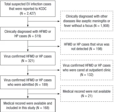
Two thousand four-hundred twenty-seven cases suspected of enteroviruses infection were registered into the Enterovirus Sentinel Surveillance System from January through December, 2009. Among them, 519 cases were HFMD or HP and enterovi-ruses were detected in 321 patients from 24 hospitals. This study
excluded 132 outpatients and requested medical record review on 189 patients who were admitted and treated at each hospi-tal. Among them, a total of 168 patients were able to undergo a medical record review (Fig. 1). Clinical data were collected by each hospital in structured manner for the period from April through August, 2010.
Virus detection
One to 3 specimens among throat swab, endotracheal aspirates, rectal swab, stool or CSF were collected depending on each pa-tient. Enteroviruses genome detection was attempted by real-time reverse transcription–PCR (RT-PCR) by using TaqMan tech-nology (Applied Biosystems, Foster City, CA, USA). A causative organism was determined in cases of detecting one or more vi-ruses among them.
Briefly, viral RNAs were extracted by using the magnetic bead– based viral nucleic acid purification protocol described by Boom et al. (12). Subsequently, 1-step real-time RT-PCR was performed by using a dual-labeled fluorogenic enteroviruses-specific probe and primers designed on the basis of previous data (13). For ge-notyping, seminested RT-PCR was used to amplify part of the viral protein 1 (VP1) gene of enteroviruses, based on the KCDC protocol for detection of pan-enteroviruses, and sequencing analysis for VP1 amplicon was performed by using the auto-matic sequencer and the DNAstar software package (14).
Case definitions
HFMD was defined as having vesiculo-papular rashes over the hands, soles, or buttocks with mouth ulcers while HP was de-fined as having oral ulcers without any skin rash. Cases with development of complications were categorized as the cases accompanied by aseptic meningitis, encephalitis, encephalo-myelitis, and polio-like syndrome (PLS). Aseptic meningitis was defined as having clinical signs or symptoms of meningitis with pleocytosis (leukocyte > 5 cells/μL) with negative bacterial cul-ture in the CSF obtained from a lumbar punccul-ture. Encephalitis was defined as having CSF pleocytosis with a change in con-sciousness. PLS was defined as having acute weakness in the extremities with a drop in muscle power or deep tendon reflex. Encephalomyelitis was defined as having encephalitis and PLS simultaneously.
Data collection
Data were collected for demographic information, past medical history, contact history of enteroviruses associated diseases, vital signs (the presence of fever, peak body temperature, etc.), symp-toms and signs at presentation during the whole course of dis-ease. Collected also were laboratory data including white blood cell and platelet count, hemoglobin, C-reactive protein (CRP), erythrocyte sedimentation rate, blood glucose and cell counts, glucose and protein in CSF, and isolated virus and subtype. Clinically diagnosed with other
diseases like aseptic meningitis or fever without a focus (N = 1,908)
HFMD or HP cases that virus was not detected (N = 198)
Medical recored were not available (N = 21)
Medical recored were available and included in this study (N = 168)
Virus confirmed HFMD or HP cases who were cared at outpatient clinic
(N = 132) Total suspected EV infection cases
that were reported to KCDC (N = 2,427)
Clinically diagnosed with HFMD or HP cases (N = 519)
Virus confirmed HFMD or HP cases (N = 321)
Virus confirmed HFMD or HP cases who were admitted (N = 189)
Fig. 1. The flow chart shows how we selected the study population in this study. EV, enterovirus; HFMD, hand, foot and mouth disease; HP, herpangina; KCDC, Korea Centers for Disease Control and Prevention.
Statistical analysis
We analyzed the data using SPSS 13.0 software (SPSS Inc., Chi-cago, IL, USA). We analyzed fever duration, peak fever, clinical symptom (vomiting, diarrhea, poor oral intake, headache), neu-rologic sign (jerking, gaze palsy, tremor, dysarthria, ataxia, gait disturbance, upper or lower extremity weakness, insomnia, sei-zure, lethargy, coma), laboratory test (white blood cell count, erythrocyte sedimentation rate, CRP, glucose) and isolated virus type as a risk factor. Normally distributed data were compared using Student’s t test; data that were not normally distributed were compared by the Mann-Whitney U test. Categorical data were tested using the chi-square test or Fisher’s exact test. A mul-tiple logistic regression analysis was used to examine the multi-variate-adjusted odds ratios for risk factors that were significant in univariate analysis. A P value less than 0.05 was considered statistically significant.
Ethics statement
The study protocol was approved by the institutional review board of The Catholic Medical Center of The Catholic Universi-ty of Korea (Approval No. XC10RNMI0022). Informed consent was waived by the board.
RESULTS
Among 189 patients from 24 hospitals subject to investigation, 168 patients (88.9%) from 21 hospitals (87.5%) underwent a med-ical record review. Among these subjects, 80 patients (47.6%) were not accompanied by complications while 88 patients (52.4 %) had complications. Of those 88 patients that developed
com-plications, 56 patients (33.3%) had aseptic meningitis, 18 pa-tients (10.7%) had encephalitis or encephalomyelitis, and 14 patients (8.3%) had PLS. Of those 18 subjects with encephalitis or encephalomyelitis, 4 patients (25%) were accompanied by cardiopulmonary complications and 2 of these 4 patients ex-pired (Table 1).
Demographic data
Of those 168 patients who were investigated, 103 patients (68.3%) were males and 65 patients (38.7%) were females with the ratio of 1.58:1. The overall median age was 28.0 months (with a range 16.5-49.5). With respect to the age at which the disease occurs, the peak incidence was shown just before and after 12 months. Ninety two point nine percent of these patients were less than 5 yr old (152/168) (Fig. 2). There was no statistically significant difference in the male-female ratio between the group with com-plications and the group without comcom-plications, but the group with complications was significantly older (P = 0.041) and heavier (P = 0.002), and had a longer hospital stay (P < 0.001) (Table 2).
Outbreak pattern
The patients showed distinctive seasonal prevalence. A death occurred in early May and the number of patients rapidly in-creased thereafter showing the peak incidence in near summer between May and July (Fig. 3).
Table 2. Demographic characteristics of each complicated versus non-complicated group of hand, foot and mouth disease or herpangina in the Republic of Korea, by age group on 2009
Characteristics Total cases Non-complicated cases (n = 88) Complicated cases (n = 80) P
Male:Female 1.58:1 1.35:1 1.84:1 0.334
Age (month)* 28.0 (16.0-48.8) 24.0 (14.3-39.8) 33.5 (18.0-56.5) 0.041
Body weight (kg)* 13.0 (11.0-17.0) 12.1 (9.8-15.7) 14.5 (12.0-19.1) 0.002
Length of hospitalization (day)* 5.0 (4.0-7.0) 4.0 (3.0-5.0) 6.0 (5.0-8.0) 0.000 *Data are presented as median (25-75 %tile range).
Table 1. Classification of various clinical syndrome of virus confirmed hand, foot and mouth disease or herpangina
Clinical syndrome No. of patients (%)
Hand, foot and mouth disease or herpangina without complication
80 (47.6) Hand, foot and mouth disease or herpangina with
complications 88 (52.4)
Aseptic meningitis only Encephalitis or encephalomyelitis without cardiopulmonary complication with cardiopulmonary complications with cardiopulmonary complications with death Polio-like myelitis 56 (33.3) 18 (10.7) 14 (8.3) 2 (1.2) 2 (1.2) 14 (8.3) Total 168 (100) Nu m be r o f c as es Age 0-5 mo6-11 mo 1 yr 2 yr 3 yr 4 yr 5 yr 6 yr 7 yr 8 yr 9 yr 10 yr 11 yr 40 35 30 25 20 15 10 5 0 Complicated group Non-complicated group
Fig. 2. Number of hand, foot and mouth disease or herpangina in the Republic of Ko-rea in 2009, by age group. mo, months; yr, years.
Clinical manifestations
Patients were admitted to the hospital with fever that lasted for the median period of 2 days prior to admission. Fever continued for the median period of 4 days. Of those 168 subjects examined, 133 patients (79.2%) had HFMD while 35 patients (20.8%) had HP. The group with complications had more incidences of HFMD than HP (P = 0.002), had a longer period of fever (P = 0.001) and a higher fever over 39°C (P = 0.028). This group more frequently showed headache (P < 0.001), vomiting (P < 0.001) and one or more neurologic signs/symptoms (P < 0.001). In the group with complications, ataxia, gait disturbance, lower extremity weak-ness, seizure, lethargy and coma, among above neurologic signs and symptoms, were shown statistically more often (Table 3).
Laboratory findings
There was no statistical difference in ratio of patients with leu-kocytosis between the group with complications and without complication (P = 0.465). The mean CRP of the group with com-plications (1.09 ± 2.05 mg/dL) was lower than that of the group without complications (1.47 ± 1.81 mg/dL) (P = 0.022). The mean serum glucose of the group with complications was 110.3 ± 30.8 mg/dL while that of the group without complications was 98.3 ± 26.4 mg/dL, showing a higher level for the former (P = 0.003)
(Table 4).
Viral characteristics
Throat swabs from 92 patients were obtained for virus isolation and 51 (55.4%) were positive for enteroviruses. Of those 154 pa-tients who had a rectal swab or stool sample for virus isolation, 146 (94.8%) were positive. However, 40 patients had a CSF sam-ple for virus isolation and only 3 patients (7.5%) were positive (Fig. 4).
Enterovirus 71 was isolated in 122 patients (72.6%), showing the largest number of cases. Other isolated viruses detected were
Table 4. Laboratory findings of each complicated versus non-complicated group of hand, foot and mouth disease or herpangina in the Republic of Korea in 2009, by age group Laboratory items Total cases Non-complicated cases (n = 88) Complicated cases (n = 80) P
White blood cell count (cells/µL)* 12,255 ± 4,636 12,847 ± 4,970 11,724 ± 4,256 0.118
Leukocytosis† 22 12 10 0.645
Erythrocyte sedimentation rate (mm/hr)* 23.5 ± 17.6 26.0 ± 20.1 18.4 ± 12.5 0.230
C-reactive protein (mg/dL)* 1.27 ± 1.95 1.47 ± 1.81 1.09 ± 2.05 0.022 Glucose (mg/dL)* 104.5 ± 29.3 98.3 ± 26.4 110.3 ± 30.8 0.003 Glucose > 100 mg/dL† 85 33 52 0.011 Glucose > 150 mg/dL† 9 4 5 0.822 Enterovirus 71† 122 46 76 0.000 Non-Enterovirus V71† 46 34 12 0.000
*These data are shown as mean ± SD; †These date are shown as number of patients.
Nu m be r o f c as es 8-M ar 22 -M ar 5-Ap r 19 -A pr 3-M ay 17 -M ay 31 -M ay 14 -J un 28 -J un 12 -J ul 26 -J ul 9-Au g 23 -A ug 6-Se p 20 -S ep 4-Oc t 18 -O ct 1-No v 15 -N ov 29 -N ov 40 35 30 25 20 15 10 5 0 Complicated group Non-complicated group 2nd fatal case 1st fatal case
Fig. 3. Distribution of hand, foot and mouth disease or herpangina in the Republic of Korea in 2009, by date. The arrows indicate the dates of onset of the two fatal cases.
Table 3. Clinical manifestations of each complicated versus non-complicated group of hand, foot and mouth disease or herpangina in the Republic of Korea in 2009, by age group
Characteristics casesTotal
Non- complicated cases (n = 88) Complicated cases (n = 80) P Fever before admission (days)* 2.0
(1.0-3.0) 2 (1.0-3.0) 2.0 (1.0-3.0) 0.206 Total fever duration (days)* 4.0
(2.0-5.0) (2.0-5.0)3.0 (3.0-5.5)4.0 0.001
Peak fever over 39°C† 30 30 39 0.028
Fever duration over 4 days† 85 30 55 0.001
Vomiting† 74 24 50 0.000
Diarrhea† 12 4 8 0.304
Poor oral intake† 113 59 54 0.088
Headache† 46 7 39 0.000 Neurologic sign† 57 7 50 0.000 Jerking† 11 3 8 0.162 Gaze palsy† 6 1 5 0.122 Tremor† 3 0 3 0.096 Dysarthria† 5 1 4 0.209 Ataxia† 5 0 5 0.030 Gait disturbance† 17 0 17 0.000
Upper extremity weakness† 1 0 1 0.524
Lower extremity weakness† 14 1 13 0.002
Insomnia† 2 0 2 0.175
Seizure† 11 1 10 0.008
Lethargy† 28 5 23 0.001
Coma† 5 0 5 0.030
*These data are shown as median (25-75 %tile range); †These date are shown as number of patients.
coxsackievirus A2, A5, A6, A12, A16, coxsackievirus B1, echovi-rus 9 and untypable enteroviechovi-rus. Among cases with enteroviechovi-rus 71 isolation, 67 cases (54.9%) belonged to the subgenogroup C4a. This was shown to be 97% of homologous to enterovirus 71 subgenogroup that was endemic in China, 2008.
Risk factor analysis
The group with complications and the group without complica-tions were compared by univariate analysis. It was found that significant risk factors for complications included a rash form of HFMD, fever longer than 4 days, peak body temperature over 39°C, vomiting, headache, neurologic sign (such as jerking, gaze palsy, tremor, dysarthria, ataxia, gait disturbance, extremity weak-ness, insomnia, seizure, lethargy, coma), glucose over than 100 mg/dL and enterovirus 71 as a causative virus. After multiple logistic analysis, headache (Odds ratio [OR], 10.75; P < 0.001) and neurologic sign (OR, 42.76; P < 0.001) were the most signif-icant factors (Table 5).
DISCUSSION
In the late 1990s, the large outbreaks of HFMD or HP caused by enterovirus 71 continuously occurred in the neighbor countries of the ROK and many deaths had occurred (4-8). Thus, a large outbreak of HFMD or HP was also a matter of concern in the ROK. According to the Enterovirus Sentinel Surveillance Sys-tem of the ROK, there was no HFMD or HP caused by enterovi-rus 71 up until 2008, but the outbreaks of HFMD or HP caused by enterovirus 71 and deaths occurred from the year 2009. Ac-cordingly, this study attempted to understand more accurately the epidemic aspect of large outbreaks of HFMD or HP that oc-curred in the ROK in 2009, and to analyze the risk factors asso-ciated with complications.
In this study, the ratio of males and females of HFMD or HP
incidences was 1.58:1 while 93.8% of all patients were under the age of five years. The largest number of patients was especially around the age of one year. Deaths occurred at around the age of one year, despite the fact that only two had expired. This re-sult concurred with that of other studies (15-18).
As the patients are younger, there are higher possibility of com-plications and death from HFMD or HP. Nevertheless, neuro-logic signs are often subtle and easily overlooked (19, 20). Thus, in times of HFMD or HP epidemic, careful observations with respect to occurrences of complications of patients are neces-sary in their early childhood. The HFMD or HP epidemic in the ROK was manifested largely in the period from May through August, 2009. It almost did not occur after October that year, which was similar to what was seen in other neighbor countries (15, 20). However, owing to the possibility of a continuous epi-demic regardless of any particular season, it is necessary to put together a well-built sentinel surveillance system and to estab-lish an early warning system for the national public in times of an epidemic.
Complications of HFMD or HP disclosed the spectrum of dis-ease severity. Complications began largely in the form of asep-tic meningitis. Then, they progressed to the state of meningo-encephalitis that manifested a change of consciousness. How-ever, some patients did not show any change of consciousness, instead of complications progressed to PLS. Well-known already was the report that enterovirus 71 caused complications in the form of not only meningoencephalitis but also PLS (21). Entero-virus 71 Entero-virus is on the rise as the most important causative Entero-virus for the PLS in the Western Pacific region where polio viruses had been eradicated (22).
It was reported that enterovirus 71 that often brought about complications would have a tissue tropism in the brain stem, cerebellum and spinal cord anterior horn cell (23). Clinical man-ifestations may change depending on the body tissues where these viruses first act upon. The possibility of complications pro-gression is high in cases of change of consciousness such as irri-tability or lethargy, as well as manifestations of neurologic symp-toms, such as ataxia, tremor, lower extremity weakness, inability to walk and decreased deep tendon reflex. Thus, careful obser-Table 5. Multivariated risk factor analysis of complications of hand, foot and mouth disease or herpangina
Risk factors (95% Confidence Interval)Odds ratio P
Peak fever ≥ 39°C 1.03 (0.39-2.74) 0.958 Fever duration ≥ 4 days 1.27 (0.48-3.36) 0.636 Rash of hand, foot and mouth disease 2.87 (0.72-11.48) 0.135
Vomiting 1.20 (0.47-3.07) 0.699 Headache 10.75 (3.56-32.52) 0.000 Neurologic sign 15.00 (4.95-45.4) 0.000 Serum glucose > 100 mg/dL 1.35 (0.56-3.24) 0.508 Enterovirus 71 1.20 (0.37-3.85) 0.766 114 (67.9%) 21 (12.5%) 1 (0.6%) 30 (17.9%) 2 (1.2%) 0 0 Rectal specimens 146/154 (94.8%) Throat specimens 51/92 (55.4%) CSF specimens 3/40 (7.5%) Fig. 4. Origins and numbers (percentage) of enterovirus positive samples on RT-PCR in the Republic of Korea in 2009. CSF, cerebrospinal fluid.
vations and effective measures for these symptoms would be necessary.
In this study, hyperglycemia and leukocytosis at admission were not statistically significant risk factors of complications. These findings were not present on admission of the expired patients in this study, only manifested after witnessing declin-ing consciousness and the worsendeclin-ing patient’s condition. The results of this study led us to believe that clinicians would not be able to predict looming complications with mere general labo-ratory findings. However, the finding of hyperglycemia or leu-kocytosis in patients with HFMD or HP may already reflect the considerable progression of complications.
Chen et al. (22) reported, akin to the results of this study, that hyperglycemia or leukocytosis were signs after the progression of the disease itself, rather than risk factors of complications. However, there were reports in which hyperglycemia and leu-kocytosis were investigated as the risk factors for fatal enterovi-rus 71 infections (19, 25).
In this study, the mean CRP was significantly lower in the com-plications group. It might reflect that inflammatory responses were low in the complications group. Owing to weak inflamma-tory responses toward viruses in patients with HFMD or HP ac-companied by complications, viral proliferation would not ef-fectively be suppressed. As a result, a severe clinical progression might ensue. Additional study is necessary for this pathophysi-ologic mechanism.
The viral diagnostic sensitivity was the highest in the stool specimens in this study. However, according to Ooi et al. (26), the diagnostic sensitivity for enterovirus 71 was the highest in the throat swab specimens. It is advisable for clinicians to take two or more specimens in order to increase diagnostic sensitiv-ity. This study showed that the enterovirus 71 subgenogroup epidemic in the ROK, 2009, was enterovirus 71 C4a. This was 97% homologous to enterovirus 71 C4a, which had been preva-lent in China, 2008 (8). Nevertheless, enterovirus 71 was not an epidemic in the ROK in 2008. Thus, the enterovirus 71 epidemic in China might have been introduced to the ROK in 2009. A re-port claimed that a gradual increase in recent international travel often introduce epidemic viruses from one country to neighbor countries (27). Thus, it is necessary to monitor not only the state of the domestic enterovirus 71 epidemic but also of other coun-tries.
The HFMD or HP patients with neurologic complications could progress to death within a few days after development of fever. However, at primary hospital, diagnosis and treatment while conducting tests searching for causative viruses would not be possible in reality. Therefore, it is of utmost importance to find risk factors associated with development of complica-tions based on clinical symptoms. In this study, rash patterns of HFMD rather than HP, fever longer than 4 days, peak body tem-perature over 39°C, vomiting, headache and neurologic signs
were associated with complications. Also, neurologic signs in-cluded ataxia, gait disturbance, lower extremity weakness, sei-zure, lethargy and coma. Ooi et al. (24) reported that fever longer than 3 days, peak temperature over 38.5°C and history of leth-argy were identified as independent risk factors for a neurologi-cal involvement. Chang et al. (19) reported that fever for longer than 3 days, peak body temperature over 39°C, headache, leth-argy, vomiting, and seizure were associated with a central ner-vous system involvement with an enterovirus 71 infection. Put-ting these various reports together up to now, higher fever, lon-ger fever duration, as well as meningeal irritation or other neu-rologic signs, are common risk factors of central nervous system complications. In times of HFMD or HP epidemic, early detec-tion and timely proper management of patients with such risk factors are essential.
Until now, there is no vaccine or antiviral agent effective against enteroviruses-caused HFMD or HP. However, there was a re-port, although additional verifications are required, that timely administration of intravenous immunoglobulin (IVIG) for se-vere enterovirus 71 patients had reduced acute mortality (28-30). Thus, attempts of close monitoring and timely IVIG infu-sion would be necessary for patients with the above mentioned risk factors.
Our study has some limitations due to its retrospective nature. Firstly, we did not investigate potentially important epidemio-logic factors likewise number of children and adults in a family, history of HFMD or HP before admission, intrafamilial or out-side contact with HFMD or HP, enrollment in a kindergarten or child care center. Secondly, each patient’s specimens were not collected at the same time from disease onset. Thirdly, we inves-tigated only hospitalized patient, therefore our result may be not applicable to primary care setting.
In conclusion, in cases of having rash patterns of HFMD rath-er than HP, fevrath-er longrath-er than 4 days, peak body temprath-erature over 39°C, vomiting, headache, neurologic signs, serum glucose over 100 mg/dL and enterovirus 71 as a causative virus might be indicative of a grave prognosis in times of HFMD or HP epi-demic. Therefore, early recognition and especially meticulous management in patients with these risk factors are requisite.
ACKNOWLEDGMENTS
Members of the Enteroviruses Complications Working Group
The following investigators and institutions participated in the Enteroviruses Complications Working Group other than those listed authors of this paper: Byung Min Choi, Department of Pediatrics, College of Medicine, Korea University, Seoul; Eun Hwa Choi, Department of Pediatrics, Seoul National University College of Medicine, Seoul; Jae Yoon Kim, Department of Pedi-atrics, National Medical Center, Seoul; Jung Yeun Hong,
Depart-ment of Pediatrics, School of Medicine, Cheju National Univer-sity, Cheju; Sung Hee Oh, Department of Pediatrics, Hanyang University School of Medicine, Seoul; Sung Ho Cha, Depart-ment of Pediatrics, College of Medicine, Kyunghee University, Seoul; Yae-Jean Kim, Department of Pediatrics, Sungkyunkwan University School of Medicine, Seoul, Korea. The authors have no conflicts of interest to disclose.
REFERENCES
1. Lum LC, Wong KT, Lam SK, Chua KB, Goh AY, Lim WL, Ong BB, Paul G, AbuBakar S, Lambert M. Fatal enterovirus 71 encephalomyelitis. J
Pedi-atr 1998; 133: 795-8.
2. Schmidt NJ, Lennette EH, Ho HH. An apparently new enterovirus
iso-lated from patients with disease of the central nervous system. J Infect Dis 1974; 129: 304-9.
3. Landry ML, Fonseca SN, Cohen S, Bogue CW. Fatal enterovirus type 71
infection: rapid detection and diagnostic pitfalls. Pediatr Infect Dis J 1995; 14: 1095-100.
4. Shekhar K, Lye MS, Norlijah O, Ong F, Looi LM, Khuzaiah R, Marzuki I, Hussein I, Wong SL, Mohan J, et al. Deaths in children during an
out-break of hand, foot and mouth disease in Peninsular Malaysia: clinical and pathological characteristics. Med J Malaysia 2005; 60: 297-304.
5. Ho M, Chen ER, Hsu KH, Twu SJ, Chen KT, Tsai SF, Wang JR, Shih SR.
An epidemic of enterovirus 71 infection in Taiwan. Taiwan Enterovirus Epidemic Working Group. N Engl J Med 1999; 341: 929-35.
6. Fujimoto T, Chikahira M, Yoshida S, Ebira H, Hasegawa A, Totsuka A, Nishio O. Outbreak of central nervous system disease associated with
hand, foot, and mouth disease in Japan during the summer of 2000: de-tection and molecular epidemiology of enterovirus 71. Microbiol Immu-nol 2002; 46: 621-7.
7. Shah VA, Chong CY, Chan KP, Ng W, Ling AE. Clinical characteristics of
an outbreak of hand, foot and mouth disease in Singapore. Ann Acad Med Singapore 2003; 32: 381-7.
8. Zhang Y, Tan XJ, Wang HY, Yan DM, Zhu SL, Wang DY, Ji F, Wang XJ, Gao YJ, Chen L, et al. An outbreak of hand, foot, and mouth disease
as-sociated with subgenotype C4 of human enterovirus 71 in Shandong, China. J Clin Virol 2009; 44: 262-7.
9. Jee YM, Cheon DS, Kim K, Cho JH, Chung YS, Lee J, Lee SH, Park KS, Lee JH, Kim EC, et al. Genetic analysis of the VP1 region of human
en-terovirus 71 strains isolated in Korea during 2000. Arch Virol 2003; 148: 1735-46.
10. Ryu W, Kang B, Hong J, Hwang S, Kim A, Kim J, Cheon DS. Enterovirus
71 infection with central nervous system involvement, South Korea. Emerg Infect Dis 2010; 16: 1764-6.
11. Ryu WS, Kang B, Hong J, Hwang S, Kim J, Cheon DS. Clinical and
etio-logical characteristics of enterovirus 71-related diseases during a recent 2-year period in Korea. J Clin Microbiol 2010; 48: 2490-4.
12. Boom R, Sol CJ, Salimans MM, Jansen CL, Wertheim-van Dillen PM, van der Noordaa J. Rapid and simple method for purification of nucleic
acids. J Clin Microbiol 1990; 28: 495-503.
13. Tan EL, Yong LL, Quak SH, Yeo WC, Chow VT, Poh CL. Rapid detection
of enterovirus 71 by real-time TaqMan RT-PCR. J Clin Virol 2008; 42: 203-6.
14. Nix WA, Oberste MS, Pallansch MA. Sensitive, seminested PCR
amplifi-cation of VP1 sequences for direct identifiamplifi-cation of all enterovirus sero-types from original clinical specimens. J Clin Microbiol 2006; 44: 2698-704.
15. Tseng FC, Huang HC, Chi CY, Lin TL, Liu CC, Jian JW, Hsu LC, Wu HS, Yang JY, Chang YW, et al. Epidemiological survey of enterovirus
infec-tions occurring in Taiwan between 2000 and 2005: analysis of sentinel physician surveillance data. J Med Virol 2007; 79: 1850-60.
16. Kung SH, Wang SF, Huang CW, Hsu CC, Liu HF, Yang JY. Genetic and
antigenic analyses of enterovirus 71 isolates in Taiwan during 1998-2005. Clin Microbiol Infect 2007; 13: 782-7.
17. Ortner B, Huang CW, Schmid D, Mutz I, Wewalka G, Allerberger F, Yang JY, Huemer HP. Epidemiology of enterovirus types causing neurological
disease in Austria 1999-2007: detection of clusters of echovirus 30 and enterovirus 71 and analysis of prevalent genotypes. J Med Virol 2009; 81: 317-24.
18. Ma E, Lam T, Chan KC, Wong C, Chuang SK. Changing epidemiology of
hand, foot, and mouth disease in Hong Kong, 2001-2009. Jpn J Infect Dis 2010; 63: 422-6.
19. Chang LY, Lin TY, Hsu KH, Huang YC, Lin KL, Hsueh C, Shih SR, Ning HC, Hwang MS, Wang HS, et al. Clinical features and risk factors of
pul-monary oedema after enterovirus-71-related hand, foot, and mouth dis-ease. Lancet 1999; 354: 1682-6.
20. Podin Y, Gias EL, Ong F, Leong YW, Yee SF, Yusof MA, Perera D, Teo B, Wee TY, Yao SC, et al. Sentinel surveillance for human enterovirus 71 in
Sarawak, Malaysia: lessons from the first 7 years. BMC Public Health 2006; 6: 180.
21. Huang CC, Liu CC, Chang YC, Chen CY, Wang ST, Yeh TF. Neurologic
complications in children with enterovirus 71 infection. N Engl J Med 1999; 341: 936-42.
22. Chen CY, Chang YC, Huang CC, Lui CC, Lee KW, Huang SC. Acute
flac-cid paralysis in infants and young children with enterovirus 71 infection: MR imaging findings and clinical correlates. AJNR Am J Neuroradiol 2001; 22: 200-5.
23. Hsueh C, Jung SM, Shih SR, Kuo TT, Shieh WJ, Zaki S, Lin TY, Chang LY, Ning HC, Yen DC. Acute encephalomyelitis during an outbreak of
entero-virus type 71 infection in Taiwan: report of an autopsy case with patho-logic, immunofluorescence, and molecular studies. Mod Pathol 2000; 13: 1200-5.
24. Ooi MH, Wong SC, Mohan A, Podin Y, Perera D, Clear D, del Sel S, Chieng CH, Tio PH, Cardosa MJ, et al. Identification and validation of clinical
predictors for the risk of neurological involvement in children with hand, foot, and mouth disease in Sarawak. BMC Infect Dis 2009; 9: 3.
25. Yang TT, Huang LM, Lu CY, Kao CL, Lee WT, Lee PI, Chen CM, Huang FY, Lee CY, Chang LY. Clinical features and factors of unfavorable
out-comes for non-polio enterovirus infection of the central nervous system in northern Taiwan, 1994-2003. J Microbiol Immunol Infect 2005; 38: 417-24.
26. Ooi MH, Solomon T, Podin Y, Mohan A, Akin W, Yusuf MA, del Sel S, Kontol KM, Lai BF, Clear D, et al. Evaluation of different clinical sample
types in diagnosis of human enterovirus 71-associated hand-foot-and-mouth disease. J Clin Microbiol 2007; 45: 1858-66.
27. Mizuta K, Abiko C, Murata T, Matsuzaki Y, Itagaki T, Sanjoh K, Sakamo-to M, Hongo S, Murayama S, Hayasaka K. Frequent importation of
en-terovirus 71 from surrounding countries into the local community of Yamagata, Japan, between 1998 and 2003. J Clin Microbiol 2005; 43: 6171-5.
28. Wang J, Yao C, Yeh C, Huang CC, Wang SM, Liu CC, Wu JM. Critical
management in patients with severe enterovirus 71 infection. Pediatr Int 2006; 48: 250-6.
29. Chang LY, Hsia SH, Wu CT, Huang YC, Lin KL, Fang TY, Lin TY.
Out-come of enterovirus 71 infections with or without stage-based
manage-ment: 1998 to 2002. Pediatr Infect Dis J 2004; 23: 327-32.
30. Wang SM, Lei HY, Huang MC, Su LY, Lin HC, Yu CK, Wang JL, Liu CC.
Modulation of cytokine production by intravenous immunoglobulin in patients with enterovirus 71-associated brainstem encephalitis. J Clin Virol 2006; 37: 47-52.