Proteases are now considered not only to be enzymes that conduct hydrolysis of peptide bonds linking amino acids, but also signaling molecules that play an important role in homeostatic regulation in mammals and numerous pathological conditions.1,2In the skin, three families of proteases have been found in the stratum
corneum (SC), including the epidermal specific serine proteases, kallikrein 5 (SC tryptic enzyme, SCTE) and kallikrein 7 (SC chymotryptic enzyme, SCCE), as well as cysteine proteases, including cathepsin C, L, and V (SC thiol protease),
Protease and Protease-Activated Receptor-2 Signaling
in the Pathogenesis of Atopic Dermatitis
Sang Eun Lee,
1,2Se Kyoo Jeong,
3and Seung Hun Lee
1,21Department of Dermatology, 2Human Barrier Research Institute, Yonsei University College of Medicine, Seoul; 3Research Division, NeoPharm Co., Ltd., Daejeon, Korea.
Proteases in the skin are essential to epidermal permeability barrier homeostasis. In addition to their direct proteolytic effects, certain proteases signal to cells by activating protease-activated receptors (PARs), the G-protein-coupled receptors. The expression of functional PAR-2 on human skin and its role in inflammation, pruritus, and skin barrier homeostasis have been demonstrated. Atopic dermatitis (AD) is a multifactorial inflammatory skin disease characterized by genetic barrier defects and allergic inflammation, which is sustained by gene-environmental interactions. Recent studies have revealed aberrant expression and activation of serine proteases and PAR-2 in the lesional skin of AD patients. The imbalance between proteases and protease inhibitors associated with genetic defects in the protease/protease inhibitor encoding genes, increase in skin surface pH, and exposure to proteolytically active allergens contribute to this aberrant protease/ PAR-2 signaling in AD. The increased protease activity in AD leads to abnormal desquamation, degradation of lipid-processing enzymes and antimicrobial peptides, and activation of primary cytokines, thereby leading to permeability barrier dysfunction, inflammation, and defects in the antimicrobial barrier. Moreover, up-regulated proteases stimulate PAR-2 in lesional skin of AD and lead to the production of cytokines and chemokines involved in inflammation and immune responses, itching sensation, and sustained epidermal barrier perturbation with easier allergen penetration. In addition, PAR-2 is an important sensor for exogenous danger molecules, such as exogenous proteases from various allergens, and plays an important role in AD pathogenesis. Together, these findings suggest that protease activity or PAR-2 may be a future target for therapeutic intervention for the treatment of AD.
Key Words: Atopic dermatitis, protease, protease-activated receptor-2 (PAR-2)
Received: August 17, 2010
Corresponding author: Dr. Seung Hun Lee, Department of Dermatology,
Yonsei University College of Medicine, Gangnam Severance Hospital, 712 Eonju-ro, Gangnam-gu, Seoul 135-720, Korea.
Tel: 82-2-2019-3361, Fax: 82-2-3463-6136 E-mail: ydshderm@yuhs.ac
∙The authors have no financial conflicts of interest.
© Copyright:
Yonsei University College of Medicine 2010 This is an Open Access article distributed under the terms of the Creative Commons Attribution Non-Commercial License (http://creativecommons.org/ licenses/by-nc/3.0) which permits unrestricted non-commercial use, distribution, and reproduction in any medium, provided the original work is properly cited.
and at least one aspartate protease, cathepsin D.3-7These proteases are tightly regulated by specific protease inhibi-tors and mediate various cellular responses in the skin, such as inflammation and immune responses, host defense, chemotaxis, cytokine expression, vascular function, tissue repair, and apoptosis.8In addition to endogenous proteases, exogenous proteases from allergens such as house dust mites, cockroaches, certain bacteria, and fungi can also signal the epidermis. A number of biological activities of proteases are mediated, at least in part, by the activation of its receptors, protease-activated receptors (PARs).9Recent works have indicated that PAR-2, as a sensor for endoge-nous as well as exogeendoge-nous proteases, plays numerous physiological and pathophysiological roles in the skin.8,10In addition, there is increasing evidence that protease and PAR-2 play an important role in the maintenance of epi-dermal permeability barrier homeostasis.7,11,12 Moreover, abnormal expression or activity of serine proteases and PAR-2 has been associated with several inflammatory skin disorders involving barrier abnormalities, including atopic dermatitis, netherton syndrome (NS), psoriasis, and peeling skin syndrome.13-18In this review, we will discuss the role of protease/PAR-2 signaling in epidermal permea-bility barrier homeostasis, as well as its contribution to the pathogenesis of atopic dermatitis (AD).
Human tissue kallikreins (KLKs) are the largest family of trypsin- or chymotrypsin-like secreted serine proteases.13 Eight KLKs, including KLK5, -6, -7, -8, -10, -11, -13, and -14 are known to be expressed in the epidermis and skin appendages, such as sebaceous gland.19Among these, only KLK7 exhibits chymotrypsin-like substrate specificity; the other KLKs exhibit trypsin-like serine proteases. It is well known that KLK5 and KLK7 are the major active KLKs in the SC, regulating the desquamation process through corneodesmosomal cleavage and lipid barrier formation by degrading lipid processing enzymes.19-21In addition, KLK5 and KLK7 have been shown to control the enzymatic processing of cathelicidin, thereby affecting its antimicro-bial activity and inflammatory responses.22 KLK14 has also been detected in its active form in the SC and is thought to be a candidate protease involved in the process of desqua-mation, however, its precise role has not been elucidat-ed.23,24KLK5 and KLK7 are stored in the form of pro-enzyme in the lamellar bodies (LBs) along with a substrate of KLK7, corneodesmosin and their inhibitor, lymphoe-pithelial Kazal-type-related inhibitor (LEKTI).25Upon release into the stratum granulosum (SG)-SC interface,
these pro-KLK zymogens are activated through a KLK proteolytic activation cascade.26KLK5 can be activated by itself or by KLK14 and then activate several other pro-KLKs; therefore KLK5 is thought to be the key protease for the initiation of the KLK cascade.24Significantly, KLK5 and KLK14 are known to activate PAR-2, thereby modulat-ing epidermal permeability barrier homeostasis, immune and inflammatory responses, skin pigmentation, itching sensations, as well as tumor surveillance.10,27KLK8 has been reported to be localized in the LBs with KLK5, KLK7 and their inhibitors and secreted in the SG-SC interface, regulating desquamation and epidermal proliferation.28,29 Recent studies have investigated factors regulating the expression of KLKs in the skin and reported that epider-mal calcium ions, vitamin D3, and retinoic acid indepen-dently regulate the expression of KLK5 and KLK7 in normal human epidermal keratinocytes.30An increase in extracellular calcium induced KLK5 and KLK7 expres-sion with induction of differentiation markers, suggesting that the expression of KLKs is induced during epidermal differentiation. In contrast, both 9-cis retinoic acid and 13-cis retinoic acid increased KLK5 and KLK7 expression, independently of keratinocyte differentiation. 1,25 (OH)2 vitamin D3, which is well known to induce cathelicidin expression, was also demonstrated to stimulate the expres-sion of KLK5 and KLK7, which are co-localized with cathelicidin.
In addition to KLKs, the two transmembrane serine protease, matriptase (MT-SP1) and prostasin have been identified in the uppermost epidermal layers and postulated to be involved in epidermal barrier formation and SC hydra-tion via epithelial sodium channel-induced intracellular calcium influx and consequent activation of calcium-dependent proteases and transglutaminases (TGMs).31 Moreover, the observation that mice deleted for matriptase showed impaired desquamation suggests that matriptase is also involved in the process of desquamation.32
A number of serine protease inhibitors are present in the skin, and regulate proteolytic activity in order to prevent excessive serine protease cascade, while also maintaining permeability barrier homeostasis. Among several serine protease inhibitors observed in the SC, such as secretory leucocyte protease inhibitor (SLPI), elafin skin-dericed antileukoproteinase (SKALP), and plasminogen activator inhibitor type 2, which are normally cross-linked to the cornified envelope, LEKTI-1, a secreted serine protease in-hibitor, is thought to be the major player in the SC. LEKTI is encoded by SPINK5 (serine protease inhibitor Kazal-type 5) and its multidomains exhibit diverse inhibitory effects toward trypsin, plasmin, subtilisin A, cathepsin G, and human neutrophil elastase.33In addition, previous in vitro studies demonstrated that LEKTI fragments could
SERINE PROTEASES AND THEIR
INHIBITORS IN SKIN
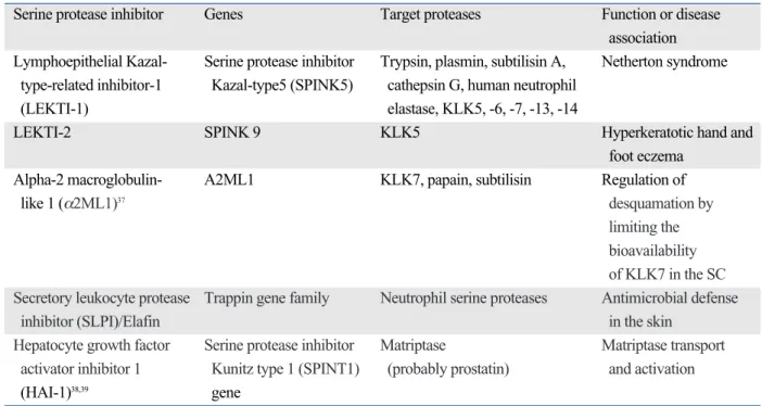
inhibit KLK5, -6, -7, -13, and -14.31LEKTI-1 is stored in specific intracellular LB cargoes, separated from the KLKs, until it is secreted and released into the SC-SG junction to colocalize with KLK5 and KLK7 and inhibit their activity. The interaction strength between LEKTI-1 and KLKs is regulated by the SC pH gradient, with its highest inhibitory capacity at a neutral pH and decreased activity in an acidic pH environment, such as in the superficial SC layer. This implies that a normal SC pH gradient regulates the controlled release of active KLKs in the superficial SC layer and prevents premature desqua-mation in the deep SC layer, where the pH is neutral.34 These findings support the fact that AD, characterized by altered skin surface pH, and NS, characterized by geneti-cally defective LEKTI, display similar phenotypic features, including premature and excessive desquamation. Recen-tly, LEKTI-2, the product of SPINK9, has been identified to be focally localized at the SG and SC at palmar and plantar sites in close proximity to KLK5.35It was found that recombinant LEKTI-2 inhibited KLK5 but not KLK7 and -14, suggesting that LEKTI-2 contributes to the regulation of the desquamation process by inhibiting KLK5 pro-teolytic activity. Other serine protease inhibitors and their roles are summarized in Table 1.36-38
PAR is a G-protein coupled receptor, characterized by a unique mechanism of self-activation following specific
proteolytic cleavage of their extracellular domains.39Until now, four PAR members have been identified. PAR-1, -3, and -4 are known to be activated by thrombin, and thereby involved in homeostasis and thrombosis, whereas PAR-2 is activated by trypsin-like serine proteases, but not by thrombin.10,40PAR-2 is known to be widely distributed throughout the mammalian body. In the skin, PAR-2 is abundantly expressed by almost all cell types, especially by keratinocytes. In addition, endothelial cells, fibroblasts, sensory neurons, and inflammatory cells such as mast cells, T lymphocytes, eosinophils, neutrophils, monocytes, macrophages, and dendritic cells are also reported to ex-press functional PAR-2.10,40Previous studies demonstrated that PAR-2 was expressed in the suprabasal layers of both human and murine epidermis, and that this expression was most prominent in the granular layer, implying that PAR-2 expression might depend on the state of epidermal differ-entiation.7,40As opposed to normal skin, the lesional skin of atopic dermatitis has been shown to express high levels of PAR-2 also in the lower epidermal layers.21,40,41PAR-2 as well as KLK14 has been shown to be widely distributed in lesional skin in rosacea, another inflammatory skin disease.21Taken together, this evidence suggests that PAR-2 expression may be induced by cutaneous inflammation. In addition, PAR-2 expression has been reported to be re-gulated by ultraviolet irradiation and involved in melano-some transfer.42
Various endogenous serine proteases including trypsin, mast cell derived tryptase, KLK5, -6, and, -14, matriptase-1 [membrane-type serine protease-matriptase-1 (MT-SPmatriptase-1)], human
Table 1. Serine Proteases and Their Inhibitor in Skin
Serine protease inhibitor Genes Target proteases Function or disease
association Lymphoepithelial Kazal- Serine protease inhibitor Trypsin, plasmin, subtilisin A, Netherton syndrome
type-related inhibitor-1 Kazal-type5 (SPINK5) cathepsin G, human neutrophil
(LEKTI-1) elastase, KLK5, -6, -7, -13, -14
LEKTI-2 SPINK 9 KLK5 Hyperkeratotic hand and
foot eczema
Alpha-2 macroglobulin- A2ML1 KLK7, papain, subtilisin Regulation of
like 1 (α2ML1)37 desquamation by
limiting the bioavailability of KLK7 in the SC Secretory leukocyte protease Trappin gene family Neutrophil serine proteases Antimicrobial defense
inhibitor (SLPI)/Elafin in the skin
Hepatocyte growth factor Serine protease inhibitor Matriptase Matriptase transport activator inhibitor 1 Kunitz type 1 (SPINT1) (probably prostatin) and activation
(HAI-1)38,39 gene
SC, stratum corneum.
airway trypsin-like protease (HAT), cathepsin G, and factor Xa have been demonstrated to activate PAR-2. In addition, some kinds of pathogenic organisms with proteolytic acti-vity such as house dust mites, cockroaches, pollens, molds or bacteria may also be exogenous activators of PAR-2. Upon activation, PAR-2, which is mainly localized to lipid raft domains under basal status, is known to be endocytosed and degraded.7,43As a G-protein coupled receptor, PAR-2 is known to have a common signaling pathway, including the activation of phospholipase C, which results in the forma-tion of ionsitol triphosphate and diacylglycerol, followed by calcium mobilization.39In vitro, PAR-2 activation by PAR-2 agonist peptide has been demonstrated to provoke transient intracellular calcium mobilization in primary kera-tinocytes, suggesting that PAR-2 could regulate the proli-feration and differentiation of keratinocytes.44,45The precise role of PAR-2 in epidermal barrier homeostasis is discuss-ed below.
KLKs and their inhibitors are co-localized in the LBs and secreted to the SG-SC junction. The enzyme activity of various proteases in the skin and the inhibitory activity of protease inhibitors are regulated by the SC pH gradient. These findings imply that proteases play an important role in epidermal permeability barrier homeostasis, and recent studies have further shown the importance of proteases and
PAR-2 signaling in the barrier function of the skin. Pro-teases exert various cellular responses, some of which may be, in part, mediated via activation of PAR-2.
Non-PAR-2-mediated function
Filaggrin processing
Filaggrin aggregates keratin intermediate filaments to form the cornified cell envelope, thereby providing the structural support and mechanically resilient skin barrier. In addition, these polypeptides are degraded into natural moisturizing factors (NMF), and contribute to water retention within the SC layers, helping to maintain skin hydration.46,47A num-ber of proteases, including PEP-1, µ-calpain, furin, prosta-tin, matriptase, and caspase-14, elastase2 (ELA2), have been demonstrated to be involved in the proteolytic pro-cessing of filaggrin and profilaggrin, which leads to epi-dermal differentiation, barrier formation and hydration.32,48-52 Recently, the two membrane-bound serine proteases matri-ptase and prostasin have been reported to be involved in the processing of profilaggrin by activating the epithelial sodium channel (ENaC) which causes a calcium influx through a voltage-gated calcium channel, thereby inducing the activation of calcium-dependent proteases.31 Matriptase, or MT-SP1 is a type II trans-membrane serine protease and is now considered to be an essential component of the pro-filaggrin-processing pathway, in accordance with the observation that neonate Matriptase/MT-SP1-deficient skin displayed a loss of mature filaggrin monomer.32 Matri-ptase is also known to be co-localized with another
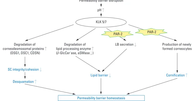
mem-Fig. 1. Role of serine proteases in the epidermal permeability barrier homeostasis. aSMase, acid sphingomyelinase; GlcCer’ase, β-glucocerebrosidase; CDSN, corneodesmosin; DSC, desmocollin; DSG, desmoglein; KLK, kallikrein; LB, lamellar body; PAR-2, protease-activated receptor-2; SC, stratum corneum.
PROTEASE/PAR-2 AND
EPIDERMAL BARRIER
brane-bound serine protease, prostatin (CAP1/PRSS8), and could activate prostatin in vitro, resulting in the initia-tion of a proteolytic cascade toward ENaC activainitia-tion.53 Desquamation
Desquamation involves the enzymatic process of degrada-tion of corneodesmosomal proteins including corneodes-mosin (CDSN), desmoglein 1 (DSG1), and desmocollin 1 (DSC1). It is well known that KLKs, especially KLK5 and KLK7, act as main players in desquamation through a pH-dependent protease signaling cascade (Fig. 1).20 KLK7 directly cleaves CDSN and DSC1 but is unable to degrade DSG1, while KLK5 can.54During KLK proteolytic activa-tion cascade, pro-KLK5 is activated by KLK14 and by KLK5 itself, and the active KLK5 then activates pro-KLK7 and pro-KLK14; thus, KLK5 is believed to be the cascade initiator.24Besides KLK5, -7, and -14, it has been reported that KLK8 is also involved in skin desquamation through a protease cascade reaction leading to the degradation of DSG1 and CDSN.29Recently, KLK6, -13, and -14 have been found to degrade DSG1 and be inhibited by LEKTI, suggesting that these KLKs are also potential desquama-tory enzymes.54The desquamation process is tightly con-trolled by the epidermal pH gradient. Skin pH regulates not only the activity of KLK but also the binding of KLK to LEKTI fragments.55At a neutral pH of the SG-SC junc-tion, KLK is tightly bound to LEKTI fragments, however, with a decrease in pH (acidic pH of the SC) the dissocia-tion of KLK from LEKTI becomes more frequent, releas-ing free KLKs into the outer layer of the SC.55However, at the acidic pH of the superficial SC layer, KLKs exhibit lower activity than at a neutral pH. This bidirectional regulation of KLKs and their inhibitors by pH is important
to maintain proper skin desquamation.13In addition to serine proteases, cystein protease, cathepsin V, and cysta-tin M/E, an inhibitor of asparaginyl endopeptidase legu-main (LGMN) and cysteine proteases also controls desqua-mation.56,57Cathepsin V has been known to degrade DSG1, DSC1, and CDSN with a higher proteolytic activity at an acidic pH, suggesting that cathepsin V is a major player in desquamation under basal conditions with a normal SC acidic pH.56Cystatin M/E regulates desquamation by inhi-bition of cathepsin V as well as LGMN, which regulates pH-dependent processing of (pro)-cathepsins. In addition, cystatin M/E regulates crosslinking of structural proteins by transglutaminase (TGM) 3 during epidermal differen-tiation by controlling cathepsin L and LGMN activities.56A recent study has shown that the cystatin M/E and cathep-sin V were expressed to a lesser degree in lesional skin of AD, suggesting that disturbance of the cystatin M/E-cathepsin pathway could contribute to abnormal skin barrier function in AD.58
Degradation of lipid processing enzyme
The permeability barrier function of the SC is provided by lipid bilayers and corneocytes. The lamellate structure of SC intercellular lipids is formed by the delivery of lipid precur-sors to the SG-SC junction by LBs and the proper process-ing of these precursors by their extracellular processprocess-ing enzymes. It has been demonstrated that serine proteases have a central role in the formation and maintenance of the epidermal lipid barrier by degrading the key lipid processing enzymes required for normal permeability barrier homeos-tasis and PAR-2-mediated manipulating LB secretion (Fig. 1).7,21Hachem, et al.21reported that increased serine protease activity provoked by sustained SC neutralization with
base application in murine skin leads to degradation of both β-glucocerebrosidase (β-GlcCer’ase) and acid sphingomye-linase (aSMase) and consequent defect of epidermal barrier function, which was reversed by coapplied SP inhibitors. In addition to this direct proteolytic effect, serine proteases could affect LB secretion via PAR-2 activation.7
Control of antimicrobial function in skin
Antimicrobial peptides (AMPs) are important molecules that comprise the innate immune defense system of the skin. Besides their antimicrobial activity, AMPs in skin have been known to have multiple functions, including modulation of host imflammatory responses and promo-tion of wound healing.59Moreover, previous study has sug-gested that AMPs are associated with permeability barrier function by showing a significant delay in permeability barrier recovery after tape stripping and structural abnor-malities in LB contents in cathelin-related antimicrobial peptide (CRAMP), the murine homologue of LL-37, knockout mice.60 Recently, proteases have been demon-strated to regulate the antimicrobial activity or inflamma-tory effect of AMPs through proteolytic degradation of these peptides. KLK5 and KLK7 were shown to be co-localized with cathelicidin and to control enzymatic acti-vation of the cathelicidin precursor (hCAP18) and also to influence processing into shorter peptides with alternate biological activity.22 The observation that the epidermal extracts of SPINK5-deficient mice show increased antimi-crobial activity as compared with the controls indicates that the processing of cathelicidin by highly active serine proteases in SPINK5-deficient mice skin augmented antimicrobial activity.22Recent study has demonstrated that the expression of KLK5 and cathelicidin was up-regulated
in the lesional skin of patients with rosacea with altered abundance and the processing of cathelicidin peptides compared to normal individuals.61It was also suggested that the high levels of abnormally processed cathelicidins observed in rosacea patients are a result of a post-transla-tional processing associated with increased serine protease activity and that these proteolytically processed forms of cathelicidin peptides trigger skin inflammation in rocasea.61 PAR-2- mediated function
Permeability barrier homeostasis
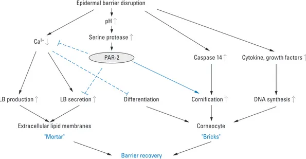
The involvement of PAR-2 in epidermal permeability barrier homeostasis by mediating signaling from serine proteases in the skin has been recently evidenced.7,11,12 Al-though controversial, recent studies have shown that PAR-2 localized in the suprabasal layers of mouse and human epidermis with high levels of PAR-2 in the stratum gra-nulosum, suggesting that PAR-2 might act as the primary sensor of barrier-initiated serine protease activity.7,62,63 Acute barrier disruption leads to an increase in SC pH from basal levels of 5.0-5.5 toward a more neutral pH, which, in turn, increases the activity of serine protease, which has an optimal activity at neutral pH.11,64This inc-rease in serine protease activity activates PAR-2 on kera-tinocytes of SG, resulting in decreased LB secretion and a consequent decrease in the formation of caveolae and lipid raft (Fig. 2).7It was reported that topical PAR-2 agonist pep-tide significantly delayed barrier recovery and inhibited LB secretion following acute barrier disruption in murine skin, whereas PAR-2 knockout mice showed increased LB secretion and accelerated barrier recovery following acute disruption as compared to wild-type littermates.7In
addi-Fig. 3. PAR-2 and Netherton syndrome. Ela-2, pancreatic elastase-2; KLK, kallikrein; LETKI, lympho-epithelial Kazal-type-related inhibitor; PAR-2, protease-activated receptor-2; SPINK-5, serine protease inhibitor kazal-type 5; TSLP, thymic stromal lymphopoietin.
tion, inhibition of PAR-2 activation, by topical application of either serine protease inhibitor or PAR-2 specific anta-gonist significantly accelerated the barrier recovery rate after acute barrier disruption.65These results implicates that PAR-2 activation might be an important signal for regulat-ing LB secretion durregulat-ing the repair response after barrier disruption. While serine protease/PAR-2 signaling negati-vely affects permeability barrier homeostasis by inhibiting the restoration of lipid barrier (mortar), it could also act as a positive regulator in the permeability barrier recovery by accelerating cornification, which then induces the forma-tion of corneocytes (bricks) (Fig. 2).11Recent studies have demonstrated that proteolytically active allergens, house dust mites and cockroaches, also activated PAR-2 and delayed barrier recovery via PAR-2 signal-mediated inhibi-tion of LB secre-inhibi-tion.12By showing the abnormal distribu-tion of calcium ions after barrier disrupdistribu-tion in skin where allergens had been applied, the authors suggest that PAR-2 signaling-mediated modulation of calcium ions in the skin could be one of the mechanisms involved in the regulation of LB secretion by PAR-2.12
Inflammation
The role of PAR-2 in the regulation of inflammation has been widely investigated. During cutaneous inflammation, potential endogenous activators of PAR-2, including leuko-cyte elastase, mast cell tryptase, proteinases of the trypsin-family produced by keratinocytes (trypsinogen-4), and proteinases of the fibrinolysis cascade such as factor VII/Xa, are released; these then activate PAR-2 on keratinocytes, endothelial cells, inflammatory cells, and dermal sensory nerves to amplify the inflammation via the up-regulation of inflammatory mediators.66It has been demonstrated that PAR-2 agonist peptide induces intercellular adhesion mole-cule-1 (ICAM-1) expression in primary human
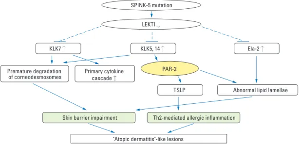
keratino-cytes via the activation of nuclear factor-kappa B.67Another study reported that stimulation of PAR-2 on keratinocytes increased the secretion of interleukin (IL)-6 and granulo-cyte-macrophage colony-stimulating factor (GM-CSF).68 In keratinocytes, PAR-2 activation also leads to a secretion of IL-8/CXCL8, promoting granulocyte and T-cell recruit-ment.69The observation that PAR-2 deficient mice show diminished ear swelling and inflammatory infiltrates in a model of contact hypersensitivity indicates that PAR-2 mediates inflammation in allergic dermatitis.70In addition, PAR-2 has been observed at increased levels in lesional skin of patients with AD, suggesting that PAR-2 plays a role in inflammatory dermatosis.15,17In addition to these proinflammatory responses, PAR-2 also has been shown to be involved in the T-helper type 2 (Th2) mediated aller-gic inflammation. Thymic stromal lymphopoietin (TSLP), a Th2-associated cytokine produced by inflammatory cells as well as epithelial cells, is known to induce Th2 cell recruitment and allergic inflammation via dendritic cell stimulation in response to allergen challenge, microbial infections, and inflammation.71TSLP transgenic mouse has been shown to develop an AD-like skin disease, which suggests an important role of TSLP in initiating and per-petuating Th2 immune responses in AD.72,73A recent study reported that SPINK5 knockout mice expressed TSLP in the epidermis at a higher rate than that of wild-type epider-mis, and demonstrated that KLK5 directly activates PAR-2, which in turn induces nuclear factor κB-mediated over-expression of TSLP. This implies that LEKTI defici-ency-induced serine protease/PAR-2 activation triggers proin-flammatory as well as proallergic inproin-flammatory res-ponses in the development of AD-like phenotype of NS (Fig. 3).74
Pruritus
Skin nerve fibers are known to have functional PAR-2,
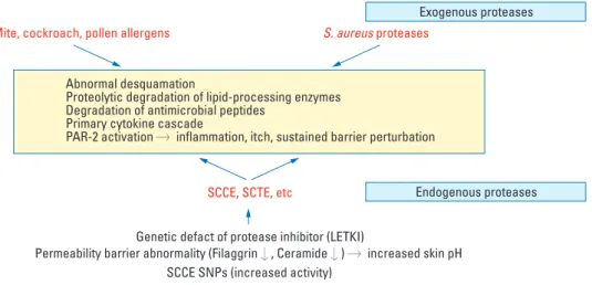
Fig. 4. Proteases and PAR-2 in the pathogenesis of atopic dermatitis. LETKI, lympho-epithelial Kazal-type-related inhibitor; PAR-2, protease-activated receptor-2; SCCE, stratum corneum chymotrypsin-like enzyme; SCTE, stratum corneum trypsin-like enzyme; SNP, single nucleotide polymorphism.
and it has been proposed that PAR-2 plays an important role in pruritus. During neurogenic inflammation, various endogenous serine proteases such as tryptase from mast cells and trypsins from keratinocytes activate PAR-2 on sensory nerve ending to release calcitonin gene-related pep-tide (CGRP) and substance P (SP). These neuropeppep-tides induce vasodilation, edema, and leukocyte recruitment, resulting in neurogenic inflammation in the local skin lesi-ons.75PAR-2 is not only expressed in the peripheral nervous system but also in the central nervous system, spinal cord and brain. Therefore, PAR-2 signaling also stimulates the release of neuropeptides from central nerve endings at the spinal cord level, thereby activating CGRP receptor and SP receptor (NK1R) to transmit itch responses to the cen-tral nervous system.76In addition, skin exposure to exoge-nous microbial proteases could also induce itch and inflam-mation via PAR-2. Recent study reported that the mice that over-expressed epidermal KLK7 displayed massive itchy behavior.77Another study demonstrated that trypsin-induced scratching behavior in mice was inhibited by a PAR-2 blocking peptide, suggesting the role of serine protease/ PAR-2 signaling in pruritus.78Moreover, PAR-2 activation is likely to be involved in pruritus of AD.79 PAR-2 and trypsin have been observed to be expressed at high rates in the lesional skin of patients with atopic dermatitis, and PAR-2 agonist peptides induce pruritus in AD patients.41In addition, the fact that KLK5, -7, -8, and -14 are present in sweat, (sweat being the most common itch-triggering factor in AD) point to the important role of PAR-2 in pruritus of AD patients.7,80PAR-2 is reported to interact synergisti-cally with transient receptor potential (TRP) vanilloid-type 1 (TRPV1), which belongs to the superfamily of TRP chan-nels, thereby amplifying itch sensation.81 These findings suggest that serine protease inhibitors or PAR-2 antago-nists might be a promising therapeutic tool for the mana-gement of itching and help break the vicious itch-scratch cycle in AD.41,75
Genetic abnormalities in the genes encoding protease/protease inhibitor
There is increasing evidence that genes related to protease/ protease inhibitor become deregulated in patients with AD, shifting the balance between proteases and protease inhi-bitors toward increased protease activity (Fig. 4). It has been de-monstrated that there is an association between AD and a four base pair (AACC) insertion in the 3’-untranslated region of KLK7 gene, which increases the half-life of KLK7 mRNA and enzymatic activity of KLK7.82The rare
AACCAACC variant of KLK7 gene showed a more signi-ficant association with AD as compared to the common AACC variant. The genetic variant of KLK7 gene was found to be more significantly relevant in patients who did not have an elevated IgE level. The enzyme KLK7 plays an important role in desquamation by cleaving corneodes-mosomal proteins. In addition, it was reported that transge-nic mice over-expressing KLK7 presented cutaneous manifestations similar to chronic AD.83 This implies that the gain-of-function mutation polymorphism in the KLK7 gene causes a premature breakdown of corneodesmo-somes, leading to excessive desquamation and impairment of the epidermal barrier in the development of AD. In addition to abnormalities in the protease-encoding genes, several genomic defects in genes encoding members of the protease inhibitors have been identified in patients with AD. LEKTI is an inhibitor of multiple serine proteases in skin and tightly regulates the enzymatic activities of serine proteases including KLK5, -6, -7, -13, and -14, thereby controlling epidermal barrier function.31
A premature stop codon mutation in the SPINK5 gene, which encodes LEKTI, is known to be associated with NS, a rare ichthyosiform dermatosis characterized by congeni-tal ichthyosiform erythroderma, severe atopic manifesa-tions, and hair-shaft abnormality.84The SPINK5 gene consists of 33 exons, encoding 15 LEKTI inhibitory do-mains with selective/specific inhibitory function.85Previous study attempting to correlate genotype with phenotype in Japanese NS patients has demonstrated that the clinical severity of NS correlates with the residual expression of LEKTI-1.86SPINK5-deficient mice have been reported to show increased proteolytic activities of KLK5 and KLK7 in the epidermis, abnormal degradation of DSG1, and resultant abnormal corneodesmosome cleavage and premature desquamation, suggesting that LEKTI is a key regulator of KLK5 and KLK7 activity and that defective SC adhesion by epidermal protease hyperactivity is the primary pathogenic event in NS.87Recently, ELA2, a novel epidermal protease, has been identified in human and mouse skin and conceived as a potential trigger in NS pathogenesis by misprocessing of filaggrin and lipid.88 Being localized to the keratohyalin granule, ELA2 directly degrades (pro-) filaggrin and disrupts the lipid lamellae formation. LEKTI was demonstrated to inhibit ELA2 acti-vity indirectly, by controlling KLK5-mediated cleavage of pro-ELA2, and indeed, ELA2 was observed to be hyperac-tive in the LEKTI-deficient epidermis of NS patients and SPINK5-deficient mice.88 Transgenic mice over-expres-sing ELA2 also mimic the clinical features of NS patients with transient ichthyosiform dermatitis and impaired lipid barrier, implying that ELA2 is a critical epidermal protease that regulates epidermal barrier homeostasis, involving the
ABERRANT PROTEASE/PAR-2 SIGNALING
IN THE PATHOGENESIS OF AD
pathogenesis of NS. Several studies reported that the poly-morphisms in the SPINK5 gene are also associated with AD.84,89Glu420Lys single-nucleotide polymorphism in the SPINK5 gene was observed to be associated with AD and six other SPINK5 gene polymorphisms were also found in Japanese patients with AD.84,89In addition, another protease inhibitor, cystatin A-encoding gene (CSTA gene) poly-morphism (+344c variant) has been reported to be asso-ciated with AD.90Cystatin A is a cysteine protease inhibi-tor and is known to inhibit the endogenous cathepsins B, -H, and -L and the exogenous proteases from house dust mites, such as dermatophagoides pteronyssinus (Der p) 1 and dermatophagoides farinae (Der f) 1. This +344c variant in CSTA gene results in the reduced levels of cystatin-A in the skin surface and sweat, allowing exogenous proteases to break down the integrity of the SC, which in turn enhances the penetration of allergens, triggering an aggravation of AD. These genetic variants in protease and protease inhi-bitor genes can be combined in some patients with AD, resulting in a more severe defect in the skin barrier.
Elevated pH
The skin has a distinct pH gradient across the SC, with a progressively more acidic pH from the deeper layer to the outer layer of the SC.91Studies have demonstrated the ‘acid mantle’ within the SC plays a critical role in regulating epi-dermal permeability barrier homeostasis, the epiepi-dermal antimicrobial barrier, and SC integrity/cohesion by regulat-ing the activity of various enzymes includregulat-ing serine pro-teases and lipid-processing enzymes.91Skin pH is known to be generated by the endogenous processes, including trans-urocanic acid production from histidine, free fatty acid pro-duction via secretory phospholipase A (sPLA2), and the sodium-proton antiporter-1 (NHE-1)-mediated H+ secre-tion.92 Numerous endogenous factors, such as moisture, sweat, sebum, anatomic site, genetic predisposition and age as well as environmental factors, such as soap/detergents, topical irritant, antibiotics, and cosmetic products, and occlusive dressings can affect skin pH.93In the lesions of patients with AD, the skin pH has been reported to be significantly elevated.94The genetically determined barrier deficiency and various environmental factors are thought to be associated with the increased skin surface pH in AD lesions.
Since Palmer, et al.95first identified the loss-of-function mutation in the gene encoding filaggrin (FLG) as a strong predisposing factor for AD, a number of additional studies have reported other mutations of FLG genes, including 20 mutations in European populations and 17 in Asian popula-tions.96From these reports, it is now conceived that muta-tions of the FLG gene are the most significant genetic factors in AD and the resultant genetically determined epidermal
barrier defect is the primary event triggering immunologic pathogenesis in AD. Filaggrin itself contributes to the epi-dermal barrier integrity by cross-linking the keratin fila-ments and the being degraded into a combined pool of highly hygroscopic amino acids, called the NMF, thereby contri-buting to the SC hydration. It is also postulated that filag-grin contributes to the formation of acid mantle within SC through the generation of urocanic acid via filaggrin-histi-dine-urocanic acid cascade.97Therefore, filaggrin deficiency in AD lesion leads to defects in the formation of cornified envelope and a decreased ability of maintaining SC hydra-tion and a concomitant elevahydra-tion of pH. The elevated pH enhances the activity of KLK5 and KLK7, which have opti-mal activity at neutral pH, resulting in the over-degradation of corneodesmosomes and a decrease in SC integrity and cohesion. In addition, the abnormally neutral skin pH inhi-bits the activity of lipid-processing enzymes, including β-GlcCer’ase, aSMase, and sPLA2, which are essential in the production of ceramide and free fatty acids and causes impaired lipid processing and defects in the lipid barrier. SC acidity is also important in epidermal antimicrobial barrier function, inhibiting the growth of pathogens. Growth of Staphylococcus aureus, which colonizes the lesional skin of AD, is normally inhibited at low skin pH; therefore the elevated pH in AD lesional skin leads to bacterial growth, resulting in allergic inflammation and aggravation of AD.94 In addition to these genetic factors, various environmental factors can increase skin surface pH in AD patients. For instance, the frequent use of soaps/detergents seems not only to aggravate AD through directly irritating the skin by removing skin surface lipids, but also raises skin pH because most detergents are alkaline; but even at neutral, overuse of soap and detergent can have a negative effect on epider-mal barrier function.
Exogenous proteases
Various contact allergens and aeroallergens are considered important factors in the initiation and aggravation of AD. Proteolytic activity from allergens has been known to play a role in the pathogenesis of allergic diseases including allergic rhinitis, asthma, and AD through inducing Th2 allergic inflammation and directly affecting the structure and function of epidermal barrier, thereby facilitating further penetration of allergens through the defective skin barrier.98House dust mite and cockroach allergens, the most important environmental factors in the pathogenesis of AD, have been shown to have proteolytic activity. House dust mite allergens have been reported to contain several cysteine and serine proteases. Serine proteases from mite allergens, Der p 3 and Der p 9, are known to activate PAR-2 on keratinocytes to produce cytokines and contribute to the pathogenesis of AD, whereas Der p 1 with cystein protease
activity stimulates inflammation via a PAR-2-independent mechanism.99 The cockroach allergens, another type of major aeroallergen, are also reported to activate PAR-2 on keratinocytes. Jeong, et al.12reported that the topical appli-cation of house dust mites and cockroach allergens to bar-rier-disrupted skin delayed barrier recovery and lamellar body secretion in murine and human skin via PAR-2 acti-vation. The topical application of cockroach allergens also showed an inhibitory effect on the epidermal calcium gra-dient change after barrier disruption, suggesting that the negative effects of cockroach allergens on barrier recovery may be due to the PAR-2 signal-induced epidermal calcium modulation. Recently, Staphylococcus aureus has also been reported to produce extracellular V8 protease, which exhi-bits a similar specificity of glutamate-specific cleavage and a similar sequence to exfoliative toxins and cause epidermal permeability barrier dysfunction in the skin of nude mice by directly degrading DSG1.100These imply that proteoly-tically active allergens could break down the skin barrier via PAR-2-mediated inhibition of lamellar body secretion or PAR-2-non-mediated mechanisms, including degrada-tion of corneodesmosomal proteins and lipid processing enzymes, triggering further allergen penetration through the disrupted epithelial barrier to aggravate Th2-mediated inflammation and possibly switch the non-IgE-associated form of AD (atopiform AD) to the typical IgE-associated form of AD. Recently, several reports have suggested that proteolytically active allergens perturb not only the SC per-meability barrier but also the tight junction (TJ), another functional barrier of the skin.101-103 The exfoliative toxin-negative Staphylococcus strains were shown to decrease the expression of TJ and atypical protein kinase C, a key player in TJ assembly with decreased transepithelial resis-tance in the human keratinocyte cell line.101Previous studies have reported that house dust mite allergen Der p 1 dis-rupts intercellular TJ via proteolytic cleavage of TJ adhesion protein, claudin-1 and occludin in airway epithelial cells.102 Pollens, which are known to be associated with allergic rhini-tis and conjunctivirhini-tis, also contain proteolytic enzymes on their surface and degrade the TJ proteins of human airway epithelial cells.103These findings suggest that proteases deriv-ed from house dust mite, cockroaches, Staphylococcus aur-eus, and pollens contribute to the pathogenesis of AD by pro-pagating the vicious cycle of protease-mediated permeabi-lity barrier defect and increased allergen penetration (Fig. 4).
Identification of the important role of PAR-2 in various AD symptoms, including inflammation, pruritus and skin
barrier impairment, has suggested that PAR-2 antagoni-zing treatment may be a potential therapeutic strategy for AD. In addition, the crucial role of mast cells and mast cell-expressed PAR-2 in various inflammatory diseases also suggests that PAR-2 antagonizing treatment could be applied to several chronic inflammatory diseases, such as asthma,104rheumatoid arthritis105and inflammatory bowel diseases (IBD).106Theoretically, several approaches could be used to antagonize PAR-2 activation, including down-regulation of protease activity, inhibition of PAR-2 expres-sion using siRNA technology, PAR-2 monoclonal antibody or PAR-2 specific antagonist; however, at this time, a practical application of PAR-2 antagonizing treatment has not yet reported.
Down-regulation of protease activity to control PAR-2 activation in skin can be achieved by either topical protease inhibitors or pH controlling agents. We recently reported that the topical application of serine protease inhibitor (soy-bean tryptic inhibitor) significantly reduced the pruritus symptoms in end-stage renal dysfunction patients, where increased PAR-2 expression in skin was observed. Impro-vement of skin barrier functions was also observed after protease inhibitor application.107While this is a relatively easier way to control PAR-2 activation, proteases are res-ponsible not only for PAR-2 activation, but also for other normal homeostatic processes such as desquamation. As a result, use of protease inhibitors may have adverse effects on the skin, which is a major drawback for practical appli-cation of protease inhibitors for topical use.
Normal skin surface pH ranges from 5.0 to 5.5, which is relatively acidic compared to the normal physiologic pH, and it is well known that pH gradually increases across the SC. This “acid mantle” is very important for the maintain-ing skin’s homeostasis, includmaintain-ing regulatmaintain-ing protease activity. In AD, increase of skin surface pH is well reco-gnized and increased proteolytic activity is also observed. In addition to the PAR-2 activation, increased pH is also considered responsible for increased susceptibility to se-condary infections in AD patients. As a result, controlling skin surface pH within normal acidic range is a very im-portant therapeutic regimen for AD patients and use of soap or alkaline detergents is strongly discouraged.
The inhibitory activity of the PAR-2 antibody on itching behavior in chronic dry skin itch in an animal model has also been recently reported. Intradermal injection of PAR-2 antibody significantly reduced spontaneous scratching behavior, which alludes the important role of PAR-2 in itching sensation. Further evidence was provided by the in-creased scratching behavior after PAR-2 activator injec-tion.108However, a therapeutic antibody against PAR-2 has not yet been reported, and practical application of PAR-2 antibody needs to be further developed.
THERAPEUTIC APPLICATION OF PAR-2
ANTAGONIZING TREATMENT
Among the several approaches for PAR-2 antagonizing treatment, use of small molecule PAR-2 specific antago-nist seems to be the most promising method. However, while several PAR-1 specific antagonists have already been reported, the lack of a specific, potent PAR-2 antagonist has hampered more detailed investigation of the role of PAR-2 in various diseases and its practical application. Recently, however, a few PAR-2 specific antagonists and their in vivo and in vitro effects have been reported. ENMD-1,068 (N1-3-methylbutyryl-N4-6-aminohexanoyl-pipera-zine) was the first reported PAR-2 specific antagonist, and its anti-inflammatory activity was evaluated using both in
vivo and ex vivo models. In a carrageenan/kaolin
(C/K)-induced arthritis animal model, intraperitoneal injection of ENMD-1,068 showed significant inhibition of joint swell-ing109and in ex vivo studies using rheumatoid arthritis syno-vium and cultured synovial fibroblasts, ENMD-1,068 also showed inhibitory effects on proinflammatory cytokine pro-duction in a dose-dependent manner.110In addition, we have shown that topical application of ENMD-1,068 on barrier disrupted skin significantly accelerated barrier recovery rate, which also suggest the potential application of ENMD-1,068 as a topical agent.12However, its low po-tency ranging low millimolar concentration has made it impractical for therapeutic application.
More recently, Kanke, et al.111have reported several pepti-domimetic compounds as having PAR-2 antagonistic activities. Among the compounds, it was reported that K-14585 {N-[1-(2, 6-dichlorophenyl)methyl]-3-(1-pyrroli- dinylmethyl)-1H-indol-5-yl)aminocarbonyl}-glycine-L-lysinyl-Lphenylalanyl-N-benzhydrylamide) showed the most potent antagonist activity against PAR-2 activating peptide induced cellular responses. Ex vivo responses, such as rat-isolated aorta relaxation, and in vivo responses, such as plasma extravasation in the dorsal skin of guinea pigs and saliva secretion in anaesthetized mice, were also inhibited by a systemic injection of K-14585. Interestingly, it was observed that while K-14585 significantly inhibited the activating peptide-induced PAR-2 activation signaling, serine protease (trypsin)-induced signaling was affected to a much lesser extent. It was suggested that there may be some structural differences between the activating peptide binding site and tethered ligand binding site; this distinc-tion need a further investigadistinc-tion. More interestingly, in a study of K-14585-induced cellular signaling, higher con-centrations of K-14585 showed agonistic activity, which still needs further investigation.112Given these results, prac-tical therapeutic application of K-14585 is currently not achievable.
Currently, the authors are investigating a new PAR-2 antagonist. In a series of in vitro and in vivo studies, the new PAR-2 antagonist, NPS-1577, has shown potential
anti-inflammatory activity as well as skin barrier improving activity and anti-hyperproliferative activity on the epidermis (paper in preparation). While detailed investigations related to the action mechanism are currently being performed, NPS-1577 could potentially be used as the first practical PAR-2 antagonist for therapeutic use in the near future.
This work was financially supported by the Ministry of Knowledge Economy (MKE) and Korea Institute for Advancement of Technology (KIAT) through the Research and Development for Regional Industry.
1. Richard I. The genetic and molecular bases of monogenic disorders affecting proteolytic systems. J Med Genet 2005;42: 529-39.
2. Ramachandran R, Hollenberg MD. Proteinases and signalling: pathophysiological and therapeutic implications via PARs and more. Br J Pharmacol 2008;153 Suppl 1:S263-82.
3. Brattsand M, Egelrud T. Purification, molecular cloning, and expression of a human stratum corneum trypsin-like serine protease with possible function in desquamation. J Biol Chem 1999;274:30033-40.
4. Hansson L, Strömqvist M, Bäckman A, Wallbrandt P, Carlstein A, Egelrud T. Cloning, expression, and characterization of stratum corneum chymotryptic enzyme. A skin-specific human serine proteinase. J Biol Chem 1994;269:19420-6.
5. Bernard D, Méhul B, Thomas-Collignon A, Simonetti L, Remy V, Bernard MA, et al. Analysis of proteins with caseinolytic acti-vity in a human stratum corneum extract revealed a yet unidenti-fied cysteine protease and identiunidenti-fied the so-called “stratum corneum thiol protease” as cathepsin l2. J Invest Dermatol 2003; 120:592-600.
6. Horikoshi T, Arany I, Rajaraman S, Chen SH, Brysk H, Lei G, et al. Isoforms of cathepsin D and human epidermal differentiation. Biochimie 1998;80:605-12.
7. Hachem JP, Houben E, Crumrine D, Man MQ, Schurer N, Roelandt T, et al. Serine protease signaling of epidermal permea-bility barrier homeostasis. J Invest Dermatol 2006;126:2074-86. 8. Rattenholl A, Steinhoff M. Role of proteinase-activated receptors
in cutaneous biology and disease. Drug Dev Res 2003;59:408-16. 9. Hansen KK, Oikonomopoulou K, Baruch A, Ramachandran R, Beck P, Diamandis EP, et al. Proteinases as hormones: targets and mechanisms for proteolytic signaling. Biol Chem 2008;389:971-82. 10. Rattenholl A, Steinhoff M. Proteinase-activated receptor-2 in the
skin: receptor expression, activation and function during health and disease. Drug News Perspect 2008;21:369-81.
11. Demerjian M, Hachem JP, Tschachler E, Denecker G, Declercq W, Vandenabeele P, et al. Acute modulations in permeability barrier function regulate epidermal cornification: role of caspase-14 and the protease-activated receptor type 2. Am J Pathol 2008; 172:86-97.
ACKNOWLEDGEMENTS
12. Jeong SK, Kim HJ, Youm JK, Ahn SK, Choi EH, Sohn MH, et al. Mite and cockroach allergens activate protease-activated receptor 2 and delay epidermal permeability barrier recovery. J Invest Dermatol 2008;128:1930-9.
13. Eissa A, Diamandis EP. Human tissue kallikreins as promiscuous modulators of homeostatic skin barrier functions. Biol Chem 2008;389:669-80.
14. Stefansson K, Brattsand M, Roosterman D, Kempkes C, Bocheva G, Steinhoff M, et al. Activation of proteinase-activated receptor-2 by human kallikrein-related peptidases. J Invest Dermatol 2008;128:18-25.
15. Descargues P, Deraison C, Prost C, Fraitag S, Mazereeuw-Hautier J, D’Alessio M, et al. Corneodesmosomal cadherins are preferential targets of stratum corneum trypsin- and chymotrypsin-like hyperactivity in Netherton syndrome. J Invest Dermatol 2006;126:1622-32.
16. Komatsu N, Suga Y, Saijoh K, Liu AC, Khan S, Mizuno Y, et al. Elevated human tissue kallikrein levels in the stratum corneum and serum of peeling skin syndrome-type B patients suggests an over-desquamation of corneocytes. J Invest Dermatol 2006;126: 2338-42.
17. Komatsu N, Saijoh K, Kuk C, Liu AC, Khan S, Shirasaki F, et al. Human tissue kallikrein expression in the stratum corneum and serum of atopic dermatitis patients. Exp Dermatol 2007;16:513-9. 18. Komatsu N, Saijoh K, Kuk C, Shirasaki F, Takehara K, Dia-mandis EP. Aberrant human tissue kallikrein levels in the stratum corneum and serum of patients with psoriasis: dependence on phenotype, severity and therapy. Br J Dermatol 2007;156:875-83. 19. Lundwall A, Brattsand M. Kallikrein-related peptidases. Cell Mol
Life Sci 2008;65:2019-38.
20. Caubet C, Jonca N, Brattsand M, Guerrin M, Bernard D, Schmidt R, et al. Degradation of corneodesmosome proteins by two serine proteases of the kallikrein family, SCTE/KLK5/hK5 and SCCE/ KLK7/hK7. J Invest Dermatol 2004;122:1235-44.
21. Hachem JP, Man MQ, Crumrine D, Uchida Y, Brown BE, Rogiers V, et al. Sustained serine proteases activity by prolonged increase in pH leads to degradation of lipid processing enzymes and profound alterations of barrier function and stratum corneum integrity. J Invest Dermatol 2005;125:510-20.
22. Yamasaki K, Schauber J, Coda A, Lin H, Dorschner RA, Schechter NM, et al. Kallikrein-mediated proteolysis regulates the antimicrobial effects of cathelicidins in skin. FASEB J 2006;20:2068-80.
23. Ekholm IE, Brattsand M, Egelrud T. Stratum corneum tryptic enzyme in normal epidermis: a missing link in the desquamation process? J Invest Dermatol 2000;114:56-63.
24. Brattsand M, Stefansson K, Lundh C, Haasum Y, Egelrud T. A proteolytic cascade of kallikreins in the stratum corneum. J Invest Dermatol 2005;124:198-203.
25. Elias PM, Cullander C, Mauro T, Rassner U, Kömüves L, Brown BE, et al. The secretory granular cell: the outermost granular cell as a specialized secretory cell. J Investig Dermatol Symp Proc 1998;3:87-100.
26. Yoon H, Laxmikanthan G, Lee J, Blaber SI, Rodriguez A, Kogot JM, et al. Activation profiles and regulatory cascades of the hu-man kallikrein-related peptidases. J Biol Chem 2007;282:31852-64. 27. Oikonomopoulou K, Hansen KK, Saifeddine M, Tea I, Blaber M,
Blaber SI, et al. Proteinase-activated receptors, targets for kalli-krein signaling. J Biol Chem 2006;281:32095-112.
28. Komatsu N, Saijoh K, Toyama T, Ohka R, Otsuki N, Hussack G,
et al. Multiple tissue kallikrein mRNA and protein expression in normal skin and skin diseases. Br J Dermatol 2005;153:274-81. 29. Kishibe M, Bando Y, Terayama R, Namikawa K, Takahashi H,
Hashimoto Y, et al. Kallikrein 8 is involved in skin desquamation in cooperation with other kallikreins. J Biol Chem 2007;282: 5834-41.
30. Morizane S, Yamasaki K, Kabigting FD, Gallo RL. Kallikrein expression and cathelicidin processing are independently con-trolled in keratinocytes by calcium, vitamin D(3), and retinoic acid. J Invest Dermatol 2010;130:1297-306.
31. Ovaere P, Lippens S, Vandenabeele P, Declercq W. The emerg-ing roles of serine protease cascades in the epidermis. Trends Biochem Sci 2009;34:453-63.
32. List K, Szabo R, Wertz PW, Segre J, Haudenschild CC, Kim SY, et al. Loss of proteolytically processed filaggrin caused by epider-mal deletion of Matriptase/MT-SP1. J Cell Biol 2003;163:901-10. 33. Mitsudo K, Jayakumar A, Henderson Y, Frederick MJ, Kang Y,
Wang M, et al. Inhibition of serine proteinases plasmin, trypsin, subtilisin A, cathepsin G, and elastase by LEKTI: a kinetic analysis. Biochemistry 2003;42:3874-81.
34. Roelandt T, Thys B, Heughebaert C, De Vroede A, De Paepe K, Roseeuw D, et al. LEKTI-1 in sickness and in health. Int J Cos-met Sci 2009;31:247-54.
35. Meyer-Hoffert U, Wu Z, Schröder JM. Identification of lympho-epithelial Kazal-type inhibitor 2 in human skin as a kallikrein-related peptidase 5-specific protease inhibitor. PLoS One 2009;4: e4372.
36. Galliano MF, Toulza E, Gallinaro H, Jonca N, Ishida-Yamamoto A, Serre G, et al. A novel protease inhibitor of the alpha2-macro-globulin family expressed in the human epidermis. J Biol Chem 2006;281:5780-9.
37. Oberst MD, Chen LY, Kiyomiya K, Williams CA, Lee MS, Johnson MD, et al. HAI-1 regulates activation and expression of matriptase, a membrane-bound serine protease. Am J Physiol Cell Physiol 2005;289:C462-70.
38. Oberst MD, Williams CA, Dickson RB, Johnson MD, Lin CY. The activation of matriptase requires its noncatalytic domains, serine protease domain, and its cognate inhibitor. J Biol Chem 2003;278:26773-9.
39. Kawabata A. PAR-2: structure, function and relevance to human diseases of the gastric mucosa. Expert Rev Mol Med 2002;4:1-17. 40. Steinhoff M, Corvera CU, Thoma MS, Kong W, McAlpine BE,
Caughey GH, et al. Proteinase-activated receptor-2 in human skin: tissue distribution and activation of keratinocytes by mast cell tryptase. Exp Dermatol 1999;8:282-94.
41. Steinhoff M, Neisius U, Ikoma A, Fartasch M, Heyer G, Skov PS, et al. Proteinase-activated receptor-2 mediates itch: a novel pathway for pruritus in human skin. J Neurosci 2003;23:6176-80. 42. Scott G, Deng A, Rodriguez-Burford C, Seiberg M, Han R, Babiarz L, et al. Protease-activated receptor 2, a receptor involved in melanosome transfer, is upregulated in human skin by ultra-violet irradiation. J Invest Dermatol 2001;117:1412-20.
43. Déry O, Thoma MS, Wong H, Grady EF, Bunnett NW. Traffick-ing of proteinase-activated receptor-2 and beta-arrestin-1 tagged with green fluorescent protein. beta-Arrestin-dependent endocy-tosis of a proteinase receptor. J Biol Chem 1999;274:18524-35. 44. Santulli RJ, Derian CK, Darrow AL, Tomko KA, Eckardt AJ,
Seiberg M, et al. Evidence for the presence of a protease-acti-vated receptor distinct from the thrombin receptor in human kera-tinocytes. Proc Natl Acad Sci U S A 1995;92:9151-5.
45. Derian CK, Eckardt AJ, Andrade-Gordon P. Differential regula-tion of human keratinocyte growth and differentiaregula-tion by a novel family of protease-activated receptors. Cell Growth Differ 1997; 8:743-9.
46. Scott IR, Harding CR, Barrett JG. Histidine-rich protein of the keratohyalin granules. Source of the free amino acids, urocanic acid and pyrrolidone carboxylic acid in the stratum corneum. Biochim Biophys Acta 1982;719:110-7.
47. McGrath JA, Uitto J. The filaggrin story: novel insights into skin-barrier function and disease. Trends Mol Med 2008;14:20-7. 48. Denecker G, Hoste E, Gilbert B, Hochepied T, Ovaere P, Lippens
S, et al. Caspase-14 protects against epidermal UVB photoda-mage and water loss. Nat Cell Biol 2007;9:666-74.
49. Resing KA, Thulin C, Whiting K, al-Alawi N, Mostad S. Characterization of profilaggrin endoproteinase 1. A regulated cytoplasmic endoproteinase of epidermis. J Biol Chem 1995;270: 28193-8.
50. Yamazaki M, Ishidoh K, Suga Y, Saido TC, Kawashima S, Suzuki K, et al. Cytoplasmic processing of human profilaggrin by active mu-calpain. Biochem Biophys Res Commun 1997;235: 652-6.
51. Pearton DJ, Nirunsuksiri W, Rehemtulla A, Lewis SP, Presland RB, Dale BA. Proprotein convertase expression and localization in epidermis: evidence for multiple roles and substrates. Exp Dermatol 2001;10:193-203.
52. Leyvraz C, Charles RP, Rubera I, Guitard M, Rotman S, Breiden B, et al. The epidermal barrier function is dependent on the serine protease CAP1/Prss8. J Cell Biol 2005;170:487-96.
53. Netzel-Arnett S, Currie BM, Szabo R, Lin CY, Chen LM, Chai KX, et al. Evidence for a matriptase-prostasin proteolytic cascade regulating terminal epidermal differentiation. J Biol Chem 2006; 281:32941-5.
54. Borgoño CA, Michael IP, Komatsu N, Jayakumar A, Kapadia R, Clayman GL, et al. A potential role for multiple tissue kallikrein serine proteases in epidermal desquamation. J Biol Chem 2007; 282:3640-52.
55. Deraison C, Bonnart C, Lopez F, Besson C, Robinson R, Jayakumar A, et al. LEKTI fragments specifically inhibit KLK5, KLK7, and KLK14 and control desquamation through a pH-dependent interaction. Mol Biol Cell 2007;18:3607-19.
56. Zeeuwen PL, Cheng T, Schalkwijk J. The biology of cystatin M/E and its cognate target proteases. J Invest Dermatol 2009; 129:1327-38.
57. Cheng T, Hitomi K, van Vlijmen-Willems IM, de Jongh GJ, Yamamoto K, Nishi K, et al. Cystatin M/E is a high affinity inhibitor of cathepsin V and cathepsin L by a reactive site that is distinct from the legumain-binding site. A novel clue for the role of cystatin M/E in epidermal cornification. J Biol Chem 2006; 281:15893-9.
58. Cheng T, Tjabringa GS, van Vlijmen-Willems IM, Hitomi K, van Erp PE, Schalkwijk J, et al. The cystatin M/E-controlled pathway of skin barrier formation: expression of its key components in psoriasis and atopic dermatitis. Br J Dermatol 2009;161:253-64. 59. Lai Y, Gallo RL. AMPed up immunity: how antimicrobial
peptides have multiple roles in immune defense. Trends Immunol 2009;30:131-41.
60. Aberg KM, Man MQ, Gallo RL, Ganz T, Crumrine D, Brown BE, et al. Co-regulation and interdependence of the mammalian epidermal permeability and antimicrobial barriers. J Invest Dermatol 2008;128:917-25.
61. Yamasaki K, Di Nardo A, Bardan A, Murakami M, Ohtake T, Coda A, et al. Increased serine protease activity and cathelicidin promotes skin inflammation in rosacea. Nat Med 2007;13:975-80. 62. Seeliger S, Derian CK, Vergnolle N, Bunnett NW, Nawroth R,
Schmelz M, et al. Proinflammatory role of proteinase-activated receptor-2 in humans and mice during cutaneous inflammation in vivo. FASEB J 2003;17:1871-85.
63. Iwakiri K, Ghazizadeh M, Jin E, Fujiwara M, Takemura T, Takezaki S, et al. Human airway trypsin-like protease induces PAR-2-mediated IL-8 release in psoriasis vulgaris. J Invest Dermatol 2004;122:937-44.
64. Feingold KR, Schmuth M, Elias PM. The regulation of per-meability barrier homeostasis. J Invest Dermatol 2007;127:1574-6. 65. Denda M, Kitamura K, Elias PM, Feingold KR.
trans-4-(Aminomethyl)cyclohexane carboxylic acid (T-AMCHA), an anti-fibrinolytic agent, accelerates barrier recovery and prevents the epidermal hyperplasia induced by epidermal injury in hairless mice and humans. J Invest Dermatol 1997;109:84-90.
66. Steinhoff M, Buddenkotte J, Shpacovitch V, Rattenholl A, Moor-mann C, Vergnolle N, et al. Proteinase-activated receptors: trans-ducers of proteinase-mediated signaling in inflammation and immune response. Endocr Rev 2005;26:1-43.
67. Buddenkotte J, Stroh C, Engels IH, Moormann C, Shpacovitch VM, Seeliger S, et al. Agonists of proteinase-activated receptor-2 stimulate upregulation of intercellular cell adhesion molecule-1 in primary human keratinocytes via activation of NF-kappa B. J Invest Dermatol 2005;124:38-45.
68. Wakita H, Furukawa F, Takigawa M. Thrombin and trypsin induce granulocyte-macrophage colony-stimulating factor and interleukin-6 gene expression in cultured normal human keratino-cytes. Proc Assoc Am Physicians 1997;109:190-207.
69. Hou L, Kapas S, Cruchley AT, Macey MG, Harriott P, Chinni C, et al. Immunolocalization of protease-activated receptor-2 in skin: receptor activation stimulates interleukin-8 secretion by keratino-cytes in vitro. Immunology 1998;94:356-62.
70. Kawagoe J, Takizawa T, Matsumoto J, Tamiya M, Meek SE, Smith AJ, et al. Effect of protease-activated receptor-2 deficiency on allergic dermatitis in the mouse ear. Jpn J Pharmacol 2002; 88:77-84.
71. Liu YJ, Soumelis V, Watanabe N, Ito T, Wang YH, Malefyt Rde W, et al. TSLP: an epithelial cell cytokine that regulates T cell differentiation by conditioning dendritic cell maturation. Annu Rev Immunol 2007;25:193-219.
72. Yoo J, Omori M, Gyarmati D, Zhou B, Aye T, Brewer A, et al. Spontaneous atopic dermatitis in mice expressing an inducible thymic stromal lymphopoietin transgene specifically in the skin. J Exp Med 2005;202:541-9.
73. Ziegler SF, Liu YJ. Thymic stromal lymphopoietin in normal and pathogenic T cell development and function. Nat Immunol 2006;7:709-14.
74. Briot A, Deraison C, Lacroix M, Bonnart C, Robin A, Besson C, et al. Kallikrein 5 induces atopic dermatitis-like lesions through PAR2-mediated thymic stromal lymphopoietin expression in Netherton syndrome. J Exp Med 2009;206:1135-47.
75. Steinhoff M, Vergnolle N, Young SH, Tognetto M, Amadesi S, Ennes HS, et al. Agonists of proteinase-activated receptor 2 induce inflammation by a neurogenic mechanism. Nat Med 2000;6:151-8.
76. Paus R, Schmelz M, Bíró T, Steinhoff M. Frontiers in pruritus research: scratching the brain for more effective itch therapy. J
Clin Invest 2006;116:1174-86.
77. Steinhoff M, Bienenstock J, Schmelz M, Maurer M, Wei E, Bíró T. Neurophysiological, neuroimmunological, and neuroendocrine basis of pruritus. J Invest Dermatol 2006;126:1705-18.
78. Costa R, Marotta DM, Manjavachi MN, Fernandes ES, Lima-Garcia JF, Paszcuk AF, et al. Evidence for the role of neurogenic inflammation components in trypsin-elicited scratching behaviour in mice. Br J Pharmacol 2008;154:1094-103.
79. Steinhoff M, Stander S, Seeliger S, Ansel JC, Schmelz M, Luger T. Modern aspects of cutaneous neurogenic inflammation. Arch Dermatol 2003;139:1479-88.
80. Komatsu N, Tsai B, Sidiropoulos M, Saijoh K, Levesque MA, Takehara K, et al. Quantification of eight tissue kallikreins in the stratum corneum and sweat. J Invest Dermatol 2006;126:925-9. 81. Amadesi S, Nie J, Vergnolle N, Cottrell GS, Grady EF, Trevisani
M, et al. Protease-activated receptor 2 sensitizes the capsaicin receptor transient receptor potential vanilloid receptor 1 to induce hyperalgesia. J Neurosci 2004;24:4300-12.
82. Vasilopoulos Y, Cork MJ, Murphy R, Williams HC, Robinson DA, Duff GW, et al. Genetic association between an AACC insertion in the 3’UTR of the stratum corneum chymotryptic enzyme gene and atopic dermatitis. J Invest Dermatol 2004;123: 62-6.
83. Hansson L, Bäckman A, Ny A, Edlund M, Ekholm E, Ekstrand Hammarström B, et al. Epidermal overexpression of stratum corneum chymotryptic enzyme in mice: a model for chronic itchy dermatitis. J Invest Dermatol 2002;118:444-9.
84. Walley AJ, Chavanas S, Moffatt MF, Esnouf RM, Ubhi B, Lawrence R, et al. Gene polymorphism in Netherton and common atopic disease. Nat Genet 2001;29:175-8.
85. Mägert HJ, Ständker L, Kreutzmann P, Zucht HD, Reinecke M, Sommerhoff CP, et al. LEKTI, a novel 15-domain type of human serine proteinase inhibitor. J Biol Chem 1999;274:21499-502. 86. Hachem JP, Wagberg F, Schmuth M, Crumrine D, Lissens W,
Jayakumar A, et al. Serine protease activity and residual LEKTI expression determine phenotype in Netherton syndrome. J Invest Dermatol 2006;126:1609-21.
87. Descargues P, Deraison C, Bonnart C, Kreft M, Kishibe M, Ishida-Yamamoto A, et al. Spink5-deficient mice mimic Nether-ton syndrome through degradation of desmoglein 1 by epidermal protease hyperactivity. Nat Genet 2005;37:56-65.
88. Bonnart C, Deraison C, Lacroix M, Uchida Y, Besson C, Robin A, et al. Elastase 2 is expressed in human and mouse epidermis and impairs skin barrier function in Netherton syndrome through filaggrin and lipid misprocessing. J Clin Invest 2010;120:871-82. 89. Kato A, Fukai K, Oiso N, Hosomi N, Murakami T, Ishii M.
Association of SPINK5 gene polymorphisms with atopic derma-titis in the Japanese population. Br J Dermatol 2003;148:665-9. 90. Vasilopoulos Y, Cork MJ, Teare D, Marinou I, Ward SJ, Duff
GW, et al. A nonsynonymous substitution of cystatin A, a cys-teine protease inhibitor of house dust mite protease, leads to decreased mRNA stability and shows a significant association with atopic dermatitis. Allergy 2007;62:514-9.
91. Mauro T. SC pH: measurement, origins, and functions. In: Eilas P, Feingold K, editors. Skin Barrier. New York: Taylor & Francis Group; 2006. p.223-30.
92. Fluhr JW, Behne MJ, Brown BE, Moskowitz DG, Selden C, Mao-Qiang M, et al. Stratum corneum acidification in neonatal skin: secretory phospholipase A2 and the sodium/hydrogen anti-porter-1 acidify neonatal rat stratum corneum. J Invest Dermatol 2004;122:320-9.
93. Schmid-Wendtner MH, Korting HC. The pH of the skin surface and its impact on the barrier function. Skin Pharmacol Physiol 2006;19:296-302.
94. Rippke F, Schreiner V, Doering T, Maibach HI. Stratum cor-neum pH in atopic dermatitis: impact on skin barrier function and colonization with Staphylococcus Aureus. Am J Clin Dermatol 2004;5:217-23.
95. Palmer CN, Irvine AD, Terron-Kwiatkowski A, Zhao Y, Liao H, Lee SP, et al. Common loss-of-function variants of the epi-dermal barrier protein filaggrin are a major predisposing factor for atopic dermatitis. Nat Genet 2006;38:441-6.
96. O’Regan GM, Sandilands A, McLean WH, Irvine AD. Filag-grin in atopic dermatitis. J Allergy Clin Immunol 2008;122:689-93.
97. Fluhr JW, Elias PM, Man MQ, Hupe M, Selden C, Sundberg JP, et al. Is the filaggrin-histidine-urocanic acid pathway essen-tial for stratum corneum acidification? J Invest Dermatol 2010; 130:2141-4.
98. Roelandt T, Heughebaert C, Hachem JP. Proteolytically active allergens cause barrier breakdown. J Invest Dermatol 2008;128: 1878-80.
99. Kato T, Takai T, Fujimura T, Matsuoka H, Ogawa T, Muraya-ma K, et al. Mite serine protease activates protease-activated receptor-2 and induces cytokine release in human keratinocytes. Allergy 2009;64:1366-74.
100. Hirasawa Y, Takai T, Nakamura T, Mitsuishi K, Gunawan H, Suto H, et al. Staphylococcus aureus extracellular protease causes epidermal barrier dysfunction. J Invest Dermatol 2010;130:614-7. 101. Ohnemus U, Kohrmeyer K, Houdek P, Rohde H, Wladykowski
E, Vidal S, et al. Regulation of epidermal tight-junctions (TJ) during infection with exfoliative toxin-negative Staphylococcus strains. J Invest Dermatol 2008;128:906-16.
102. Wan H, Winton HL, Soeller C, Tovey ER, Gruenert DC, Thomp-son PJ, et al. Der p 1 facilitates transepithelial allergen delivery by disruption of tight junctions. J Clin Invest 1999;104: 123-33. 103. Runswick S, Mitchell T, Davies P, Robinson C, Garrod DR.
Pollen proteolytic enzymes degrade tight junctions. Respirology 2007;12:834-42.
104. Kawabata A, Kawao N. Physiology and pathophysiology of proteinase-activated receptors (PARs): PARs in the respiratory system: cellular signaling and physiological/pathological roles. J Pharmacol Sci 2005;97:20-4.
105. Ferrell WR, Lockhart JC, Kelso EB, Dunning L, Plevin R, Meek SE, et al. Essential role for proteinase-activated receptor-2 in arthritis. J Clin Invest 2003;111:35-41.
106. Hyun E, Andrade-Gordon P, Steinhoff M, Vergnolle N. Pro-tease-activated receptor-2 activation: a major actor in intestinal inflammation. Gut 2008;57:1222-9.
107. Kim H, Jeong S, Jeong M, Ahn J, Moon S, Lee S. The relation-ship of PAR2 and pruritus in end stage renal disease patients and the clinical effectiveness of soybean extracts containing moisturizer on epidermal permeability barrier in end stage renal disease patients. J Invest Dermatol 2010;130:S56.
108. Akiyama T, Carstens MI, Carstens E. Enhanced scratching evoked by PAR-2 agonist and 5-HT but not histamine in a mouse model of chronic dry skin itch. Pain 2010 Aug 13. (Epub ahead of print).
109. Kelso EB, Lockhart JC, Hembrough T, Dunning L, Plevin R, Hollenberg MD, et al. Therapeutic promise of proteinase-acti-vated receptor-2 antagonism in joint inflammation. J Pharmacol
Exp Ther 2006;316:1017-24.
110. Kelso EB, Ferrell WR, Lockhart JC, Elias-Jones I, Hembrough T, Dunning L, et al. Expression and proinflammatory role of proteinase-activated receptor 2 in rheumatoid synovium: ex vivo studies using a novel proteinase-activated receptor 2 antagonist. Arthritis Rheum 2007;56:765-71.
111. Kanke T, Kabeya M, Kubo S, Kondo S, Yasuoka K, Tagashira
J, et al. Novel antagonists for proteinase-activated receptor 2: inhibition of cellular and vascular responses in vitro and in vivo. Br J Pharmacol 2009;158:361-71.
112. Goh FG, Ng PY, Nilsson M, Kanke T, Plevin R. Dual effect of the novel peptide antagonist K-14585 on proteinase-activated receptor-2-mediated signalling. Br J Pharmacol 2009;158:1695-704.