저작자표시-비영리-변경금지 2.0 대한민국 이용자는 아래의 조건을 따르는 경우에 한하여 자유롭게 l 이 저작물을 복제, 배포, 전송, 전시, 공연 및 방송할 수 있습니다. 다음과 같은 조건을 따라야 합니다: l 귀하는, 이 저작물의 재이용이나 배포의 경우, 이 저작물에 적용된 이용허락조건 을 명확하게 나타내어야 합니다. l 저작권자로부터 별도의 허가를 받으면 이러한 조건들은 적용되지 않습니다. 저작권법에 따른 이용자의 권리는 위의 내용에 의하여 영향을 받지 않습니다. 이것은 이용허락규약(Legal Code)을 이해하기 쉽게 요약한 것입니다. Disclaimer 저작자표시. 귀하는 원저작자를 표시하여야 합니다. 비영리. 귀하는 이 저작물을 영리 목적으로 이용할 수 없습니다. 변경금지. 귀하는 이 저작물을 개작, 변형 또는 가공할 수 없습니다.
Evaluation of buccolingual inclination
of posterior teeth in skeletal Class III
with or without asymmetry
Jaechan Ahn
Department of Dental Science
The Graduate School, Yonsei University
Evaluation of buccolingual inclination
of posterior teeth in skeletal Class III
with or without asymmetry
Directed by Professor Kyung-Ho Kim
The Master’s Thesis
submitted to the Department of Dentistry
and the Graduate School of Yonsei University
in partial fulfillment of the requirements for the degree of
Master of Dental Science
Jaechan Ahn
감사의 글
이 논문이 완성되기 까지 학문적 지도와 따뜻한 배려로 저를 이끌어 주 신 김경호 지도 교수님께 진심으로 감사 드리며, 바쁘신 와중에도 큰 관심 과 함께 부족한 논문을 세심하게 살펴주신 이지연 교수님과 정주령 교수님 께 깊이 감사 드립니다. 부족한 저에게 교정학에 입문할 수 있는 기회를 주시고, 교정과 수련 기간 내내 따뜻한 관심과 가르침과 함께 교정의사로서의 마음가짐과 태도를 일깨 워 주신 이지연 교수님과 김정훈 교수님께 늘 감사 드립니다. 연세대학교 대 학원에서 배움의 기회를 주신 박영철 교수님, 백형선 교수님, 황충주 교수님, 유형석 교수님, 이기준 교수님, 차정열 교수님, 최윤정 교수님께 감사한 마음 을 전합니다. 그리고 바쁜 와중에도 통계 부분에 대해 많은 자문을 해주신 국민건강보험 일산병원 의학 연구소 임현선 선생님께 감사드립니다. 의국 생활 동안 아껴주시고 많은 것을 가르쳐 주신 임준, 김진석, 김경원 선배님께 감사 드리며, 더 챙겨주지 못해 미안한 안상인, 황동민, 이은환 선 생에게도 고마운 마음을 전합니다. 그리고 수련기간 동안 함께 하며 일상을 공유한 모든 일산병원 의국원들에게도 고맙다는 말을 전하고 싶습니다. 마지막으로, 현재의 제가 있기 까지 변함없는 사랑과 지원으로 저를 키 워주신 부모님과 하나뿐인 동생에게 감사하고 사랑한다는 말을 전하고 싶 습니다. 2015년 6월 저자 씀TABLE OF CONTENTS
List of Tables ··· ii
List of Figures ···iii
Abstract (English) ···iv I. Introduction ···1
II. Materials and Methods ···4
1. Sample ···4
2. Cone beam computed tomography and software ···6
3. Assessment of asymmetry on 3-dimensional image ···6
4. Measurement of buccolingual inclination of posterior teeth ···9
5. Statistical analysis ··· 12
III. Results ··· 14
1. Error of the method ··· 14
2. Buccolingual inclination of posterior teeth in groups ··· 14
3. Comparison of the buccolingual inclination among groups ··· 17
4. Relationship between asymmetry and buccolingual inclination ··· 17
IV. Discussion ··· 20
V. Conclusion ··· 26
References ··· 28
LIST OF TABLES
Table 1. Age and sex distribution of the patients ···5
Table 2. Descriptive statistics for skeletal characteristics ···5
Table 3. Landmarks and reference planes ···7
Table 4. Long axes of teeth and reference lines for angular measurements ··· 10
Table 5. Comparison of the buccolingual inclination between deviated side and non-deviated side in each group ··· 15
Table 6. Comparison of the buccolingual inclination among groups ··· 16
Table 7. Univariate and multivariate linear regression analyses and correlation coefficients between asymmetry and buccolingual inclination ··· 19
LIST OF FIGURES
Figure 1. Landmarks and reference planes on 3D image ···8
Figure 2. Measurement of maxillary canting at each tooth ···9
Figure 3. Method to measure buccolingual inclination of maxillary teeth ··· 11
ABSTRACT
Evaluation of buccolingual inclination
of posterior teeth in skeletal Class III
with or without asymmetry
Jaechan Ahn, D.D.S.
Department of Dentistry
The Graduate School, Yonsei University
(Directed by Prof. Kyung-Ho Kim, D.D.S., M.S.D., Ph.D.)
The aim of this study was to evaluate the buccolingual inclination of posterior teeth in skeletal Class III patients with or without asymmetry based on CBCT analysis, which was compared with that in skeletal Class I patients. Also, this study assessed the relationship between asymmetry and buccolingual inclination of posterior teeth.
A total of 63 skeletal Class III patients (32 males, 31 females) in age of 22.4 ±
4.00 were selected and divided into 2 groups according to Menton (Me) deviation from midsagittal plane: skeletal Class III, symmetry; Me deviation < 2mm (group S) and skeletal Class III, asymmetry; Me deviation > 4mm (group AS). The control group (group I) was consisted of 25 skeletal Class I patients (11 males, 14 females) in age of 22.7 ± 5.31. The buccolingual inclination of posterior teeth (premolars and molars) was measured in relation to Frankfurt horizontal (FH) plane in maxillary teeth and tangent line to inferior border of mandible in mandibular teeth. The
buccolingual inclination of skeletal Class III patients was compared with that of skeletal Class I patients and significant differences among groups were examined. Also, the influence of asymmetry on the buccolingual inclination of posterior teeth was examined. The results are as followings,
1. The maxillary teeth had a tendency to be buccally inclined and the mandibular teeth lingually inclined from premolars through molars in all groups.
2. Group S showed more buccal inclination of maxillary premolars and molars, and more lingual inclination of mandibular first premolar and second molar compared to those of group I (p<0.05).
3. Group AS had significant difference in buccolingual inclination of posterior teeth between on the deviated side and non-deviated side (p<0.001). On the deviated side, maxillary teeth were more buccally inclined and mandibular teeth were more lingually inclined than the non-deviated side.
4. In Group AS, all teeth on the deviated side showed more buccally inclined maxillary teeth and lingually inclined mandibular teeth than those in other groups (p<0.05). On the non-deviated side, maxillary teeth had no significant difference with group I (p>0.05) and only mandibular first molar was more buccally inclined than that of group I (p<0.05).
5. In group AS, Me deviation had negative correlation with the buccolingual inclination of all mandibular teeth on the deviated side (p<0.05), and canting of maxilla had negative correlation with only buccolingual inclination of maxillary second premolar on the non-deviated side (p<0.05).
There was transverse dental compensation in skeletal Class III patients with asymmetry, especially on the deviated side, and Me deviation had correlation with the dental compensation of mandibular teeth on the deviated side.
Evaluation of buccolingual inclination
of posterior teeth in skeletal Class III
with or without asymmetry
Jaechan Ahn, D.D.S.
Department of Dentistry
The Graduate School, Yonsei University
(Directed by Prof. Kyung-Ho Kim, D.D.S., M.S.D., Ph.D.)
I. INTRODUCTION
The buccolingual inclination of posterior teeth is an important factor in ideal occlusion as it was introduced by Andrews (Andrews, 1972). It is a fundamental factor in determination of the prescription of straight wire appliance (Andrews, 1976) and the American Board of Orthodontics (ABO) Objective Grading System contains buccolingual inclination as one of the evaluation criteria (Casko et al., 1998).
In orthodontic treatment of skeletal Class III patients with or without asymmetry, we face many patients with transverse discrepancy. Treatment planning depends on the goal of orthodontic treatment : camouflage or orthognathic surgery. In making decisions whether to expand or constrict either upper or lower arch, the buccolingual inclination of posterior teeth and transverse dental compensation should be understood.
Various landmarks and reference planes to measure the buccolingual inclination were introduced by previous studies. They evaluated the inclination using tangent point of buccal crown contour and occlusal plane with cast models (Vardimon and Lambertz, 1986) or 3D cast models (Nouri et al., 2014; Sjogren et al., 2010). However, the crown might not indicate the inclination of whole teeth, including the roots (Germane et al., 1989; Janson et al., 2004), and the crown inclination cannot be a standard of judgment or diagnosis. The information about the inclination of posterior teeth considering the roots and skeletal base is required to correct or decompensate the inclination of posterior teeth in skeletal Class III patients with or without asymmetry.
Traditionally, lateral cephalogram is available to assess the faciolingual inclination of whole teeth in central incisors. For the buccilingual inclination of posterior teeth, posteroanterior caphalogram may show the inclination of molars, including the roots, but superimposition of anatomic structures and teeth reduces the visibility and there are limitations to decide a reference plane (Major et al., 1994, 1996). At present, it has been able to see the roots in 3-dimensions and view the individual tooth in any plane by the development and use of cone beam computed tomography (CBCT). This lets us evaluate the mesiodistal angulation and the buccolingual inclination of whole teeth including roots (Tong et al., 2012). Several recent studies that evaluated the buccolingual inclination of teeth defined the axial plane as the occlusal plane or the archwire plane (Miner et al., 2012; Tong et al., 2012). However, those planes are likely to be influenced by orthodontic tooth movement and may not be constant. It was found that Frankfort horizontal (FH) plane
was a reliable horizontal reference plane (Zebeib and Naini, 2014), and the inferior border of the mandible was reproducible and reliable for measuring inclination of mandibular teeth (Shewinvanakitkul et al., 2011).
There are not enough knowledge about the inclination of posterior teeth including roots and dental compensation, and only few studies evaluated those of both maxillary and mandibular posterior teeth.
The aim of this study was to evaluate the buccolingual inclination of posterior teeth in skeletal Class III malocclusion with or without asymmetry based on CBCT analysis, and to compare the buccolingual inclination with that in skeletal Class I. Also, this study assessed the relationship between asymmetry and buccolingual inclination of posterior teeth.
II. MATERIALS AND METHODS
1. Sample
The CBCT scans of patients who visited Dept. of Orthodontics, Gangnam Severance Dental Hospital from 2011 through 2014 were examined.
The 83 skeletal Class III subjects were selected as following criteria: (1) Adult over 18 years of age who did not receive orthodontic treatment (2) ANB less than 0° (3) fully erupted permanent premolars and molars without ectopic eruption (4) no missing or extracted teeth (5) no crowns or cuspal restorations of posterior teeth (6) no systemic disease (7) no cleft lip or palate and temporomandibluar joint disease.
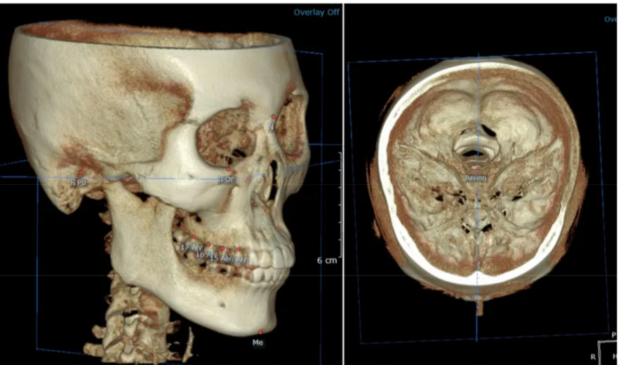
They were divided into 2 groups according to the amount of Menton (Me) deviation in relation to the midsagittal plane, which was defined as the plane perpendicular to FH plane and passing through Nasion and Basion, assessed by 3D analysis (Table 3, Figure 1) (OnDemand 3D, Cybermed Co., Seoul, Korea) : skeletal Class III, symmetry; Me deviation < 2mm (group S) and skeletal Class III, asymmetry; Me deviation > 4mm (group AS) (Haraguchi et al., 2002; Masuoka et al., 2007).
The 20 borderline patients of Me deviation between 2mm and 4mm were excluded from the study. Group S included 30 patients (17 males, 13 females) and group AS included 33 patients (15 males, 18 females).
The control group (group I) was consisted of 25 patients (11 males, 14 females) as following criteria : (1) 1° <ANB< 4° and Me deviation < 2mm (2) anterior
crowding less than 4mm (3) no buccal or lingual crossbite of molars, other criteria same as above. Table 1 reports the sex and age distribution in the sample and Table 2 shows the skeletal characteristics of the subjects.
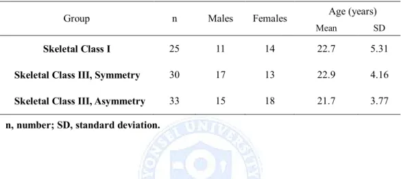
Table 1. Age and sex distribution of the patients
Group n Males Females Age (years)
Mean SD
Skeletal Class I 25 11 14 22.7 5.31
Skeletal Class III, Symmetry 30 17 13 22.9 4.16
Skeletal Class III, Asymmetry 33 15 18 21.7 3.77
n, number; SD, standard deviation.
Table 2. Descriptive statistics for skeletal characteristics
SD, standard deviation; ANB, A point-Nasion-B point angle; Me, Menton; S, Skeletal Class III, symmetry; AS, Skeletal Class III, asymmetry; I, skeletal Class I.
Data analyzed by one-way ANOVA and multiple comparison with Bonferroni test at significance level of p<0.001.
Skeletal Class I
(n=25)
(Mean ± SD)
Skeletal Class III, symmetry (n=30)
(Mean ± SD)
Skeletal Class III, asymmetry (n=33) (Mean ± SD) Multiple comparison ANB (°) 2.6 ± 0.89 -1.9 ± 1.53 -1.7 ± 1.60 S = AS < I Me deviation (mm) 1.2 ± 0.64 1.0 ± 0.51 8.4 ± 3.09 S = I < AS
2. Cone beam computed tomography and software
The CBCT scans were taken (PaX-Zenith 3D, Vatech, Gyeonggi-Do, Korea) under following conditions : 120 kV, 10 mA, voxel size 0.3mm. The scanned images were saved as digital imaging and communication in medicine (DICOM) files and reconstructed with 3-dimensional analysis software program (OnDemand 3D, Cybermed Co., Seoul, Korea).
3. Assessment of asymmetry on 3-dimensional image
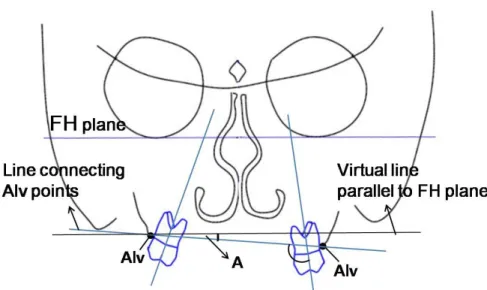
Landmarks and reference planes are defined in Table 3 and Figure 1. The definitions were modified from previous studies that assessed facial asymmetry with 3D images (Baek et al., 2012; Park et al., 2006). The asymmetry of all subjects was assessed on 3D image. Maxillary canting was measured as the angle between the FH plane and the line connecting alveolar point (Alv) of both sides (Figure 2). As the canting of maxilla is caused in 3-dimensions, it was measured at each location of tooth to assess the influence of canting on inclination. Me deviation was measured as the distance between the Me and midsagittal plane.
Table 3. Landmarks and reference planes
Landmarks Definition
N (Nasion) Middle point between frontal bone and nasal bone Ba (Basion) Most anterior point of foramen magnum
Or (Orbitale) Lowest point of lower margin of orbit Me (Menton) Most inferior point on symphysis of mandible Po (Porion) Most superior of external auditory meatus Alv (Alveolar point)
The point of alveolar bone at buccal ridge of premolar or buccal groove of molar
Reference planes
FH (Frankfurt horizontal) plane Plane passing through right Porion, left Porion and midpoint of left and right Orbitale Midsagittal plane Plane perpendicular to FH plane, passing through Nasionand Basion
Figure 2. Measurement of maxillary canting at each tooth. Maxillary canting was measured as the angle of (A°) between the FH plane and the line connecting Alv of both sides. It was performed on each location of the tooth. FH, frankfurt horizontal; Alv, alveolar point.
4. Measurement of buccolingual inclination of posterior teeth
The buccolingual inclination of maxillary and mandibular posterior teeth (premolars and molars) was measured with CBCT data. The CBCT scan was reconstructed as following reference planes to ensure the 2-dimensional coronal slices to be consistently oriented: (1) the axial plane was defined as FH plane; (2) the sagittal plane was defined as patient’s midsagittal plane. Then, the tooth to be measured was located in the axial view. In the sagittal view, coronal slice was obtained according to the long axis of the teeth: a plane passing through the mesial cusp tip and mesial root apex in molar; a plane passing through the cusp tip and root apex in premolar. In the coronal view, measurements of buccolingual inclination were
taken between the long axis of the tooth and the FH plane for maxillary teeth, and the long axis of the tooth and the inferior border of mandible for mandibular teeth. The long axes of teeth and the reference lines of maxilla and mandible are shown in Table 4 and the technique for measuring buccolingual inclination of posterior teeth is described in Figure 3 and 4.
If any amount of Me deviation existed, we distinguished the deviated side and non-deviated side in measuring the buccolingual inclination. The deviated side was defined as the side toward which the Me was deviated in relation to the midsagittal plane.
Table 4. Long axes of teeth and reference lines for angular measurements
Maxilla Definition Long axis,
multi-rooted premolar and molar
The line connecting the groove between the buccal and palatal cusps and the furcation of the roots
Long axis,
single-rooted premolar
The line connecting the groove between the buccal and palatal cusps and the root apex
Reference line FH plane in the coronal slice shown as a line
Mandible Definition
Long axis, premolar and molar The line connecting the groove between the buccal and lingual cusps and the root apex
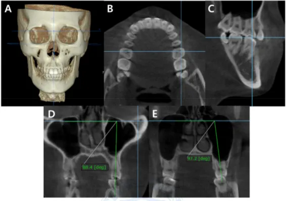
Figure 3. Method to measure buccolingual inclination of maxillary teeth. (A) Ensure that the axial plane to be oriented as FH plane. (B) Locate molar in the axial view. (C) In the sagittal view, position the line to obtain the coronal view. (D,E) In the coronal view, the horizontal reference line shown is parallel to the FH plane. Measure the inclination between the long axis of tooth and the reference line (FH plane): (D) premolar, (E) molar.
Figure 4. Method to measure buccolingual inclination of mandibular teeth. (A) Locate molar in the axial view. (B) In the sagittal view, position the line to obtain the coronal view. (C,D) In the coronal view, measure the inclination between the long axis of tooth and the reference line which is tangent to the inferior border of mandible: (C) premolar, (D) molar.
5. Statistical analysis
One-way analysis of variance (ANOVA) was applied to detect significant differences in skeletal characteristics among groups and multiple comparisons were performed with Bonferroni test. All measurements of buccolingual inclination were repeated after a 2 week interval on CBCT images of 20 randomly selected patients.
The intra-class correlation coefficient (ICC) was used to test the intra-examiner reliability and the reproducibility of the measurements was assessed.
The means and standard deviations were calculated for each measurement. Paired t-tests were used to examine the differences of inclination between the deviated side and the non-deviated side in each group. One-way ANOVA was used to check for statistically significant differences among 3 groups and multiple comparisons were performed with Bonferroni test. Univariate and multivariate linear regression analyses were carried out to detect the influence of asymmetry on buccolingual inclination of posterior teeth and standardized correlation coefficients were computed between the buccolingual inclination and variables of asymmetry. All statistical analyses were performed with SAS version 9.2 (SAS Institute Inc., Cary, NC, USA) and p value less than 0.05 was regarded to be statistically significant.
III. RESULTS
1. Error of the method
The intra-examiner reproducibility was assessed by the intraclass correlation coefficient for repeated measurements. It showed high reliability with range from 0.992 to 0.998 (p < 0.001).
2. Buccolingual inclination of posterior teeth in groups
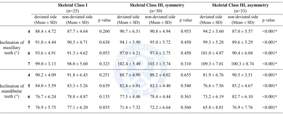
There was no significant difference of buccolingual inclination between deviated and non-deviated sides in group I and group S (p>0.05). In group AS, the buccolingual inclination of both sides were significantly different from each other (p<0.001) (Table 5). On the deviated side, maxillary teeth were more buccally inclined and mandibular teeth were more lingually inclined than the non-deviated side. As regarding there were no differences in both sides, the measurements of both sides were combined for subsequent analyses in group I and group S (Table 6).
In all groups, from premolars through molars, there was a tendency to be buccally inclined in maxillary teeth and to be lingually inclined in mandibular teeth (Table 6).
Table 5. Comparison of the buccolingual inclination between deviated side and non-deviated side in each group
Skeletal Class I
(n=25)
Skeletal Class III, symmetry
(n=30)
Skeletal Class III, asymmetry
(n=33) deviated side (Mean ± SD) non-deviated side (Mean ± SD) p value deviated side (Mean ± SD) non-deviated side (Mean ± SD) p value deviated side (Mean ± SD) non-deviated side (Mean ± SD) p value Inclination of maxillary teeth (°) 4 88.4 ± 4.72 87.7 ± 4.64 0.260 90.7 ± 6.51 90.8 ± 4.94 0.953 94.2 ± 5.60 87.0 ± 5.57 <0.001* 5 91.0 ± 4.44 90.5 ± 4.71 0.638 94.1 ± 5.90 95.0 ± 5.72 0.450 99.3 ± 5.28 89.6 ± 5.29 <0.001* 6 93.6 ± 4.91 91.3 ± 4.62 0.053 97.0 ± 4.21 97.4 ± 3.75 0.450 101.0 ± 4.87 90.4 ± 6.04 <0.001* 7 99.0 ± 3.13 98.0 ± 5.60 0.323 102.4 ± 5.49 103.3 ± 5.74 0.310 109.3 ± 7.01 100.3 ± 8.74 <0.001* Inclination of mandibular teeth (°) 4 90.2 ± 4.09 91.8 ± 6.43 0.251 88.7 ± 4.90 88.2 ± 4.02 0.655 81.9 ± 6.76 90.5 ± 3.51 <0.001* 5 84.0 ± 5.59 83.3 ± 5.26 0.639 82.4 ± 6.01 83.1 ± 4.40 0.540 76.6 ± 7.56 85.2 ± 4.67 <0.001* 6 76.7 ± 6.24 78.8 ± 4.87 0.135 77.5 ± 4.46 78.4 ± 4.44 0.363 73.2 ± 6.19 82.7 ± 6.10 <0.001* 7 76.9 ± 5.75 77.1 ± 6.20 0.855 71.4 ± 7.32 72.2 ± 6.64 0.560 65.8 ± 8.01 76.9 ± 7.76 <0.001* Paired t-test was conducted to compare the values of deviated side and non-deviated side in each group.
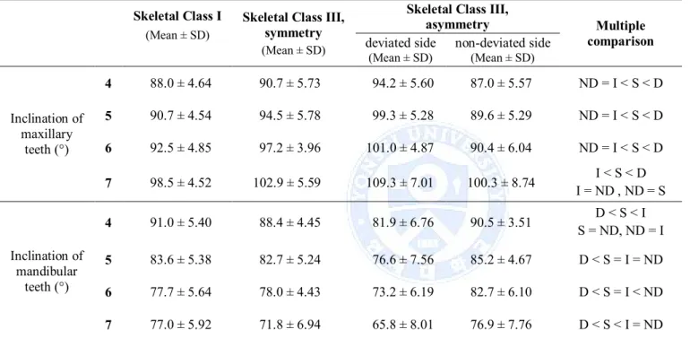
Table 6. Comparison of the buccolingual inclination among groups
Skeletal Class I (Mean ± SD)
Skeletal Class III, symmetry (Mean ± SD)
Skeletal Class III,
asymmetry Multiple comparison deviated side (Mean ± SD) non-deviated side (Mean ± SD) Inclination of maxillary teeth (°) 4 88.0 ± 4.64 90.7 ± 5.73 94.2 ± 5.60 87.0 ± 5.57 ND = I < S < D 5 90.7 ± 4.54 94.5 ± 5.78 99.3 ± 5.28 89.6 ± 5.29 ND = I < S < D 6 92.5 ± 4.85 97.2 ± 3.96 101.0 ± 4.87 90.4 ± 6.04 ND = I < S < D 7 98.5 ± 4.52 102.9 ± 5.59 109.3 ± 7.01 100.3 ± 8.74 I = ND , ND = SI < S < D Inclination of mandibular teeth (°) 4 91.0 ± 5.40 88.4 ± 4.45 81.9 ± 6.76 90.5 ± 3.51 S = ND, ND = ID < S < I 5 83.6 ± 5.38 82.7 ± 5.24 76.6 ± 7.56 85.2 ± 4.67 D < S = I = ND 6 77.7 ± 5.64 78.0 ± 4.43 73.2 ± 6.19 82.7 ± 6.10 D < S = I < ND 7 77.0 ± 5.92 71.8 ± 6.94 65.8 ± 8.01 76.9 ± 7.76 D < S < I = ND
SD, standard deviation; I, skeletal Class I; S, skeletal Class III, symmetry; D, deviated side of skeletal Class III, asymmetry; ND, non-deviated side of skeletal Class III, asymmetry.
3. Comparison of the buccolingual inclination among groups
The results in Table 6 illustrates the comparison among three groups. There were significant differences in the buccolingual inclination of maxillary posterior teeth between the group I and group S (p<0.05), suggesting that the maxillary teeth were more buccally inclined in group S. The mandibular first premolar and second molar of group S differed significantly from those of group I (p<0.05), which implies that the teeth were more lingually inclined, whereas the mandibular second premolar and first molar did not.
All measurements on the deviated side of group AS were significantly different from those of group I and group S (p<0.05), suggesting that the maxillary teeth were more buccally inclined and mandibular teeth were more lingually inclined on the deviated side. However, on the non-deviated side, only mandibular first molar showed more buccal inclination than that of group I (p<0.05) and the other teeth had no significant difference from the group I (p>0.05). In comparison with group S, the inclination of maxillary premolars, maxillary first molar and mandibular molars on the non-deviated side of group AS showed significant difference (p<0.05), suggesting that palatal inclination of maxillary teeth and buccal inclination of mandibular teeth.
4. Relationship between asymmetry and buccolingual inclination
In group AS, the relationship between variables of asymmetry such as Me deviation and canting of maxilla and variables of buccolingual inclination such as the inclination of deviated side and non-deviated side was examined with univariate and multivariate linear regression analyses (Table 7). The canting of maxilla measured at each tooth was matched to each buccolingual inclination of tooth.
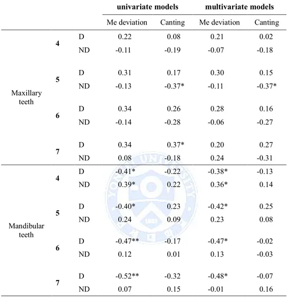
It was detected that Me deviation had significant negative correlation with all mandibular teeth on the deviated side and positive correlation with mandibular first premolar on the non-deviated side (p<0.05). On the other hand, Me deviation had no influence on maxillary teeth (p>0.05).
The canting of maxilla had no significant correlation with the inclination of mandibular teeth. It was found to have significant negative correlation with the inclination of maxillary second premolar on the non-deviated side (p<0.05).
Table 7. Univariate and multivariate linear regression analyses and correlation coefficients between asymmetry and buccolingual inclination
Me, Menton; D, deviated side; ND, non-deviated side. *p<0.05;**p<0.01
univariate models multivariate models
Me deviation Canting Me deviation Canting
Maxillary teeth 4 D 0.22 0.08 0.21 0.02 ND -0.11 -0.19 -0.07 -0.18 5 D 0.31 0.17 0.30 0.15 ND -0.13 -0.37* -0.11 -0.37* 6 D 0.34 0.26 0.28 0.16 ND -0.14 -0.28 -0.06 -0.27 7 D 0.34 0.37* 0.20 0.27 ND 0.08 -0.18 0.24 -0.31 Mandibular teeth 4 D -0.41* -0.22 -0.38* -0.13 ND 0.39* 0.22 0.36* 0.14 5 D -0.40* 0.23 -0.42* 0.25 ND 0.24 0.09 0.23 0.08 6 D -0.47** -0.17 -0.47* -0.02 ND 0.12 0.01 0.13 -0.03 7 D -0.52** -0.32 -0.48* -0.07 ND 0.07 0.15 -0.01 0.16
IV. DISCUSSION
The assessment of buccolingual inclination of posterior teeth is important for detailed treatment plan, and CBCT scans which can visualize the long axis of teeth including roots are required to evaluate the inclination of posterior teeth. There were previous studies that evaluated the buccolingual inclination of molars using CT or CBCT scans and the method to measure the inclination was introduced in which the occlusal plane and the inferior border of mandible were used as reference planes (Masumoto et al., 2001; Miner et al., 2012; Mitra and Ravi, 2011; Nojima et al., 2007; Shewinvanakitkul et al., 2011; Tsunori et al., 1998). However, occlusal plane might be variable and not constant because it can be affected by tooth movement. This study used the FH plane and the inferior border of mandible, which were not influenced by dentition, as reference planes.
This study evaluated the buccolingual inclination of premolars and molars of skeletal Class I patients and it was found that while the mandibular second premolar and molars were lingually inclined, the maxillary premolars, first molar and mandibular first premolar were uprighted to some extent and maxillary second molar was buccally inclined (Table 6). It doesn’t coincide with the conventional buccolingual inclination of posterior teeth, derived from buccal crown contour, which is a negative value that indicates the crown has palatal or lingual torque. Andrews (1972) reported that the lingual inclination from premolars through molars was constant in upper teeth and progressively increased in lower teeth. In this study, there was a tendency of maxillary teeth to be buccally
inclined and mandibular teeth to be lingually inclined from premolars through molars in all groups.
The anteroposterior skeletal discrepancy had significant influence on the buccolingual inclination. In group S, all maxillary posterior teeth were more buccally inclined with statistical significance and the mandibular first premolar and second molar were lingually inclined with statistical significance than those of the group I (p<0.05) (Table 6). There have been studies about dental compensation of patients with Class III malocclusion, however, most of them have been focused on incisors (Kim and Baek, 2013; Kim et al., 2014; Troy et al., 2009). Though some studies noticed the transverse dental compensation depending on anteroposterior skeletal discrepancy, Slaj et al. (2010) didn’t evaluate the buccolingual inclination but the intermolar width on dental casts and Kim et al. (2012) evaluated dental decompensation during presurgical orthodontic treatment, not the initial state. This study found that there was transverse dental compensation of posterior teeth in group S and it could be considered in treatment planning. If patients with skeletal Class III malocclusion are planned to be treated with orthodontic camouflage, dental expansion of maxillary arch might not ensure the favorable condition of supporting alveolar bone and post-treatment occlusal stability. As there was more compensation in maxillary teeth than mandibular teeth, in case of presurgical orthodontic treatment, constriction of maxillary arch should be considered first and expansion of mandibular arch might be performed on those teeth required.
There was a statistical significant difference between the deviated side and the non-deviated side in group AS (p<0.001) (Table 5). The transverse skeletal
discrepancy had significant influence on the inclination of all maxillary and mandibular posterior teeth.
This study found that the transverse skeletal discrepancy had influence on the buccolingual inclination of posterior teeth and had more impact on the deviated side than the non-deviated side. All maxillary and mandibular teeth on the deviated side in group AS were significantly different from those in both group I and group S (p<0.05) (Table 6), which suggests that the maxillary teeth were more buccally inclined and mandibular teeth were more lingually inclined. On the deviated side in group AS, it can be inferred that the maxillary teeth were most buccally inclined and mandibular teeth were most lingually inclined among all groups because the inclination was affected by dental compensation due to both anteroposterior and transverse discrepancy in same direction. In contrast, on the non-deviated side in group AS, all posterior teeth except the mandibular first molar had no significant difference from those in group I (p>0.05). Several teeth (maxillary premolars, first molar and mandibular molars) on the non-deviated side of group AS showed significant difference when compared with those in group S (p<0.05). The posterior teeth on the non-deviated side in group AS might be affected by dental compensation due to anteroposterior discrepancy as bucally inclined maxillary teeth and lingually inclined mandibular teeth. In addition, they might be influenced by dental compensation due to transverse discrepancy as palatally inclined maxillary teeth and buccally inclined mandibular teeth, which resulted in no significant difference between the non-deviated side of group AS and group I.
In skeletal Class III patients with asymmetry, Kusayama et al. (2003) evaluated dental casts and Nojima et al. (2007) analyzed CT scans, and reported that the
inclination of first molars had significant difference between on the deviated and deviated side. However, they only found the difference between deviated and non-deviated sides and it was impossible to compare with normative data because there were no control groups including skeletal Class I patients. This study found the dental compensation of inclination of posterior teeth in skeletal Class III patients with asymmetry showed different patterns between deviated and non-deviated side. As there was a control group composed of skeletal Class I patients, we could find that the dental compensation by anteroposterior and transverse discrepancy resulted in intense tilting of posterior teeth on the deviated side and the inclination of posterior teeth on the non-deviated side was similar to that of skeletal Class I.
It can be inferred that during presugical orthodontic treatment of skeletal Class III patients with facial asymmetry, the deviated side needs more dental decompensation than the non-deviated side. The inclinations of all posterior teeth on the deviated side need to be corrected : palatal inclination required for maxillary teeth and buccal inclination for mandibular teeth. As the mandibular first molar was the only tooth that was significantly different between non-deviated side in group AS and group I, mandibular first molar on the non-deviated side needs lingual inclination during presurgical orthodontic treatment. Other teeth on the non-deviated side can be considered that they have normal inclinations as skeletal Class I. The correction of buccolingual inclination on the deviated side should be achieved using biomechanical methods, such as unilateral constriction transpalatal arch or expansion lingual arch, modification of bracket prescription or intermaxillary elastic on the deviated side.
In group AS, the correlation between skeletal asymmetry and buccolingual inclination was evaluated by univariate and multivariate linear regression models. Me deviation had significant negative correlations with the buccolingual inclination of all mandibular teeth on the deviated side (p<0.05), suggesting that the lingual inclination of mandibular teeth on the deviated side increased with Me deviation. It is consistent with the result above that the deviated side is more affected by asymmetry. Me deviation also had significant positive correlation with first premolar on the non-deviated side (p<0.05) and no significant correlation with maxillary teeth.
It was detected that the canting of maxilla had no relation with the bucculingual inclination of mandibular posterior teeth (p>0.05). However, the canting of maxilla had significant negative correlation with the inclination of maxillary second premolar on the non-deviated side (p<0.05).
Shewinvanakitkul et al. (2011) reported the method measuring inclination used in this study was reliable, but it was discussed that the approach to each tooth was not perpendicular to buccolingual plane of tooth. This study also had similar limitations that the sagittal slice of CBCT was obtained parallel to the midsagittal plane, not parallel to the mesiodistal surface of each tooth (Figure 2 and 3). If the sagittal slice of CBCT were obtained parallel to the mesiodistal surface of tooth, the inferior border of mandible could not be seen clearly. As the buccolingual inclination and mesiodistal angulation of tooth is combined in 3-dimension (Garcia-Figueroa et al., 2008), Tong et al. (2012) introduced a root vector analysis program which is available to measure angulation and inclination in 3-dimensions, but it was discussed that it can’t be used
for patients with malocclusions, especially asymmetries. As this study included many patients with asymmetry, it could not be applied.
In group AS, we didn’t separate the sample by posterior crossbite, and about a half of the patients showed posterior crossbite on the deviated side. However, in interpreting the results, no significant relationship between posterior crossbite and buccolingual inclination was found. With regard to posterior crossbite, larger sample size and the evaluation of the width of maxilla and mandible are needed in further study.
This study found that there was transverse dental compensation of posterior teeth according to anteroposterior and transverse skeletal discrepancies. The diagnosis of patients with skeletal discrepancy should include the evaluation of buccolingual inclination of posterior teeth, which should be considered in treatment planning (Burstone, 1998). Both presurgical orthodontic treatment for orthognathic surgery and camouflage orthodontic treatment require the understanding of buccolingual inclination of posterior teeth and dental compensation. Therefore, the analysis of the buccolingual inclination of posterior teeth including roots with CBCT is recommended to establish the diagnosis of skeletal discrepancy and dental compensation.
V. CONCLUSION
The buccolingual inclination of posterior teeth in skeletal Class III patients with or without asymmetry was evaluated by CBCT scans and compared with that in skeletal Class I patients. Also, the relationship between asymmetry and buccolingual inclination of posterior teeth was assessed. The findings are as followings,
1. The maxillary teeth had a tendency to be buccally inclined and the mandibular teeth to be lingually inclined from premolars through molars in all groups.
2. Group S showed more buccal inclination of maxillary premolars and molars, and more lingual inclination of mandibular first premolar and second molar compared to those of group I (p<0.05).
3. Group AS had significant difference in buccolingual inclination of posterior teeth between on the deviated side and non-deviated side (p<0.001). On the deviated side, maxillary teeth were more buccally inclined and mandibular teeth were more lingually inclined than the non-deviated side.
4. In Group AS, all teeth on the deviated side showed more buccally inclined maxillary teeth and lingually inclined mandibular teeth than those in other groups (p<0.05). On the non-deviated side, maxillary teeth had no significant difference
with group I (p>0.05) and only mandibular first molar was more buccally inclined than that of group I (p<0.05).
5. In group AS, Me deviation had negative correlation with the buccolingual inclination of all mandibular teeth on the deviated side (p<0.05), and canting of maxilla had negative correlation with only buccolingual inclination of maxillary second premolar on the non-deviated side (p<0.05).
There was transverse dental compensation in skeletal Class III patients with asymmetry, especially on the deviated side, and Me deviation had correlation with the dental compensation of mandibular teeth on the deviated side.
REFERENCES
Andrews LF: The six keys to normal occlusion. Am J Orthod 62(3): 296-309, 1972. Andrews LF: The straight-wire appliance, origin, controversy, commentary. J Clin
Orthod 10(2): 99-114, 1976.
Baek C, Paeng JY, Lee JS, Hong J: Morphologic evaluation and classification of facial asymmetry using 3-dimensional computed tomography. J Oral
Maxillofac Surg 70(5): 1161-1169, 2012.
Burstone CJ: Diagnosis and treatment planning of patients with asymmetries. Semin
Orthod 4(3): 153-164, 1998.
Casko JS, Vaden JL, Kokich VG, Damone J, James RD, Cangialosi TJ, et al.: Objective grading system for dental casts and panoramic radiographs. American Board of Orthodontics. Am J Orthod Dentofacial Orthop 114(5): 589-599, 1998. Garcia-Figueroa MA, Raboud DW, Lam EW, Heo G, Major PW: Effect of buccolingual
root angulation on the mesiodistal angulation shown on panoramic radiographs.
Am J Orthod Dentofacial Orthop 134(1): 93-99, 2008.
Germane N, Bentley BE, Jr., Isaacson RJ: Three biologic variables modifying faciolingual tooth angulation by straight-wire appliances. Am J Orthod
Dentofacial Orthop 96(4): 312-319, 1989.
Haraguchi S, Takada K, Yasuda Y: Facial asymmetry in subjects with skeletal Class III deformity. Angle Orthod 72(1): 28-35, 2002.
Janson G, Bombonatti R, Cruz KS, Hassunuma CY, Del Santo M, Jr.: Buccolingual inclinations of posterior teeth in subjects with different facial patterns. Am J
Kim DK, Baek SH: Change in maxillary incisor inclination during surgical-orthodontic treatment of skeletal Class III malocclusion: comparison of extraction and nonextraction of the maxillary first premolars. Am J Orthod
Dentofacial Orthop 143(3): 324-335, 2013.
Kim SJ, Kim KH, Yu HS, Baik HS: Dentoalveolar compensation according to skeletal discrepancy and overjet in skeletal Class III patients. Am J Orthod
Dentofacial Orthop 145(3): 317-324, 2014.
Kim YI, Choi YK, Park SB, Son WS, Kim SS: Three-dimensional analysis of dental decompensation for skeletal Class III malocclusion on the basis of vertical skeletal patterns obtained using cone-beam computed tomography. Korean J
Orthod 42(5): 227-234, 2012.
Kusayama M, Motohashi N, Kuroda T: Relationship between transverse dental anomalies and skeletal asymmetry. Am J Orthod Dentofacial Orthop 123(3): 329-337, 2003.
Major PW, Johnson DE, Hesse KL, Glover KE: Landmark identification error in posterior anterior cephalometrics. Angle Orthod 64(6): 447-454, 1994. Major PW, Johnson DE, Hesse KL, Glover KE: Effect of head orientation on
posterior anterior cephalometric landmark identification. Angle Orthod 66(1): 51-60, 1996.
Masumoto T, Hayashi I, Kawamura A, Tanaka K, Kasai K: Relationships among facial type, buccolingual molar inclination, and cortical bone thickness of the mandible. Eur J Orthod 23(1): 15-23, 2001.
Masuoka N, Muramatsu A, Ariji Y, Nawa H, Goto S, Ariji E: Discriminative thresholds of cephalometric indexes in the subjective evaluation of facial asymmetry. Am J Orthod Dentofacial Orthop 131(5): 609-613, 2007.
Miner RM, Al Qabandi S, Rigali PH, Will LA: Cone-beam computed tomography transverse analysis. Part I: Normative data. Am J Orthod Dentofacial Orthop 142(3): 300-307, 2012.
Mitra S, Ravi MS: Evaluation of buccolingual inclination of posterior teeth in different facial patterns using computed tomography. Indian J Dent Res 22(3): 376-380, 2011.
Nojima K, Yokose T, Ishii T, Kobayashi M, Nishii Y: Tooth axis and skeletal structures in mandibular molar vertical sections in jaw deformity with facial asymmetry using MPR images. Bull Tokyo Dent Coll 48(4): 171-176, 2007.
Nouri M, Abdi AH, Farzan A, Mokhtarpour F, Baghban AA: Measurement of the buccolingual inclination of teeth: manual technique vs 3-dimensional software. Am J Orthod Dentofacial Orthop 146(4): 522-529, 2014.
Park SH, Yu HS, Kim KD, Lee KJ, Baik HS: A proposal for a new analysis of craniofacial morphology by 3-dimensional computed tomography. Am J
Orthod Dentofacial Orthop 129(5): 600.e623-634, 2006.
Shewinvanakitkul W, Hans MG, Narendran S, Martin Palomo J: Measuring buccolingual inclination of mandibular canines and first molars using CBCT.
Orthod Craniofac Res 14(3): 168-174, 2011.
Sjogren AP, Lindgren JE, Huggare JA: Orthodontic study cast analysis--reproducibility of recordings and agreement between conventional and 3D virtual measurements. J Digit Imaging 23(4): 482-492, 2010.
Slaj M, Spalj S, Pavlin D, Illes D, Slaj M: Dental archforms in dentoalveolar Class I, II and III. Angle Orthod 80(5): 919-924, 2010.
Tong H, Enciso R, Van Elslande D, Major PW, Sameshima GT: A new method to measure mesiodistal angulation and faciolingual inclination of each whole tooth with volumetric cone-beam computed tomography images. Am J
Orthod Dentofacial Orthop 142(1): 133-143, 2012.
Troy BA, Shanker S, Fields HW, Vig K, Johnston W: Comparison of incisor inclination in patients with Class III malocclusion treated with orthognathic surgery or orthodontic camouflage. Am J Orthod Dentofacial Orthop 135(2): 146.e141-149; discussion 146-147, 2009.
Tsunori M, Mashita M, Kasai K: Relationship between facial types and tooth and bone characteristics of the mandible obtained by CT scanning. Angle Orthod 68(6): 557-562, 1998.
Vardimon AD, Lambertz W: Statistical evaluation of torque angles in reference to straight-wire appliance (SWA) theories. Am J Orthod 89(1): 56-66, 1986. Zebeib AM, Naini FB: Variability of the inclination of anatomic horizontal reference
planes of the craniofacial complex in relation to the true horizontal line in orthognathic patients. Am J Orthod Dentofacial Orthop 146(6): 740-747, 2014.
국문 요약
비대칭 유무에 따른 골격성 III급 부정교합 환자의
구치부 협설측 치축 분석
연세대학교 대학원 치의학과
(지도교수 김 경 호)
안 재 찬
본 연구에서는 CBCT 분석을 통해 비대칭 유무에 따른 골격성 III급 부정교합 환자의 구치부의 협설측 치축을 골격성 I급 환자와 비교하였으며, 비대칭과 구치부 협설측 치축간의 연관성을 평가하였다. 총 63명의 골격성 III급 부정교합 환자 (남 : 32명, 여 : 31명)가 선정 되었으며 연구 대상의 평균 나이는 22.4 ± 4.00세 였다. 대상자는 이부 변 위량에 따라 두 그룹으로 나누었으며 이부변위는 정중시상면에서 이부까지 의 거리로 측정되었다. 이부 변위 2mm 미만인 환자를 비대칭이 없는 골 격성 III급 군, 이부 변위 4mm 초과인 환자를 비대칭을 동반한 골격성 III 급 군으로 정하였고 이부 변위가 2mm 이상 4mm 이하인 자는 제외하였 다. 대조군은 골격성 I급이며 경미한 총생을 보이는 총 25명의 정상교합자 (남 : 11명, 여 : 14명)로 이루어졌으며 평균 나이는 22.7 ± 5.31세 였다.CBCT 영상에서 상악은 FH plane, 하악은 하악골 하연을 기준평면으 로 소구치와 대구치의 협설측 치축이 측정되었다. 각 군간의 협설측 치축 을 비교하여 치성보상을 평가하였고, 비대칭이 구치부의 협설측 치축에 미 치는 영향을 조사하여 다음과 같은 결과를 얻었다. 1. 골격성 I급과 골격성 III급 군 모두에서 협설측 치축은 제 1 소구치부터 제 2 대구치까지 후방으로 갈수록 상악 치아는 더욱 협측 경사 되었고, 하악 치아는 더욱 설측 경사되는 양상을 나타냈다. 2. 비대칭이 없는 골격성 III급 군에서 골격성 I급 군에 비해 상악의 모든 소구치와 대구치는 협측 경사되었고 하악 제 1 소구치와 제 2 대구치는 설측 경사되었다 (p<0.05). 3. 비대칭을 동반한 골격성 III급 군에서 이환측과 비이환측 간의 협설측 치축 차이는 통계적으로 유의할만한 차이가 있었다 (p<0.001). 상악은 이환측 치아가 더 협측 경사된 치축을, 하악은 이환측 치아가 더 설측 경사된 치축을 나타냈다. 4. 비대칭을 동반한 골격성 III급 군에서 골격성 I급, 비대칭이 없는 골격성 III급 군과 비교 시 모든 이환측 치아들이 상악은 협측 경사, 하악은 설 측 경사를 보였다 (p<0.05). 비이환측은 골격성 I급 군과 비교 시 상악
은 차이가 없었으며 (p>0.05), 하악은 제 1 대구치만이 협측 경사를 보 였다 (p<0.05). 5. 비대칭을 동반한 골격성 III급 군에서 하악 이부의 변위는 하악 치아의 모든 이환측 치아의 협설측 치축과 음의 상관성을 가졌으며 (p<0.05), 상악의 기울기는 단지 비이환측 상악 제 2 소구치의 협설측 치축과 음 의 상관관계를 가졌다 (p<0.05). 비대칭을 동반한 골격성 III급 부정교합 환자의 이환측 치아에서 횡적 인 치성보상이 주로 일어났으며, 하악 이부의 변위가 하악 이환측 치아의 치성보상과 상관관계를 보였다. 핵심 되는 말: 협설측 치축, CBCT, 골격성 III급 부정교합, 비대칭