The First Case of Familial Mediterranean Fever Associated
with Renal Amyloidosis in Korea
Kyo Yeon Koo,
1* Se Jin Park,
2* Ji Young Wang,
1Jae Il Shin,
2Hyeon Joo Jeong,
3Beom Jin Lim,
3and Jin-Sung Lee
1,2Departments of 1Clinical Genetics, 2Pediatrics, and 3Pathology, Yonsei University College of Medicine, Seoul, Korea.
Received: April 1, 2011 Revised: December 16, 2011 Accepted: December 22, 2011 Corresponding author: Dr. Jin-Sung Lee, Department of Clinical Genetics, Yonsei University College of Medicine, 50 Yonsei-ro, Seodaemun-gu, Seoul 120-752, Korea.
Tel: 82-2-2228-2540, Fax: 82-2-362-0755 E-mail: [email protected]
*Kyo Yeon Koo and Se Jin Park contributed equally to this work.
∙ The authors have no financial conflicts of interest.
© Copyright:
Yonsei University College of Medicine 2012
This is an Open Access article distributed under the terms of the Creative Commons Attribution Non-Commercial License (http://creativecommons.org/ licenses/by-nc/3.0) which permits unrestricted non-commercial use, distribution, and reproduction in any medium, provided the original work is properly cited.
Familial Mediterranean fever (FMF) is an auto-inflammatory disease characterized by periodic episodes of fever and recurrent polyserositis. It is caused by a dysfunction of pyrin (or marenostrin) as a result of a mutation within the MEFV gene. It occurs most-ly in individuals of Mediterranean origin; however, it has also been reported in non-Mediterranean populations. In this report, we describe the first case of FMF in a Kore-an child. As eight-year-old boy presented recurrent febrile attacks from Kore-an unknown cause, an acute scrotum and renal amyloidosis. He also showed splenomegaly, lymphadenopathy, pleural effusion, ascites and elevated acute phase reactants. After
MEFV gene analysis, he was diagnosed as FMF combined with amyloidosis.
Key Words: Familial Mediterranean fever, amyloidosis, marenostrin
INTRODUCTION
Familial Mediterranean fever (FMF) is a hereditary auto-inflammatory disease, caused by mutation(s) in the MEFV gene.1-4 It manifests as repetitive fever
epi-sodes combined with one or more symptoms of sterile arthritis or serositis.1-4 FMF
is endemic to those of Mediterranean descent; however, sporadic cases have been reported in non-Mediterranean persons beyond this region.4-6 Especially, reports of
FMF are uncommon in the Far East although few reports and mutation studies have been conducted in Japanese patients.6 Accordingly, FMF is not familiar to
cli-nicians in this area and might be misdiagnosed as other auto-inflammatory diseas-es. The Tel-Hashomer criteria can help with clinically diagnosing the disease; however, the same criteria are also indicative of inflammatory diseases. The
MEFV gene for FMF has been cloned and DNA analysis can be useful in
diagnos-ing FMF when patient(s) do not wholly fulfill the diagnostic criteria. Herein, we describe the first case of FMF confirmed by molecular analysis of the MEFV gene in a Korean patient who showed recurrent clinical symptoms.
CASE REPORT
with 24-hour collected urine. Different markers for autoim-mune diseases such as anti-nuclear antibody, and anti-car-diolipin antibody were negative and complement levels as well as immunoglobulin concentrations were all within normal range.
After admission, he complained of abdominal pain and abdominal ultrasonography revealed slightly increased re-nal parenchymal echogenicity with splenomegaly (11 cm) and no hydrocele in both scrotal sacs. Abdominal computed tomography showed enlargement of bilateral kidneys; a well-enhancing mass lesion in the right common iliac chain; small bowel mesentery, suspected as lymphadenopathy; a small amount of ascites; and pleural effusion. Echocardiog-raphy showed minimal pericardial effusion with decreased deceleration time of the mitral valve (80-100 ms), which suggested a poor prognosis.
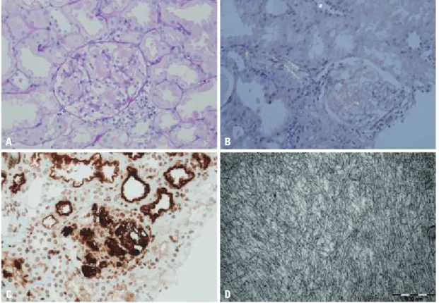
Renal biopsy was performed for evaluation of the massive proteinuria, and the pathologic diagnosis was confirmed as amyloidosis type AA (Fig. 1). Tests for monoclonal gam-mopathy to rule out AL amyloidosis, including serum/urine protein electrophoresis, immunofixation electrophoresis, and serum κ-, λ-free light chain assays, were all negative. Cochicine therapy was started with a dose of 0.025 mg/kg/ day under the impression of FMF and his clinical symp-two weeks and sudden onset of scrotal swelling that
oc-curred two days prior to admission. Since one year before the admission, he had repetitive, acute, self-limited epi-sodes of fever at varying intervals with an elevated level of acute phase reactants. Despite thorough evaluation, the rea-son for the fever was unable to be determined. After an ini-tial examination, he had been followed up under the im-pression of juvenile rheumatoid arthritis even though he did not demonstrate joint symptoms. Family history revealed that his father died due to myocardial infarction at the age of 40 and there was no history of renal disease, periodic fe-ver or auto-inflammatory disease.
Upon physical examination, his fever was 38.4°C, blood pressure was 108/75 mm Hg, and body weight increased from 23 kg to 26 kg in last two weeks prior to admission. Pretibial pitting edema and swollen abdomen were also ob-served without rash. Laboratory findings on admission showed inflammatory conditions of very high C-reactive protein (140 mg/L) and high erythrocyte sedimentation rate (120 mm/hr). Anemia (8.4 g/dL), hypoalbuminemia (1.0 g/ dL), and markedly elevated fibrinogen (1029 mg/dL) were observed as well. Massive nephrotic-range proteinuria (urine protein/creatinine ratio was 14.49, total microprotein 12297 mg/day, microalbumin 10249 mg/day) was observed
Fig. 1. The mesangium is expanded by pinkish amorphous material (PAS, ×400) (A). This material shows apple green birefringence under
the polarized microscopy after Congo red staining (×200) (B) and immunoreactivity to the amyloid A antibody (×200) (C). An electron mi-croscopy reveals haphazardly arranged non-branching fibrils measuring 8-10 nm in diameter. (×50000) (D).
A
C
B
ferential diagnosis, other genes, including MVK for HIDS,
NLRP3 for MWS and TNFRSF1A for FPF, that cause
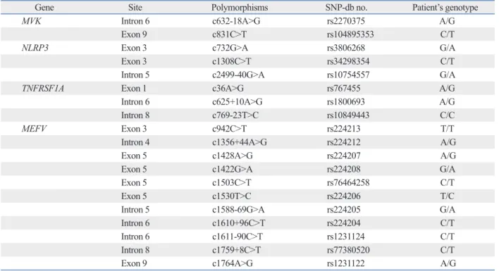
simi-lar phenotypes were also analysed. Only several SNPs were identified in the analysis of MVK, NLRP3 and TNFRSF1A genes (Table 1).
DISCUSSION
FMF is a hereditary auto-inflammatory disease character-ized by periodic episodes of fever and polyserositis at irreg-ular intervals.1-4 In 1960, Fox and Morrelli described the
first case of FMF in a patient of non-Mediterranean ethnic origin.7 Thereafter, sporadic cases among Ashkenazi Jews,
Germans and Anglo-Saxons have been continuously report-toms gradually improved.
In order to make the clinical diagnosis, the clinical enti-ties of periodic fever and systemic inflammatory reactions combined with amyloidosis were considered, which were indicative of FMF (OMIM no. 249100), familial periodic fever (FPF, TRAPS; OMIM no. 142680), hyperimmuno-globulinemia D with periodic fever syndrome (HIDS; OMIM no. 260920), and Muckle-Wells syndrome (MWS; OMIM no. 191900). The patient was able to be diagnosed as FMF since he showed typical attacks fulfilling three ma-jor criteria (pleuritis, fever and incomplete abdominal at-tack) of the Tel-Hashomer criteria. To verify the underlying cause of AA amyloidosis, DNA analysis of the MEFV gene was initially performed and two point mutations (p. Pro369Ser, p.Arg408Gln) were identified (Fig. 2). For
dif-Fig. 2. Results of the DNA analysis of the MEFV gene.
Table 1. SNPs Identified in the Patient after Analysis of the MVK, NLRP3, TNFRSF1A and MEFV Genes
Gene Site Polymorphisms SNP-db no. Patient’s genotype
MVK Intron 6 c632-18A>G rs2270375 A/G
Exon 9 c831C>T rs104895353 C/T
NLRP3 Exon 3 c732G>A rs3806268 G/A
Exon 3 c1308C>T rs34298354 C/T
Intron 5 c2499-40G>A rs10754557 G/A
TNFRSF1A Exon 1 c36A>G rs767455 A/G
Intron 6 c625+10A>G rs1800693 A/G
Intron 8 c769-23T>C rs10849443 C/C
MEFV Exon 3 c942C>T rs224213 T/T
Intron 4 c1356+44A>G rs224212 A/G
Exon 5 c1428A>G rs224207 A/G
Exon 5 c1422G>A rs224208 G/A
Exon 5 c1503C>T rs76464258 C/T
Exon 5 c1530T>C rs224206 T/C
Intron 5 c1588-69G>A rs224205 G/A
Intron 6 c1610+96C>T rs224204 C/T
Intron 6 c1611-90C>T rs1231124 C/T
Intron 8 c1759+8C>T rs77380520 C/T
Exon 9 c1764A>G rs1231122 A/G
p.Pro369Ser (c.1150C>T)
A A G C C T A A G C C C C C A G C C C C T G G C C A C C G G G T G C G C C C C A T
icantly affect interaction between pyrin and PSTPIP1. How-ever, 5 of 22 patients carrying these mutations fulfilled Tel-Hashomer criteria for clinical diagnosis of FMF. Therefore, other genes causing periodic fever syndrome were analysed with negative results.
In addition to pathologic mutations in MEFV gene, envi-ronmental factors and interactions of other genes are em-phasized in the expression of the clinical phenotype.13 It
may be important factors in FMF patients with autosomal dominant inheritance carrying heterozygote mutation in
MEFV gene. The case in this report fulfilled the
Tel-Hashom-er critTel-Hashom-eria for clinical diagnosis of FMF and carried two mutations in the MEFV gene without any pathologic muta-tions in the MVK, NLRP3 and TNFRSF1A genes. Even though the functional significance of P369S/R408Q has not been clearly elucidated, we assume that interactions with other genes may play some role in the phenotypic expres-sion of these mutations. In a study on Turkish patients, the frequency of P369S was 7.6% and the genotype of P369S/
E148Q was present in 15%.14
Clinical diagnosis of FMF is not easy, especially, in pa-tients of non-Mediterranean ethnic origin. Our case showed severe symptoms including renal amyloidosis at an early age. The most common reason for renal amyloidosis in chil-dren in the Mediterranean area15 is FMF. There is also a
pos-sibility of another gene(s) responsible for the disease. About 40% of the patients with clinical symptoms of FMF show negative results upon DNA analysis. More data on the muta-tion spectrum of the MEFV gene and phenotypic character-istics will increase the further understanding of the function of the gene and the molecular pathogenesis of the disease.
REFERENCES
1. Gedalia A. Hereditary periodic fever syndromes. In: Kliegman RM, Behrman RE, Jenson HB, Stanton BF, editors. Nelson text-book of Pediatrics. 18th ed. Philadelphia: W.B. Saunders Compa-ny; 2007. p.1029-31.
2. Katsenos S, Mermigkis C, Psathakis K, Tsintiris K, Polychro-nopoulos V, Panagou P, et al. Unilateral lymphocytic pleuritis as a manifestation of familial Mediterranean fever. Chest 2008;133: 999-1001.
3. Cekin AH, Dalbudak N, Künefeci G, Gür G, Boyacioğlu S. Fa-milial Mediterranean fever with massive recurrent ascites: a case report. Turk J Gastroenterol 2003;14:276-9.
4. Onen F. Familial Mediterranean fever. Rheumatol Int 2006; 26:489-96.
5. Konstantopoulos K, Kanta A, Tzoulianos M, Dimou S, Sotsiou F, Politou M, et al. Familial Mediterranean fever phenotype II in
ed including Asians. Reasons for the differences in the prevalence of the disease in different ethnic groups are still unknown.1,4-7 The influence of environmental factors or a
selective advantage of the heterozygote has been suggested in those from the Mediterranean area.8
In this report, the first case of familial Mediterranean fe-ver in a Korean patient was described. The patient showed symptoms of renal amyloidosis, which leads to renal failure and increased mortality. Because of the rarity of the disease in Korea, FMF was not considered until he was diagnosed as having AA amyloidosis. The diagnosis of FMF is still based on clinical manifestations listed in the Tei-Hashomer criteria.2 Since colchicine therapy is essential for prevention
and recovery from amyloidosis and other complications of FMF, early diagnosis is important.1-4 For this reason, the
clinical symptoms of abdominal pain (90%), arthritis or ar-thralgia (85%), and chest pain (20%) need to be carefully evaluated when these symptoms are combined with period-ic fever. In addition, serositis, such as perperiod-icarditis, as well as ascites and an acute scrotum are rarely afflicted. Recurrent attacks of triad symptoms, such as fever, arthritis and sero-sitis as well as abnormal acute phase reactants in conjunc-tion with an excellent response to colchicine were also sug-gestive of FMF.
Recently, molecular genetic testing should be considered for diagnostic confirmation.1-4,7-10
The gene responsible for FMF, called MEFV, is mapped to chromosome 16p13.3, which encodes a 781 amino acid pro-tein named pyrin (or marenostrin), and transmitted in an au-tosomal recessive or dominant pattern. Pyrin is expressed in the cytoplasm of mature neutrophils and monocytes and is thought to trigger the biosynthesis of neutrophil-specific tran-scription factors.1,2 More than 50 mutations have been
de-scribed and five mutations (M694V, V726A, M694I, M680I,
E148Q) are the most common mutations observed.1,3,4,11
Even though molecular DNA analysis is a powerful tech-nique for the diagnosis of the disease, it should be noted that only 60% of patients showed pathologic mutations within the
MEFV gene.1,2 Two point mutations (P369S, R408Q) that
were observed in our patient, were not frequently detected in Western populations, including the Middle East.8 However,
these mutations were reported in Japanese patients with the same genotype as our patient, carrying two mutations in
cis-; however their phenotypic effects remain
controver-sial.12 Ryan, et al.13 studied the functional significance of
P369S/R408Q mutations and concluded that these muta-tions are related with variable phenotypes and do not
signif-ranay M, et al. Phagocytic activity in familial Mediterranean fever. Yonsei Med J 2000;41:441-4.
12. Sugiura T, Kawaguchi Y, Fujikawa S, Hirano Y, Igarashi T, Kawa-moto M, et al. Familial Mediterranean fever in three Japanese pa-tients, and a comparison of the frequency of MEFV gene muta-tions in Japanese and Mediterranean populamuta-tions. Mod Rheumatol 2008;18:57-9.
13. Ryan JG, Masters SL, Booty MG, Habal N, Alexander JD, Bar-ham BK, et al. Clinical features and functional significance of the P369S/R408Q variant in pyrin, the familial Mediterranean fever protein. Ann Rheum Dis 2010;69:1383-8.
14. Caglayan AO, Demiryilmaz F, Ozyazgan I, Gumus H. MEFV gene compound heterozygous mutations in familial Mediterranean fever phenotype: a retrospective clinical and molecular study. Nephrol Dial Transplant 2010;25:2520-3.
15. Bilginer Y, Akpolat T, Ozen S. Renal amyloidosis in children. Pe-diatr Nephrol 2011;26:1215-27.
Greece. Isr Med Assoc J 2001;3:862-3.
6. Araki H, Onogi F, Ibuka T, Moriwaki H. [A Japanese family with adult-onset familial Mediterranean fever and periodic episodes of high fever and abdominal pain]. Nihon Shokakibyo Gakkai Zasshi 2010;107:427-31.
7. Dormer AE, Hale JF. Familial Mediterranean fever: a cause of pe-riodic fever. Br Med J 1962;1:87-9.
8. Yepiskoposyan L, Harutyunyan A. Population genetics of familial Mediterranean fever: a review. Eur J Hum Genet 2007;15:911-6. 9. Baş F, Kabataş-Eryilmaz S, Günöz H, Darendeliler F, Küçükemre
B, Bundak R, et al. Type 1 diabetes mellitus associated with auto-immune thyroid disease, celiac disease and familial Mediterranean fever: case report. Turk J Pediatr 2009;51:183-6.
10. Matos TC, Terreri MT, Petry DG, Barbosa CM, Len CA, Hilário MO. Autoinflammatory syndromes: report on three cases. Sao Paulo Med J 2009;127:314-6.