저작자표시-비영리-변경금지 2.0 대한민국 이용자는 아래의 조건을 따르는 경우에 한하여 자유롭게 l 이 저작물을 복제, 배포, 전송, 전시, 공연 및 방송할 수 있습니다. 다음과 같은 조건을 따라야 합니다: l 귀하는, 이 저작물의 재이용이나 배포의 경우, 이 저작물에 적용된 이용허락조건 을 명확하게 나타내어야 합니다. l 저작권자로부터 별도의 허가를 받으면 이러한 조건들은 적용되지 않습니다. 저작권법에 따른 이용자의 권리는 위의 내용에 의하여 영향을 받지 않습니다. 이것은 이용허락규약(Legal Code)을 이해하기 쉽게 요약한 것입니다. Disclaimer 저작자표시. 귀하는 원저작자를 표시하여야 합니다. 비영리. 귀하는 이 저작물을 영리 목적으로 이용할 수 없습니다. 변경금지. 귀하는 이 저작물을 개작, 변형 또는 가공할 수 없습니다.
A Dissertation
for the Degree of Master of Philosophy
Studies on effect of
graphene-based materials on
chondrogenic differentiation of
human adipose-derived stromal
cells
그래핀기반 물질을 이용한
인간 지방유래 기질세포의 연골 분화 효과 연구
August, 2019
By
Sumin Park
Biomodulation Major
i
ABSTRACT
Cartilage is a tissue consisting of chondrocytes and extracellular matrix (ECM), which is highly elastic, buffers against a given force, and the friction coefficient of joint cartilage is very low, helping the joints to move in a state of little friction. But cartilage is a kind of expendable body part that wears out as much as it is used, and it is also damaged by inflammation, trauma, and aging. Cartilage has very limited self-renewal because there are no blood vessels, nerves, or lymphatic vessels. Thus, cartilage cannot easily stop when it starts to damage, and damaged cartilage has many limitations about regenerating into cartilage with normal function and structure. Also, the damaged cartilage area becomes more vulnerable to mechanical pressure, so it is easily broken and worn, resulting in larger defects. As such, a number of cartilage-related diseases develop and progresses faster than other tissues. Some typical diseases include degenerative arthritis, which causes pain due to cartilage wear, and others include rheumatoid arthritis, achondroplasia, pyogenic arthritis, and chondrosarcoma. Furthermore, these cartilage-related diseases can lead to bone-related complications if they persist for a long time. These diseases can lead to a reduction in the quality of life and life expectancy of patients, and the loss of substantial medical costs. Therefore, it is very important to regenerate damaged
ii
cartilage early, but there are many deficiencies in current cartilage treatment. Although many researchers continue to suggest ways to treat cartilage-related diseases, they are still far from normal and perfect cartilage regeneration.
Up until now, studies have been actively conducted to regenerate damaged cartilage tissue using an ideal biomaterial that can mimic and replace cartilage tissue with various cells. Among them, cells that are widely used in the field of regenerative medicine are human adipose-derived stromal cells (hASCs). This cell has a great advantage that it can be easily obtained from any tissue of the human body, and it is suitable as a material for a cell therapy agent because it does not take much time for cell proliferation and can be repeatedly collected. And recently, graphene (G), a carbon-based material, is emerging as a biomaterial in tissue engineering and regenerative medicine. In addition, a variety of graphene-based materials (GBMs) have also been recognized for their research value as biomaterials. GBMs are derivatives of G. Most GBMs are made through the process of oxidation. GBMs that have the advantage of oxidation are more applied in various fields than G. In particular, GBMs play a role as biomaterials due to their various functional groups, biocompatibility, mechanical stability, and other characteristics. And GBMs have been extensively studied in the field of tissue regeneration and repair. However, since G was discovered in 2004 and is a new material only
iii
about 20 years old, there is a limit to the lack of prior research related to cartilage. Therefore, cartilage regeneration studies related to GBMs are essential. And because accurate standard indicators for GBMs have not yet been created, characterization and comparison of various GBMs is critical. Based on an understanding of the characteristics of GBMs, this study was conducted to apply to cartilage regeneration studies.
The GBMs used in this study were graphene oxide (GO), nano-graphene oxide (nGO) and graphene quantum dot (GQD). All are oxidized GBMs. These are the forms in which parts of the surface have been replaced with oxygen as graphene is oxidized, and the great advantage is that the binding force of each layer of graphite is reduced and distributed well in the solution. Since it can be synthesized in the solution phase, it can be mass-produced and overcome the disadvantages of graphene, which is a very expensive material and has a very high production cost. And because it is easier to attach new functional groups to oxygen than to carbon, it is possible to make more functionalized graphene derivatives. As hydrophobic graphene turns into hydrophilic, oxidized GBMs, affinity with cells increases, and it has the advantage of being easily mixed into culture medium.
iv
as biomaterials, I analyzed the characteristics of each GBMs using Fourier Transform Infra-Red (FT-IR), X-ray Photoelectron Spectroscopy (XPS), and Electrophoretic Light Scattering (ELS). Functional group analysis confirmed that all GBMs were oxidized by showing binding to carboxyl groups, hydroxyl groups and epoxy groups common to surface of GBMs. Through C1s spectra analysis, the degree of oxidation can be determined by the binding and binding ratio of carbon and oxygen. It was found that oxidation is high in the order of nGO, GO, and GQD. Hydrodynamic radius analysis was used to confirm the hydrodynamic radius of each material. This means the radius of the particles in the solution. Therefore, the size of each particle in the solution can be known, and the order in which it is large was confirmed to be GO, nGO and GQD. Finally, Zeta potential analysis was used to confirm the dispersion stability of the particles in solution. The larger the negative value, the higher the dispersion stability. Generally, over –30 means having good stability, so I found
that GO and nGO had better dispersion stability than GQD. Therefore, the dispersion stability is high in the order of nGO, GO, and GQD. These data show that all three GBMs are oxidized and help to understand and compare the physical and chemical properties of each particle.
v
to chondrogenic differentiation of hASCs. Although studies on cartilage regeneration using various cells and biomaterials have been actively studied, there are still no standardized therapies related to cartilage as various problems such as potential side effects on regenerative therapy and verification of effects. So the study was conducted to investigate the effect on the cartilage differentiation of hASCs by applying GBMs, which have recently emerged as biomaterials in the field of regenerative medicine, to cartilage research. First, I conducted a study on the characterization of hASCs. Microscopic photographs showed that hASCs had fibroblastic morphology, and the ability to differentiate into osteocyte, adipocyte and chondrocyte was confirmed through three different dyeing reagents. And based on the GO with the largest particle size, I confirmed the concentration suitable for chondrogenic differentiation of hASCs. As a result, it was confirmed that a concentration of 10 μg
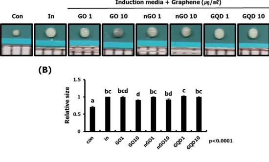
/ ml or less was suitable. Thus, GO, nGO, and GQD at concentrations of 1 and 10 μg / ml were applied to hASCs. As a result, the size of
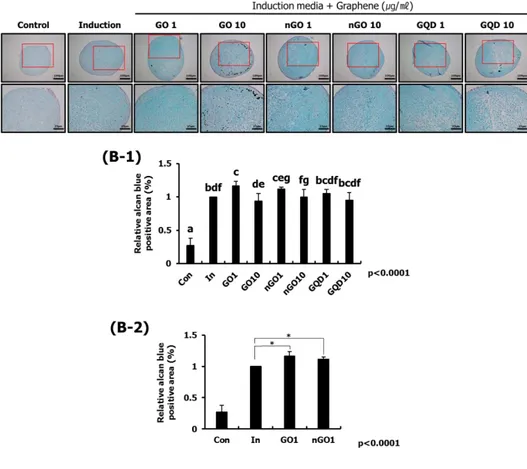
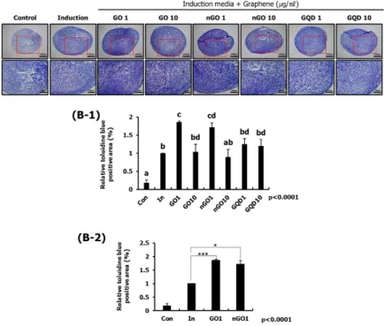
the chondrocyte pellet was not different from the induction group. However, it was confirmed that Glycosaminoglycans (GAGs) were significantly increased in the 1 μg / ml group of GO and 1 μg / ml of
nGO group compared with the induction group through alcian blue staining and toluidine blue staining. These results suggest that GO and nGO, the oxidized graphene, support Chondrogenic differentiation
vi of hASCs.
The main purpose of this study was to determine the effect of GBMs on chondrogenic differentiation of hASCs. Particularly, it was confirmed that the effect of chondrogenic differentiation is different according to the particle size and degree of oxidation of GBMs. These studies will help to understand GBMs, which are bio-new materials, and will greatly contribute to the development of bio-new cartilage therapies using these materials.
Keywords: Cartilage, Chondrogenic differentiation, Human adipose derived stromal cells, Graphene, Graphene oxide, nano-Graphene oxide, Graphene Quantum Dot, Graphene derivatives, Oxidation, Tissue engineering, Cartilage regeneration
vii
CONTENTS
ABSTRACT ... i CONTENTS ... vii LIST OF FIGURES ... ix LIST OF ABBREVIATIONS ... x PART I : GENERAL INTRODUCTION & LITERATURE REVIEW 1 CHAPTER 1 : General Introduction ... 2 CHAPTER 2 : Literature Review ... 9 1. Cartilage ... 10 1.1. Definition ... 10 1.2. Composition ... 10 1.3. Cartilage-related diseases ... 15 2. Tissue engineering ... 22 2.1. Definition ... 22 2.2. Materials ... . 22 2.2.1 Cells ... . 23 2.2.2 Biomaterials ... . 26viii
3. Cartilage regeneration ... 29
PART II : EFFECT OF GRAPHENE-BASED MATERIALS ON CHONDROGENIC DIFFERENTIATION OF HUMAN ADIPOSE-DERIVED STROMAL CELLS ... 31
CHAPTER 1 : Effect of graphene-based materials on chondrogenic differentiation of human adipose-derived stromal cells ... 32
1. Introduction ... 33
2. Materials and Methods ... 39
3. Results ... 44
4. Discussion ... 58
PART III : GENERAL DISCUSSION & CONCLUSION ... 61
1. General discussion and conclusion ... 62
REFERENCES ... 66
ix
LIST OF FIGURES
Figure 1. FT-IR spectra ... 45
Figure 2. XPS spectra ... 46
Figure 3. Hydrodynamic radius ... 47
Figure 4. Zeta potential ... 48
Figure 5. Characterization of hASCs ... 50
Figure 6. Optimization of concentration of GO effective in chondrogenic differentiation of hASCs ... 52
Figure 7. Observation of chondrogenic differentiation of hASCs by pellet size ... 54
Figure 8. Observation of chondrogenic differentiation of hASCs by alcian blue staining ... 55
Figure 9. Observation of chondrogenic differentiation of hASCs by toluidine blue staining... 57
x
LIST OF ABBREVIATIONS
GBMs : Graphene-based materials TE : Tissue engineering G : Graphene GO : Graphene oxidenGO : nano-Graphene oxide
GQD : Graphene Quantum Dot
FT-IR : Fourier Transform Infra-Red
XPS : X-ray Photoelectron Spectroscopy
ELS : Electrophoretic Light Scattering
hASCs : human adipose-derived stromal cells
ECM : Extracellular matrix
GAGs : Glycosaminoglycans
1
PART I
GENERAL INTRODUCTION
&
2
CHAPTER 1
Cartilage consists of chondrocytes that are specially differentiated cells distributed between cartilage matrix. The cartilage matrix, called extracellular matrix (ECM), consists of collagen, glycosaminoglycan, proteoglycan, and water (Sophia Fox, Bedi et al. 2009). And the only cell type that exists within the cartilage is the chondrocyte, which is located in the cartilage lacuna, an empty space in the cartilage. Chondrocyte synthesizes and secretes ECM so that cartilage tissue can perform normal function, and it plays a role to make and maintain cartilage (Phull, Eo et al. 2016). The main function of cartilage is to allow the joints to move smoothly with little friction, with abundant elasticity and resistance to pressure, and with buffers absorbing a given energy (Bhosale and Richardson 2008). These functions continue to weaken whenever the cartilage is impacted. During the lifetime, the size of the cartilage decreases markedly by more than half of its initial size (Hudelmaier, Glaser et al. 2001, Wu, Cai et al. 2013, Richardson, Kalamegam et al. 2016). Furthermore, when cartilage degenerates or begins to suffer trauma, the recovery process is very slow or nonexistent. Since the gel-like matrix does not have nerves, lymphatics and blood vessels, cartilage has little self-healing ability (Kuettner 1992). In addition, since cartilage tissue has a deficiency of undifferentiated cells and a very low cell density, cartilage self-renewal is almost impossible by reducing the possibility of chondrocytes contributing to self-renewal (Toh, Liu et al. 2005, Fisher, Tessaro et al. 2017). Cartilage is easily damaged by trauma or inflammation compared to other tissues, and
when it grows older it begins to regress due to aging. Typical cartilage-related diseases include degenerative arthritis and rheumatoid arthritis (Musumeci, Castrogiovanni et al. 2014, Pap and Korb-Pap 2015). Others include anotia, microtia, bronchial stenosis, and tumors. Most of these diseases are difficult to cure because the exact cause and treatment of the disease is not yet known (Lichtenstein and Hall 1952, Berdon, Baker et al. 1984, Ciorba and Martini 2006, Baluch, Nagata et al. 2014, Cugno and Bulstrode 2019). In general, the treatment for patients usually involves obtaining cartilage from other parts of the patient, but there is a limit because the amount of cartilage that can be obtained from our body is limited for autotransplantation (Temenoff and Mikos 2000). The method of transplanting cells is also insufficient because of the low survival rate of transplanted cells (Deng, Zhu et al. 2018). Therefore, tissue engineering regeneration studies related to cartilage are very important compared to other tissues.
Tissue engineering (TE) is a new field created by combining basic concepts and technologies in life science, medicine and engineering. It is the study of artificially regenerating damaged tissues. To regenerate tissues with the desired function, we need the cells that are the basis for tissue reconstruction, the biomaterials for cell proliferation, and the microenvironment in which the cells can grow, and they need to be properly controlled (Dhandayuthapani, Yoshida et al. 2011). The support used in TE is called biomaterials.
Biomaterials are inert materials that have no viability to replace or regenerate damaged tissue (Jaganathan, Supriyanto et al. 2014). The tissues and organs of the human body are very difficult to make artificially because various kinds of cells are highly organized (Song and Ott 2011, Vrana, Lavalle et al. 2013, Wang, Ao et al. 2016). However, techniques that can repair or replace damaged tissue using TE have already been applied in many medical fields (Atala 2004, Hendow, Guhmann et al. 2016).
Human adipose-derived stromal cells (hASCs) are widely used in TE and regenerative medicine (Jung, Kleineidam et al. 2015, Tabatabaei Qomi and Sheykhhasan 2017). The cells do not raise ethical issues in their extraction and use, and they have the great advantage that they can be easily obtained from any tissue of the body (Brett, Tevlin et al. 2017). In addition, it is suitable as a material for TE because it does not require much time for cell proliferation and can be repeatedly harvested (Verseijden, Jahr et al. 2009, Baer and Geiger 2012, Kleineidam, Sielker et al. 2018). Finally, since it has the ability to differentiate into adipocytes, osteocytes and chondrocytes, it can be usefully applied as a material for TE (Wang, Lai et al. 2014, Brett, Tevlin et al. 2017).
Graphene (G) is a carbon-based flat monolayer, in which the carbon is arranged like a honeycomb hexagonal net (Guazzo, Gardin et al. 2018). G has the most diverse characteristics among existing
materials and is an excellent semi-metallic nanomaterial. The thickness is as thin as about 0.2 nm, and thus the transparency is high, the mechanical strength is strong, and the stretch ability is also good. In addition, it has high thermal conductivity, unique optical properties, extreme chemical stability, and wide surface area (Lee, Kim et al. 2013, Shin, Li et al. 2016, Madni, Noreen et al. 2018). So it has been studied a lot recently as a biomaterial in TE (Ku, Lee et al. 2013, Lee, Kim et al. 2013). However, since graphene has hydrophobic, it is difficult to apply it to the field of TE (Feng and Liu 2011, Banerjee 2018). Thus, a number of graphene derivatives were developed to apply graphene more widely in the field of TE (Kumar and Chatterjee 2016, Romero, Soto et al. 2017).
Graphene oxide (GO) is an important graphene derivative that is the most widely applied (Nurunnabi, Parvez et al. 2015, Li, Liu et al. 2017). GO is a highly oxidized form of graphene obtained by treating graphite with many powerful oxidizing agents. Compared to G, oxygen is present on the GO surface, which also improves hydrophilic property and dispersion in solutions (Bianco, Cheng et al. 2013, Li, Zeng et al. 2014, Priyadarsini, Mohanty et al. 2018, Dideikin and Vul 2019). GO affects both cell proliferation and differentiation and improves the properties of the scaffold. Also, the use of an appropriate concentration of GO that is not too high is useful as biomaterials for TE (Nishida, Miyaji et al. 2014). Because of these advantages, GO is being studied and applied in various fields of TE
(Lee, Lim et al. 2011, Chen, Pang et al. 2012, Kim, Choi et al. 2013, La, Park et al. 2013, Nishida, Miyaji et al. 2014, Yoon, Bhang et al. 2014, Shadjou and Hasanzadeh 2016).
nano-graphene oxide(nGO) is a nano-sized GO that is made using ultrasonication (Goncalves, Vila et al. 2013). nGO has become smaller size than GO, so more research can be attempted in various biological fields (Sun, Liu et al. 2008, Dong, Zhao et al. 2010, Goncalves, Vila et al. 2013, Yang, Gong et al. 2013, Kalluru, Vankayala et al. 2016, Yang, Feng et al. 2016). Particularly, as the particle size became smaller, the solubility and biocompatibility were improved (Shi, Chen et al. 2014).
Graphene quantum dot (GQD) is small particles of 10 nm or less made from strong acid treatment and high heat treatment on carbon fiber (Peng, Gao et al. 2012, Bacon, Bradley et al. 2014). GQD is emerging as bio-new materials with excellent fluorescence, very small particle size, biocompatibility and effective drug delivery capabilities (Shen, Zhang et al. 2012, Zhang, Bai et al. 2012, Shang, Zhang et al. 2014, Su, Shen et al. 2015, Tan, Li et al. 2015, Wang, Zeng et al. 2015, Zheng, Ananthanarayanan et al. 2015). In addition, there are reports that GQD has a positive effect on the self‐renewal and differentiation of mesenchymal stem cells (Qiu, Li et al. 2016).
As such, G and GBMs have potential for biomaterials in the regenerative medicine and tissue engineering fields and are closely related to tissue regeneration. However, since the biomaterials called G were discovered in 2004, there is a lack of previous studies to be considered. In particular, studies related to cartilage are scarce. Therefore, it is essential to study cartilage repair and regeneration using hASCs and GBMs. So, in Chapter 3, I have analyzed the physical and chemical properties of GBMs and studied how they affect chondrogenic differentiation of hASCs using GBMs.
CHAPTER 2
1. Cartilage
1.1. Definition
Cartilage is a highly specialized connective tissue that maintains a remarkable strength even with a very thin thickness (Randolph, Anseth et al. 2003). This tissue is the second most rigid tissue in the human body and has a unique internal structure. Cartilage can be defined as an internal cell supporting tissue, which
typically consists of high content fibrous protein and
mucopolysaccharide (Cole 2011). It is a viscoelastic material that helps the body move flexibly and helps to buffer a given force (Farokhi, Jonidi Shariatzadeh et al. 2019). The ultrastructure composition and complex tissue of cartilage can play an important role in minimizing friction in the joint surface (Wirth and Rudert 1996). Therefore, the cartilage has abundant elasticity, resistance to pressure, and the ability to absorb a given energy, allowing joints to move smoothly with little friction (Bhosale and Richardson 2008, Veronesi, Maglio et al. 2014).
1.2. Composition
Cartilage consists of extracellular matrix (ECM) that make up most of the cartilage and highly specialized cells called
chondrocyte, which distributes at a very low density (Cohen, Foster et al. 1998, Vinatier, Mrugala et al. 2009).
ECM is mainly composed of water, collagen and proteoglycans, and there are also small amounts of non-collagenous proteins and glycoproteins (Woo and Buckwalter 1988, Buckwalter and Mankin 1997). ECM of mature cartilage consists mostly of type II collagen, along with proteoglycans and hyaluronic acids and small amounts of types V, VI, IX, X, XI and XII collagen. Most components of cartilage contribute to the mechanical properties of the cartilage, since they are highly viscous. In particular, collagen-type II fibers account for about 50 percent or more of all the proteins that make up the cartilage, and greatly affect the tensile strength and stiffness of the cartilage (Sophia Fox, Bedi et al. 2009). Hyaluronic acid provides a temporary moisturizing environment to facilitate cell migration and thus contribute significantly to cell separation (Zhu, Wang et al. 2017). Proteoglycan, combined with collagen, regulates the polymerization degree of collagen, contributes to the compressive load resistance of cartilage and the firmness of tissue (James and Uhl 2001). In addition, water, which accounts for up to 80% of the total weight of cartilage, combines with proteoglycan to engage in the transport of metabolites of chondrocytes, and actively contributes to the lubrication and elasticity of joints (Dijkgraaf, de Bont et al. 1995).
And only cell type present in cartilage is chondrocyte (Kuettner 1992). Chondrocytes are located in the lacunae, an empty space in the ECM, and play a role in creating and maintaining cartilage (Musumeci, Loreto et al. 2013). Chondrocytes account for only 1% to 10% of cartilage.Due to the low density of chondrocytes, cartilage has low overall metabolic activity. Their mission is to synthesize and degrade ECMs such as collagen and proteoglycans (Wirth and Rudert 1996, Alford and Cole 2005). First, cell division occurs in chondroblasts, but it no longer divides when cartilage growth stops. Chondroblasts form ECM, which gradually differentiate into chondrocyte as they form more ECM (Poole 1997). Chondrocytes establish their own specialized microenvironment, and perform metabolic activities in it (van der Kraan, Buma et al. 2002, Gao, Liu et al. 2014). And this microenvironment prevents the transfer of chondrocytes to adjacent areas and trap cells (Woo and Buckwalter 1988). Therefore, chondrocytes rarely make cell-to-cell contact for direct signal transmission between cells. Also, since the potential for replication is limited, the survival of chondrocytes depends on the optimal chemical and mechanical environment. Chondrocytes receive limited nutrient supply from the synovial fluid through diffusion and depend on anaerobic metabolism. The size of chondrocytes is getting bigger from the epidermis layer to the deep layer (Temenoff and Mikos 2000). Chondrocytes function to synthesize and secrete ECM so that cartilage tissue can perform normal function. And metabolic activity of chondrocytes is closely related to age. During the growth
period, there is active metabolism and cell proliferation. However, after the growth period, proliferation of chondrocytes is stopped and metabolism activity is decreased, but it continues. Cartilage regeneration and remodeling rely on chondrocytes and their metabolism, including the synthesis of ECM molecules such as collagen, proteoglycan, and degradative enzymes (Mollano, Martin et al. 2002, Smith, Carter et al. 2004, Loeser 2009).
Cartilage consists mainly of four zones of superficial zone, middle zone, deep zone and calcified zone depending on depth. Each zone has different main components and functions and also different density and shape of the chondrocytes that are distributed (Fetter, Leddy et al. 2006, Antons, Marascio et al. 2018). The thin superficial zone under the perichondrium accounts for about 10% to 20% of the thickness of the cartilage. This zone is mainly filled with collagen fiber of type II and IX, arranged parallel to the articular surface (Muir, Bullough et al. 1970, Venn 1978). And it has a relatively high water content and high cartilage cell density compared to other zones. A large number of chondrocytes are located parallel to the surface, and the shape of the cells is flat (Hunziker, Quinn et al. 2002). The main function of this zone is to protect and maintain deeper zones. It is also responsible for most tensile properties of the cartilage because it is the area in contact with synovial fluid, and can resist the compression forces imposed on the cartilage (Wu, Kirk et al. 2008, Mansfield, Bell et al. 2015). Below the superficial zone is the middle zone. The
middle zone is located between the superficial zone and the deep zone, and serves as an anatomical and functional bridge. This zone accounts for about 40-60% of the total cartilage volume and is composed mainly of proteoglycans and thicker collagen fibers (Grogan, Duffy et al. 2013). The deep zone accounts for about 30% of the total cartilage volume. It consists of the thickest collagen fiber, the highest proteoglycan content, and the lowest moisture content (Lorenzo, Bayliss et al. 1998). Collagen fibers in this zone are arranged perpendicular to the articular surface. Therefore, it plays the role of providing the maximum resistance against the compression force (Kuiper and Sharma 2015). The chondrocytes are parallel to the collagen fibers and are arranged perpendicular to the articular surface and have a columnar shape (Dowthwaite, Bishop et al. 2004). Finally, the calcified zone is the deepest zone where the cartilage matrix is calcified. This zone is highly mineralized and connected to the subchondral bone (Koszyca, Fazzalari et al.1996, Wang, Ying et al. 2009, Zhang, Wang et al. 2012). And it is separated from other zones by a wavelike tidemark. The tidemark separates the calcified zone from the noncalcified zone because it exists between the deep zone and the calcified zone (Havelka, Horn et al. 1984, Oegema, Carpenter et al. 1997). The main function of this zone is to deliver the mechanical stress and biological stimuli received by cartilage to the bone (Zhang, Wang et al. 2012). The main constituent of this zone is hydroxyapatiete, which accounts for about 70% (Zizak, Roschger et al. 2003). In this zone, the density of the cells is low, chondrocytes
are hypertrophic (Sophia Fox, Bedi et al. 2009). Cells are found in hydroxyapatite-deposited cartilage matrix.
Cartilage does not have nerves, blood vessels, or lymphatic vessels, unlike most tissues (Poole, Kojima et al. 2001). Therefore, there is no stimulation from the nerve, and monocytes or immunoglobulin approach is difficult. The possibility of self-renewal of cartilage is rare because it has limitations on general immune response (Buckwalter and Mankin 1998, Shah, Shah et al. 2010, Huey, Hu et al. 2012). Lack of blood supply limits not only the cells available for tissue regeneration, but also the nutrients and factors (Darling and Athanasiou 2003).
1.3. Cartilage-related diseases
With the unique biological and mechanical properties of cartilage, repair or regeneration of damage is very limited (Buckwalter and Mankin 1998). The recovery process of cartilage damaged by trauma, inflammation is either very slow or almost nonexistent (Hunter 1995, Wu, Cai et al. 2013). In addition, the natural regressive process of aging cannot be stopped. Therefore, cartilage-related diseases are inevitable senile diseases for anyone (Bachmann, Basad et al. 1999, Han, Grodzinsky et al. 2011, Li, Wei et al. 2013, Brittberg, Gomoll et al. 2016, Richardson, Kalamegam et al. 2016). Some of the major diseases include degenerative arthritis,
rheumatoid arthritis, and other diseases include achondroplasia, pyogenic arthritis, andchondrosarcoma.
Degenerative arthritis refers to a disease in which cartilage is worn out and bones that exist under the cartilage are exposed, which can harden and cause deformation of the joint (McIlwraith and Vachon 1988, Felson 2010). Gradual damage or degenerative changes in the cartilage can lead to inflammation and pain in the joints (Barrett 2014, Joshi, Jain et al. 2014). In addition, new bones are formed abnormally at the site where cartilage is broken down (Li, Yin et al. 2013). The sequence and correlation of these cartilage and bone changes are currently unknown. Degenerative arthritis is the most common inflammatory disease of the joint (Jonsson, Olafsdottir et al. 2016, Robinson, Lepus et al. 2016). Common symptoms are local pain in the joint area where arthritis occurs, usually without systemic symptoms. This characteristic differs greatly from rheumatoid arthritis. And degenerative arthritis can be classified into two types depending on the cause. Primary or idiopathic arthritis is generally known to be affected by age, gender, genetic factors, obesity, certain joints, etc., although the exact cause is not known (Chodosh, Morton et al. 2005, Blagojevic, Jinks et al. 2010, Chen, Shen et al. 2017). Secondary or rapid arthritis is arthritis caused by trauma, illness, and deformities that can damage cartilage (Kettelkamp, Hillberry et al. 1988, Volpin, Dowd et al. 1990, Felson, Niu et al. 2013, Jonsson, Olafsdottir et al. 2016). Typically, it is
damaged by bacterial arthritis or tuberculous arthritis, or occurs after severe shock or trauma (Shirtliff and Mader 2002, Mathews, Weston et al. 2010, Al-Sayyad and Abumunaser 2011). However, the distinction between primary and secondary is not clear, since there may be cases where the cause cannot be determined even if diagnosed as secondary. And depending on the body part, the cause is slightly different. In the case of the spine, it can be caused by occupationally repeated work or lifestyle, and hip joints are the cause of avascular necrosis and dysplasia (Milgram 1983). In the case of ankle or wrist, fracture or damage to surrounding ligaments is the main cause (Harrington 1979, Watson and Ryu 1986). Most are diseases that occur a lot in old age (Wood, Brock et al. 2013), but aging itself is not the cause. In recent years, the number of patients suffering from arthritis is increasing, and the number of young patients is increasing (Vavken and Samartzis 2010). As the disease progresses, other tissues and related musculoskeletal conditions are also affected, so prevention and treatment of this disease is important(Dekker, Boot et al. 1992). However, there is no definitive treatment that can completely stop degenerative arthritis. To prevent this disease, there is a way to prevent damage from progressing quickly through rehabilitation, and to treat it with painkillers or anti-inflammatory drugs (Yu and Hunter 2015, Yusuf 2016).Despite this non-surgical treatment, if the symptoms do not improve and the joints continue to change, surgical treatment will be performed. In general, arthroscopic removal of the vitreous in the joint,
synovectomy (Ogilvie-Harris and Weisleder 1995, Blahut 2003), osteotomy (Coventry 1965), arthroplasty (Morrey 1992), and arthrodesis(Lauge-Pedersen 2003) are some of them (Burks 1990, Aichroth, Patel et al. 1991). However, it is known that any joint that has already undergone degenerative changes of cartilage by any treatment method cannot be restored to normal functioning joints.
Rheumatoid arthritis is a chronic inflammatory disease that exact cause has yet to be identified. So far, it has been hypothesized that people with genetic predisposition may be immune responses that are exposed to infectious agents such as smoking or periodontitis (Carmona, Cross et al. 2010). Rheumatoid arthritis is the second most common disease after osteoarthritis in chronic arthritis, the most common of inflammatory arthritis (Chou and Chu 2018). It is generally known that inflammation of the synovial membrane surrounding the joints caused by autoimmune abnormalities leads to disability. Synovial membrane is a thin membrane that is located in joints and plays a role in helping joints move smoothly by generating synovial fluid (Otero and Goldring 2007). Immunity is a function that protects our body from bacteria that invade from the outside (Warrington, Watson et al. 2011), and lymphocytes play an important role in immunity (Balakrishnan and Adams 1995). However, when lymphocytes erroneously recognize a part of our body as a bacterium that invades from the outside, various diseases arise. Such diseases are called autoimmune diseases (Warrington, Watson et al. 2011).
Rheumatoid arthritis is also an autoimmune disease in which lymphocytes attack the synovial membrane and damage the surrounding joints and bones (Hitchon and El-Gabalawy 2011). If this damage continues, inflammatory synovial membrane tissue grows, causing disruption of the shape and function of bone and cartilage (Aletaha and Smolen 2018, Calabresi, Petrelli et al. 2018). Furthermore, the disease can severely affect major organs such as the lungs, heart, and blood vessels, and in severe cases it can shorten the patient's life span (Khurana and Berney 2005, Beirith, Ikino et al. 2013, Shaw, Collins et al. 2015). A typical symptom of the disease is the symmetrical development of arthritis in small joints of the hands and feet. Also, the disease occurs over a period of days to months and can occur at any age, but most commonly occurs between the ages of 35 and 50 years. This disease is more common in women than in men (Donahue, Gartlehner et al. 2008). The earlier treatment starts, the better the treatment results, but the greater the difficulty in identifying early diseases (Boutry, Morel et al. 2007, Ma and Xu 2013, Rein and Mueller 2017). A typical early symptoms is that the joint becomes stiff when it wakes up in the morning, and the symptom that the joint does not stretch properly lasts for more than one hour. Also, joints of hands and feet are swollen and sore (Iqbal and Rattu 2019).Treatment for this disease includes non-drug treatment, such as proper exercise and balance of rest and nutrition (Forestier, Andre-Vert et al. 2009), drug treatment (Wilsdon and Hill 2017), and surgical treatment (Straub and Ranawat 1969). The main
treatment among these is drug treatment using anti-inflammatory drugs (Van Vollenhoven, Fleischmann et al. 2012). If the disruption of the joint is severe even after drug treatment, artificial arthroplasty or arthrodesis is performed (Lee and Choi 2012, Trieb 2014). Most rheumatoid arthritis progresses with repeated improvement and deterioration, and there is still no way to prevent or cure it.
In addition, there are achondroplasia (Wright and Irving 2012), pyogenic arthritis (Nolla, Lora-Tamayo et al. 2015), and chondrosarcoma (Bloch, Jian et al. 2009). Achondroplasia is a disorder that has a disability in the ability of cartilage to form bones. In the process of cartilage being replaced with bone, the cartilage becomes impaired and bone growth is not achieved properly (Ornitz and Legeai-Mallet 2017). In most cases, the cause of the disease occurs randomly, without any special reason, without family history. And although it is inherited, about 90% of patients are caused by genetic mutations of unknown origin. The main symptom is characterized by a megalencephaly, short arms and legs, abnormal finger shape, spinal abnormality, and a small height (Pauli 2019). The disease is accompanied by physical pain from complications, but does not impair or detract from mental abilities. Typical therapies include growth hormone therapy and the surgical treatment of the Ilizarov technique (Horton, Hall et al. 2007). But since both have side effects, they are not the perfect remedy.
Pyogenic arthritis is an inflammation of the joints caused by bacterial invasion into the pus, with more than 70% of patients occurring during the first 1-5 months of life (Schaad, McCracken et al. 1980). Hematogenous infection is the most common and the most common causative organism is Staphylococcus aureus. Typical symptoms are not injured, but the joints are sore or hot, and joint edema or redness may be observed. Damage to the joint is a damage to the growth plate, which poses a risk of leaving a fatal disorder, requiring emergency treatment. Therefore, adults are dangerous, but especially dangerous to children. Early treatment is important because inflammation can affect joints and surrounding bone, leading to bone loss, growth failure, and bone deformation. Treatment method is to remove the pus and dead tissue inside the joint by performing arthroscopy immediately after diagnosis (Dubost, Soubrier et al. 2000). Immediately after surgery, intravenous antibiotics should be administered (Meier, Wirth et al. 2017). It is important to observe for a certain period of time whether the inflammation level remains normal after stopping antibiotics.
Chondrosarcoma is a malignant tumor of the cartilage. It usually develops as a cause of primary, in which case it occurs in normal bone without any preceding lesions (Limaiem and Sticco 2019). A typical cartilage breed grows relatively slowly, and spreads slowly (Gelderblom, Hogendoorn et al. 2008). The cause of the chondrosarcoma is not known precisely, so there is no
well-established preventive measure. Treatment options include surgery, anti-cancer drug treatment and radiation therapy.
2. Tissue engineering
2.1. Definition
Tissue engineering (TE) was defined in the early 1990s as "an interdisciplinary field which applies the principles of engineering and life sciences toward the development of biological substitutes that restore, maintain, or improve tissue function" (Langer and Vacanti 1993). TE is the study of transplanting tissue cultured in vitro so that it can perform normal functions, and is not yet fully explored (Griffith and Naughton 2002). And this study is a combination of basic concepts and techniques of life sciences, medicine, and engineering. By applying three different sciences, the replacement of damaged tissues is transplanted to maintain, enhance, and restore biological functions (Shafiee and Atala 2017). And TE can overcome the disadvantages of organ transplantation: the lack of donors and the need for immunosuppressive therapies (Caddeo, Boffito et al. 2017). The materials needed for TE are three things: cells, biomaterials, and an environment suitable for cells (Nainar, Vicki et al. 2014, Park, Shin et al. 2018).
2.2.1, Cells
To create an engineered tissue in vitro, cells must be used to populate matrices and create a matrix that is similar to the native tissue (Barthes, Ozcelik et al. 2014). Usually, primary cells from patients were used for successful transplantation (Koh and Atala 2004). However, this method has limitations because it requires secondary surgery for cell collection and the collected cells may be in a diseased state. Therefore, TE studies using mesenchymal stromal cells derived from various tissues and embryonic stem cells are increasing (Metallo, Azarin et al. 2008, Spencer, Gimble et al. 2011).
Human embryonic stem cells (hESCs) are derived from the inner cell mass of the blastocyst-stage embryos (Stoltz, de Isla et al. 2015). These cells can proliferate while maintaining their undifferentiated state for a long time during the culturing process and have the ability to differentiate into various somatic cell types (Sykova and Forostyak 2013). Thus, these cells have been expected to provide selective cell types as infinite materials for future cell therapy (Thomson, Itskovitz-Eldor et al. 1998). For about 20 years after the first appearance of hESCs, stem cell biology has made considerable progress. However, the controversy over the use of hESCs in regenerative medicine or TE studies has continued to
increase over time (Sherley 2004). Because the present technology cannot properly control the differentiation of stem cells and solve ethical problems (King and Perrin 2014). There are many reports of hyperproliferation and formation of teratomas after transplantation of these cells (Seminatore, Polentes et al. 2010). Therefore, there are many problems to be solved in TE research using hESCs.
Human mesenchymal stromal cells (hMSCs) are used to overcome the limitations of clinical studies on hESCs. Mesenchymal 'stromal' cells are more suitable than mesenchymal 'stem' cells because of their limited self-renewal capacity compared to pluripotent embryonic stem cells (Yin, Zhu et al. 2019). hMSCs and hMSC-derived products are capable of numerous clinical applications with regenerative potential and potent trophic properties, and their potential as a future therapeutic tool has been extensively evaluated (Marion and Mao 2006, Davies, Walker et al. 2017). hMSCs are usually extracted from placental tissue, umbilical cord, Wharton's jelly, dental tissues, skin, thymus, spleen, trabecular bone, peripheral blood, adipose tissue, endometrium, amniotic fluid and bone marrow (Tuan, Boland et al. 2003). The most commonly used cells are bone marrow-derived stromal cells and adipose-derived stromal cells (Potier, Noailly et al. 2010, Tabatabaei Qomi and Sheykhhasan 2017).
Human bone marrow is a complex tissue containing a
hematopoietic stromal cells and mesenchymal stromal cells (Kawai, Tsujigiwa et al. 2018). In particular, human bone marrow-derived mesenchymal stromal cells (hBMSCs) have been extensively studied as a cell source for regenerative medicine application (Petersen, Bowen et al. 1999, Okamoto, Yajima et al. 2002, Aghi and Chiocca 2005, Hu, Yang et al. 2008). It was first discovered in the late 1960s that bone marrow contains cells with proliferation capacity and the ability to differentiate into bone or cartilage (Friedenstein, Chailakhjan et al. 1970). Since then, many studies have shown that these cells can also be differentiated into other tissues (Pittenger, Mackay et al. 1999, Egusa, Schweizer et al. 2005). In addition, since these cells have high proliferate rates on in vitro, numerous cells can be easily obtained and the risks of rejection and pathogen transmission are low even when these cells are transplanted into a patient. And numerous clinical results demonstrate that hBMSCs reach senescence without forming teratomas (Stenderup, Justesen et al. 2003). Therefore, hBMSCs with multipotential capability are a promising source of application for various cell therapies (Krebsbach, Kuznetsov et al. 1999, Hu, Xu et al. 2011, Sassoli, Zecchi-Orlandini et al. 2012, Kasashima, Yashiro et al. 2015, Polymeri, Giannobile et al. 2016).
Human adipose-derived stromal cells (hASCs) can also improve the treatment of a wide range of diseases (Badimon, Onate et al. 2015, Sridhar, Gottipamula et al. 2018). hASCs, along with
hBMSCs, are good cell materials for TE, but have many advantages over hBMSCs (Tabatabaei Qomi and Sheykhhasan 2017). Compared to hBMSCs, hASCs can be isolated in an easier way, thus reducing the risk of sampling.It is usually easily obtained from lipo-aspirated adipose tissues, and more stromal cells can be obtained compared to bone marrow tissue (Sheykhhasan, Qomi et al. 2015). In addition, hASCs are suitable for repair and regeneration of various tissues due to their high proliferation capacity and ability to differentiate into various cells (Fathi and Farahzadi 2016). Soft tissue of the human body consists mainly of adipose tissue, so it is also suitable for the treatment of large soft tissue defects (Hong, Peptan et al. 2006). The biggest advantage is that you can get as many abundant amounts as you need and maintain cell characteristics at any age (Tabatabaei Qomi and Sheykhhasan 2017). It also has immunosuppressive properties that allow transplantation regardless of the match of human leukocyte antigens between the host and the donor (Mundra, Gerling et al. 2013). Therefore, hASCs are being applied to various fields of TE and regenerative medicine. (Seo, Suh et al. 2005, Amos, Kapur et al. 2010, Scuderi, Ceccarelli et al. 2013, Zielins, Luan et al. 2015).
2.2.2, Biomaterials
Suitable biomaterials are needed for successful tissue regeneration (Perez, Won et al. 2013). Biomaterials are widely used
to improve the quality of life of patients by replacing and restoring the function of traumatic or degenerated tissues or organs (Tathe, Ghodke et al. 2010, Patel and Gohil 2012). Biomaterials can be divided into naturally derived biomaterials and artificially synthesized biomaterials. It is important to properly use the two materials because they have different advantages and disadvantages.
The most representative of naturally derived biomaterials is collagen. Collagen is a protein that plays an important role in providing strength and structural stability to tissues including skin, blood vessels, tendons, cartilage, and bones (Cen, Liu et al. 2008). It is the most abundant protein in the body. Collagen has excellent mechanical strength, cell binding capacity, stable biocompatibility and biodegradability (Nimni and Harkness 1988). In particular, naturally derived biomaterials such as collagen can enhance cell adhesion and migration from the surrounding environment, induce extracellular matrix formation, and facilitate tissue repair, thereby favoring transplantation at the site of the defect (Hortensius and Harley 2016). Thus, since the 1970s, many researchers have started using collagen as a material in regenerative medicine research in various connective tissues (Song, Yeon Kim et al. 2006). At the same time, it became easier to obtain medical collagen, improved techniques for processing collagen, and new collagen products were successfully produced (Pachence 1987). Collagen, which is suitable for biomaterials, has rapidly increased in biochemical applications and has expanded into
many biotechnology fields including polymer scaffolds (Pachence 1996, Van Wachem, Van Luyn et al. 1996, Friess 1998). However, since collagen is a protein, it is still difficult to sterilize without changing its structure (Parenteau-Bareil, Gauvin et al. 2010).
The most representative of artificially synthesized biomaterials is based biomaterials. In particular, carbon-based nanomaterials have been actively studied in various fields of biomedical engineering due to their unique chemical and physical properties (Samavedi, Poindexter et al. 2014). Research on medical carbon and carbon based materials has been actively conducted in Europe since the mid-1970s. These studies have succeeded in introducing carbon into a variety of medical fields. The advantages of carbon such as biocompatibility, sterilization ability, chemical inactivity, sufficient mechanical properties, and excellent tribological properties were used to solve the problems of the existing metal implants (Huettner and Claes 1990). However, artificially synthesized biomaterials have relatively low biocompatibility and tissue remodeling capability compared to naturally derived biomaterials because they are not normally similar to native tissues in the body. Nonetheless, using the properties of carbon can overcome these shortcomings. Carbon-based biomaterials can form a chemically strong shared bond with other carbon-based materials and various elements, resulting in an infinite number of applications, and also the ability to create a variety of carbon-based biomaterials
according to the synthesis method. Efforts to make carbon-based biomaterials with these diverse structures and functions continue (Nasir, Hussein et al. 2018). Particularly, various studies based on graphene and carbon nanotubes are increasing (Polizu, Savadogo et al. 2006, Cha, Shin et al. 2013).
3. Cartilage regeneration
Cartilage has little self-repair ability due to the low replication capacity of chondrocytes and the absence of blood vessels. Therefore, it is especially vulnerable to various diseases (Tuan 2004). In particular, osteoarthritis is a cartilage destructive disease that affects more than 27 million adults in the United States(Neogi 2013). However, the treatment options for cartilage are still limited and the treatment is very difficult. Current cartilage treatment methods are mainly drilling, autologous chondrocyte implantation, and osteochondral autografting (Patil and Tapasvi 2015, Niemeyer, Albrecht et al. 2016, Gao, Goebel et al. 2018). Even with these treatments, damaged cartilage is difficult to regenerate into cartilage with normal structure and function. This is because the cartilage that is regenerated is mainly fibrous cartilage that contains a lot of collagen, which is weak in strength and elasticity (Tuan, Chen et al. 2013). Then, even if treated, the joint cannot move smoothly and can lead to early degeneration (Martin, Patel et al. 2019). For joints with more severe cartilage damage, joint replacement is often performed
using metal implants. While this current treatment reduces pain and increases mobility, there is a growing need for treatment options to restore the original biological properties of cartilage. In the field of cartilage tissue engineering, many studies are being conducted to make biologically compatible synthetic cartilage structures (Hoshi, Fujihara et al. 2013). Successful cartilage tissue engineering is important to use artificial supports with good biocompatibility and suitable cells. Other factors include growth factors, bioactive molecules and biomaterial scaffolds (Wang, Yuan et al. 2017, Deng, Zhu et al. 2018). Appropriate application of these may suggest a new approach to cartilage regeneration research.
PART II
EFFECT OF
GRAPHENE-BASED MATERIALS ON
CHONDROGENIC DIFFERENTIATION OF
HUMAN ADIPOSE-DERIVED STROMAL
CELLS
CHAPTER 1
EFFECT OF
GRAPHENE-BASED MATERIALS ON
CHONDROGENIC DIFFERENTIATION OF
HUMAN ADIPOSE-DERIVED STROMAL
CELLS
1. Introduction
Cartilage is a very important tissue that helps move joints without friction. Since cartilage regeneration ability is very limited, it is vulnerable to aging, damage, and disease. Furthermore, cartilage-related diseases are no longer age-cartilage-related diseases.There are many patients who suffer from diseases such as trauma and arthritis at a young age (Felson, Lawrence et al. 2000, Walter, Ossendorff et al. 2019). Pediatric arthritis, such as juvenile idiopathic arthritis, autoinflammatory disorders, and rheumatoid arthritis, cause irreversible structural damage to the cartilage and bone and cause loss of joint function (Andersson Gare 1999, Gurion, Lehman et al. 2012, Giancane, Consolaro et al. 2016). Although cartilage destruction can be stopped, it can never fully recover (Apparailly 2017). And cartilage-related diseases are limited in treatment because most of them are unknown causes (van Vollenhoven 2009, Fabre and Apparailly 2011, Onishi, Utturkar et al. 2012). That is why research related to cartilage regeneration is very important.
Tissue engineering (TE) for cartilage requires biomaterials to support cells and cells to repair cartilage defects (Vacanti and Langer 1999) . There have been many attempts with various cells and various biomaterials, but they have yet to succeed in restoring
the structure and function of native cartilage (Steadman, Rodkey et al. 2001, Bernhard and Vunjak-Novakovic 2016, Lietman 2016).
Graphene (G) is the basic structure of graphite, a sheet in which carbon atoms are arranged like honeycomb structures(Batzill 2012, Young, Kinloch et al. 2012). G has advantages such as electrical conductivity, mechanical strength, thermal conductivity, chemical stability and optical properties (Rao, Subrahmanyam et al. 2011, Wang and Shi 2015). Also, G is the thinnest substance ever produced, and is the simplest form of carbon. G is gaining huge popularity and attention from the science world because of its physical, mechanical and thermal characteristics. Due to these characteristics, various studies have been reported in combination with the biology (Pumera 2011, Pinto, Goncalves et al. 2013). However, due to the hydrophobicity of G, there are major limitations in industrial applications and biological applications (McCallion, Burthem et al. 2016, Bai 2017). It cannot be mass-produced because it cannot be dispersed in the liquid phase (Dong, Chen et al. 2018), and the manufacturing cost is very high, and it takes a relatively long time and is not efficient(Pu, Wang et al. 2012, Jia, Plentz et al. 2019). Thus, various methods of synthesizing G have been developed in a short period of time (Shams, Zhang et al. 2015, Coroş, Pogăcean et al. 2019, Lee, Hiew et al. 2019). A derivative is a similar compound obtained by chemically changing only a part of a compound (Sturala,
Luxa et al. 2018). Among the most commonly used ones made by oxidized graphene using a strong oxidizer are graphene oxide (GO), and other things include nano-graphene oxide (nGO), graphene quantum dot (GQD). Since oxidized G is hydrophilic, dispersion stability in solution is good. In addition, since it is possible to synthesize in liquid phase, mass production is possible, which is efficient (Sun and Fugetsu 2013). Because of these characteristics, they are being applied in a variety of fields (Skoda, Dudek et al. 2014, Khan, Kausar et al. 2016, Daniyal, Liu et al. 2019). G and various graphene-based materials (GBMs) are being applied in many fields (Aïssa, Memon et al. 2015, Sood, Lund et al. 2015, Zhu, Ji et al. 2017, Nag, Mitra et al. 2018). Especially, there is a lot of research in biotechnology (Hamzah, Selvarajan et al. 2017, Reina, Gonzalez-Dominguez et al. 2017, Suvarnaphaet and Pechprasarn 2017). A number of studies have been reported on G and GBMs applied to bone tissue engineering as biomaterials (Dubey, Bentini et al. 2015, Shadjou and Hasanzadeh 2016, Cheng, Wan et al. 2018, Shadjou, Hasanzadeh et al. 2018) . They can also promote neural tissue regeneration (Akhavan 2016, Gardin, Piattelli et al. 2016, Aydin, Gurcan et al. 2018, Bramini, Alberini et al. 2018, Bei, Yang et al. 2019). In addition,haematopoiesis, myogenesis and adipogenesis can be promoted, enabling potential applications of TE (Chaudhuri, Bhadra et al. 2015, Garcia-Alegria, Iliut et al. 2016, Wu, Ding et al. 2017). While many studies have been carried out that GBMs induce and promote cell differentiation, there is still a lack of regenerative
research on GO, nGO and GQD. Especially, there is a great lack of TE research related to cartilage.
GO is an oxidized form of graphene and have the advantage of being hydrophobic and well distributed in liquids. Therefore, it can be easily dispersed in water or other organic solvents, and the desired properties can be obtained in oxygen functional groups by using various organic chemical reactions (Compton and Nguyen 2010, Dreyer, Park et al. 2010). Having hydrophilicity makes it easier to apply in the biotechnology field because it mixes well with water and increases cell affinity. It can also be easily transformed into a specific use. With these characteristics, it is possible to apply to various fields such as functional composite materials, biomedical and bio-imaging applications, energy conversion & storage system and catalytic reaction (Chen, Feng et al. 2012). And it is also studied a lot in the field of TE (Li, Zeng et al. 2014).
nGO is obtained from the GO by converting micrometric lateral dimension of the sheets to a nanometric size (Liu, Robinson et al. 2008). So nGO is chemically similar to GO and has a different particle size. There are no distinct standard indicators of oxidation graphene GO and nGO, but they can be distinguished by the size of particles. Even if they are chemically similar, the size of the particles
is different, so the fields that can be applied and the extent to which they are affected are also different. For example, GO and nGO have a non-toxic effect and can be widely used in medical research, but it has been reported that the particles in nGO exhibit better antibacteriality compared to GO (Gupta, Rajaura et al. 2015). So it is important to understand that GO and nGO are different materials. Therefore, nGO has the advantages of small size, intrinsic optical properties, large surface area, and low cost. And it is rapidly emerging as a multifunctional biomaterial in cancer therapies because it can serve as a drug delivery and carrier due to useful non-covalent interactions with aromatic drug molecules (Sun, Liu et al. 2008, Tian, Wang et al. 2011, Feng, Yang et al. 2013, Rahmanian, Hamishehkar et al. 2014, Tao, Zhu et al. 2018). In addition, many studies related to biomedical imaging and biomedical sensors have been reported (Kim, Ryoo et al. 2013, Jang, Kang et al. 2018, Behera and De 2019, Gurunathan, Kang et al. 2019). Because of these features, nGO is a new material that has a lot of potential, mainly for biological and medical applications.
GQD is defined as graphene sheets with single, double, and fewer than 10 layers and with lateral dimensions less than 100 nanometers. GQD has many advantages, including very small size, excellent photostability, highly adjustable photoluminescence, biocompatibility, electrochemiluminescence, ease of function and
chemical inactivation (Barreiro, van der Zant et al. 2012, Zhou, Xu et al. 2016, Elvati, Baumeister et al. 2017). Due to these various properties, it is a versatile material for fields such as bio-imaging (Zhu, Zhang et al. 2011, Wu, Tian et al. 2013, Wang, Zeng et al. 2015), nano-medicine(Sun, Liu et al. 2008, Wang, Xia et al. 2013), and bio-sensing (Wang, Zhang et al. 2013).
Therefore, GBMs have unlimited potential as biomaterials to help regenerate damaged tissue. In addition, GBMs affect cell proliferation and differentiation and are recognized as excellent biocompatibility. So, using GBMs, which have great potential for medical applications, I studied how they affect the chondrogenic differentiation of hASCs.
2. Materials and methods
Preparation of GO and nGO Flakes
GO flakes were prepared by Hummer’s method Shortly, 5 g graphite (99.9995 % purification, purchased from Alfa Aesar) was poured into 169 ml sulfuric acid (95%, purchased from Sigma Aldrich), followed by adding NaNO3. Then, the oxidative reagent, KMnO4 was slowly poured into the mixed solution under ice bath to keep the solution temperature below 10 °C. After stirring for 5 days in ambient conditions, 15 ml H2O2 was poured into the mixed solution, and mild stirring was maintained for 1 more day. For purification, distilled water was added into the reaction solution, and centrifuged at 5,000 rpm for 30 min. The supernatant liquid of the GO solution was removed and the rinsing process was repeated more than 15 times to eliminate the residual salts and acids completely. The GO flakes were freeze dried (Bondiro, Ilshinbiobase, Korea) for 3 days and stored in a desiccator. To prevent the precipitation of GO flakes in media, 0.1% GO solution was centrifuged at 500 rpm for 5 min. For this study, large sized GO flakes were removed and the supernatant liquid was used.
nGO flakes were prepared by mechanically breaking of 0.01% GO solution in an ice bath with a prove-type sonicator (Q500, Qsonica,
U.S.) for 12 hours. After that, the GO solution was filtered with 450 nm and 200 nm sized membrane filters in sequence. The final product were freeze dried and stored in desiccator
Preparation of GQD
1.2 g of chopped pitch carbon fiber were added into a mixture of a concentrated sulfuric acid (95% H2SO4, purchased from Sigma Aldrich, 240 ml) and nitric acid (70% HNO3, purchased from Sigma Aldrich, 80ml). The solution were pretreated by sonication and pre-stirring for 1 hour at a room temperature to mix them well. After that, the solution were heated in a silicon oil bath at a temperature of 100 °C for 3 days. The bright brown mixture solution were naturally cooled down, followed by adding of 3 L distilled water. The diluted solution were dialyzed by a dialysis bag with 2000 Da of molecular weight cut-off (MWCO) for 3 days.
FT-IR spectra analysis
After preparing the KBr pallet, and then liquid samples of GO, nGO and GQD were loaded and thermally cured and measured by FT-IR spectrophotometer (Nicolet 6700, Thermo Scientific, U.S.).
XPS spectra analysis
0.1% of GO, nGO, and GQD liquid samples were dropped on a SiO2 substrate, and dried, and then measured by XPS spectrometer (XPS, AXIS-HSi, KRATOS).
Hydrodynamic analysis
At room temperature, 0.1% GO, nGO, and GQD liquid samples were measured by light scattering instrument (Zetasizer Nano Z, Malvern Panalytical).
Zeta potential analysis
0.05% GO, nGO, GQD liquid samples were measured by light scattering instrument (Zetasizer Nano Z, Malvern Panalytical).
Isolation of human adipose-derived stromal cells
Human adipose tissue were obtained with informed consent using a guideline approved by the Institutional Review Boards of Seoul national university. Human adipose tissue was collected after obtaining consent to hASCs separation from five random patients who underwent gynecological surgery for laparoscopic surgery. In the
operating room, adipose tissue in the intestinal reservoir was immediately transferred to the PBS containing the FBS and then hASCs was isolated.
Induction of differentiation
To confirm ability of differentiation of hASCs in vitro, osteogenic, adipogenic and chondrogenic differentiation were induced by standard protocols
Osteogenic differentiation
A Basic culture medium for osteogenic differentiation was low
glucose DMEM supplemented with 100 μM dexamethasone, 10 mM
β-glycerophosphate and 50 μM ascorbiate-2-phosphate. At the end of culture for 2 weeks, the cells were fixed with 4% (v/v) paraformaldehyde and alizarin red s staining was conducted.
Adipogenic differentiation
A Basic culture medium for adipogenic differentiation was F12 glucose DMEM supplemented with 0.5 mM isobutyl-methylxanthine, 200 μM indomethacin, 1 μM dexamethasone and 10 ㎍/㎖ human insulin. At the end of culture for 2 weeks, the cells were fixed with 4% (v/v) paraformaldehyde and oil red o staining was conducted.
Chondrogenic differentiation
A Basic culture medium for chondrogenic differentiation was high
glucose DMEM supplemented with 10 ng/㎖ TGF-β 1, 1x
Insulin-Transferrin-Selenium, 10-7 M dexamethasone, 1.25 mg/㎖ BSA, 50 ㎍/㎖ ascorbic acid, 5.33 ㎍/㎖ linoleic acid and 40 ㎍/㎖ L-proline. At the end of culture for 2 weeks, the pellets were embedded in paraffin block and alcian blue and nuclear fast red staining was conducted.
3. Results
Characterization of graphene-based materials
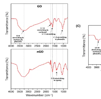
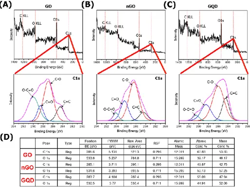
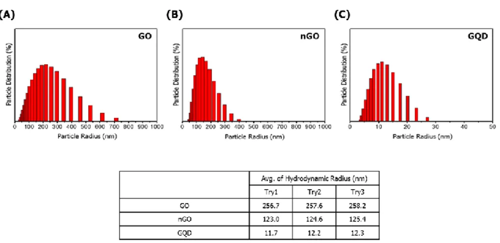
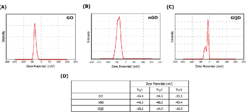
The physical and chemical properties of GBMs were analyzed. As shown in Figure 1, functional groups of GBMs were measured by FT-IR spectrophotometer. GO, nGO, and GQD all have bonds with carboxyl group, hydroxyl group and epoxy group. So, I confirmed that GBMs are all oxidized (Figure 1).To compare the degree of oxidation, the ratio and binding of oxygen and carbon in GBMs were measured by XPS spectrometer. Based on carbon double bond, the degree of oxidation is high in the order nGO, GO, and GQD (Figure 2). And hydrodynamic radius of GBMs was confirmed by light scattering instrument. Because hydrodynamic radius means particle size in solution, I confirmed that the order in which the size of the particles is large was GO, nGO, and GQD (Figure 3). Finally, dispersion stability of GBMs in solution was confirmed by light scatting instrument. Since a higher negative value means better dispersion stability, I confirmed that the dispersion stability is good in order of nGO, GO and GQD (Figure 4).
Figure 1. FT-IR spectra Using FT-IR spectrophotometer, functional groups on the surface of GBMs were analyzed. GBMs are all oxidized. (A) GO (B) nGO (C) GQD
Figure 2. XPS spectra The degree of oxidation of GBMs was analyzed using XPS spectrometer. These results showed C1s spectra of GBMs. The degree of oxidation is high in the order of nGO, GO, and GQD. (A) GO (B) nGO (C) GQD (D) XPS data
Figure 3. Hydrodynamic radius The hydrodynamic radius of GBMs was analyzed using light scatting instrument. These were used to determine the size of particles in a solution. The order in which particles are large is GO, nGO, and GQD. (A) GO (B) nGO (C) GQD (D) ELS data
Figure 4. Zeta potential of GBMs The zeta potential of GBMs was analyzed using light scatting instrument. These data were used to confirm the dispersion stability. The order of good dispersion stability is nGO, GO, GQD. (A) GO (B) nGO (C) GQD (D) ELS data
Characterization of human adipose-derived stromal cells
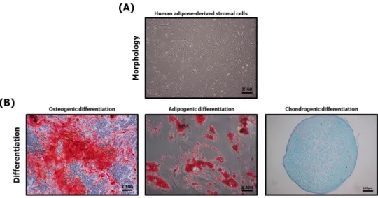
For the characterization of hASCs fibroblastic morphology was identified through microscopic photographs and differentiation into osteocytes, adipocytes and chondrocytes was confirmed through three different dyeing agents. Therefore, hASCs have fibroblastic morphology and ability of differentiation into osteocytes, adipocytes and chondrocytes (Figure 5).
Figure 5. Characterization of human adipose-derived stromal cells Using microscope, the fibroblastic morphology of hASCs was confirmed (A), and the differentiation ability to osteocytes, adipocytes and chondrocytes was confirmed using three different dyeing agents. All differentiation induction period was 2 weeks. (B).
Determination of concentration of GO suitable for chondrogenic differentiation of hASCs
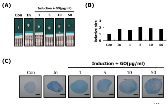
Based on the GO with the largest particle size, I confirmed the concentration suitable for chondrogenic differentiation of hASCs. Chondrogenic differentiation study of hASCs was conducted for two weeks in order to identify optimal induced conditions. Size of the chondrocyte pellets were found to have no effect on all concentrations of GO. To determine the degree of chondrogenic differentiation, the content of GAGs was measured using alcian blue staining. As a result, it was confirmed that a concentration of 10 μg / ml or less was suitable. (Figure 6).
Figure 6. Optimization of concentration of GO effective in chondrogenic differentiation of hASCs Using various concentrations of GO, the chondrogenic differentiation of hASCs was identified. Effect of GO was confirmed through the size of the chondrocyte pellets (A, B), and the effect of chondrocyte differentiation was confirmed by the content of GAGs in the chondrocyte pellet using alcian blue staining. After alcian blue staining, hASCs was monitored under an inverted microscope. Bar=100μm (C). As a result, it was confirmed that a concentration of 10 μg / ml or less was suitable.