116 Acta Orthopaedica 2011; 82 (1): 116–118
Management of severe tibial bony defects with double metal blocks
in knee arthroplasty—a technical note involving 9 cases
Seung-Wook Baek and Choong H Choi
Department of Orthopaedic Surgery, Hanyang University College of Medicine, South Korea Correspondence : [email protected]
Submitted 10-06-17. Accepted 10-09-28
Open Access - This article is distributed under the terms of the Creative Commons Attribution Noncommercial License which permits any noncommercial use, distribution, and reproduction in any medium, provided the source is credited.
DOI 10.3109/17453674.2010.548031
Severe bony defects during either primary or revision knee arthroplasty are common. If the defect is > 10 mm in its greatest depth, metal augmentation or a bone graft should be considered. We report the operative technique of metal aug-mentation using double metal blocks for severe uncontained proximal tibial defects.
Surgical technique and postoperative protocol Through an anterior midline skin incision and medial parapa-tellar approach, the knee joint is exposed (Figures 1 and 2). Soft tissue is released to obtain balancing of varus or valgus deformities. The intramedullary alignment instrumentation is used to prepare the femoral side.
The extramedullary tibial alignment guides are applied to the tibia. Once proper axial alignment is verified, the proxi-mal end of the tibia can be cut at usually 10 mm depth from the unaffected tibial condyle. The tibial surface is then pre-pared to accept the tibial base tray. The sclerotic base of the defect is cut to expose a flat, cancellous bony surface and the concave, irregular defect is converted to a flat one by mini-mal bone removal with a saw. The tibial bone defect is then
assessed, and an appropriate-sized metal block is selected. A cutting guide for the block is assembled and a matching bone resection carried out. Care must be taken not to over-resect the bone, since the tibial blocks should be inserted in a tight manner. The positions of the central keels on the baseplate are prepared on the tibial surface.
The trial tibial component with the block and intramedullary stem is assembled and inserted. A trial reduction of the pros-thesis is done, and alignment and stability with patellar track-ing is assessed. After lavage, ustrack-ing pulsed normal saline and drying out of the prepared surfaces, blood and fat are kept out of the cement-prosthesis interface. The real components are assembled and cemented. We use one mix of PMMA cement with gentamicin to cement the tibial and femoral components separately.
Metal blocks of 10 mm + 10 mm or 10 mm + 5 mm are used on the medial aspect of the tibial component to compensate for bone defects. Both the tray and the block have a waffled surface that allows interdigitation of cement.
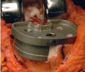
The first block is attached to the tibial tray with screws. After that, the next block is cemented to the first one (Figure
Figure 1. Preoperative radiographs with marked medial tibial bone
Acta Orthopaedica 2011; 82 (1): 116–118 117
3). The intramedullary stems on the tibial components are commonly used to protect the peripheral bone from stress. Finally, the real prosthesis with cemented tibial stem and with 10 mm + 10mm or 10 mm + 5 mm double metal augmentation are cemented into place.Downsized metal blocks can be used when the tibial blocks protrude over the cortical rim because of the natural taper of the proximal tibia (Figure 4).
Once the cement is cured and excess is removed, a polyeth-ylene implant is inserted and the knee is reduced. Final checks of soft tissue tension, alignment, patellar tracking, and range of knee movements is made before wound closure.
A continuous passive motion is started within 24 h postop-eratively, progressing slowly in flexion. Passive knee exten-sion is encouraged by placing the patient’s foot on a pillow while in bed. Weight bearing with the aid of crutches or a cane starts on the fifth or sixth postoperative day.
Patients
Between 2004 and 2007, we carried out metal augmentation of tibial defects in 9 patients during either primary or revision knee arthroplasty, using the operative technique described. All the procedures were carried out by a single surgeon. Primary diagnosis were osteoarthritis in 4 cases, rheumatoid arthritis in 1, aseptic loosening in 2, and septic loosening in 1 case. Patients’ mean age at operation was 65 (51–80) years. The mean follow-up period was 5 (2.5–6) years.
During the follow-up period, all the patients had a pain-free knee. The mean range of motion was 127° (120–135), and there was no radiographic evidence of prosthesis loosening or subsidence (Figures 5 and 6).
Discussion
The indication for augmentation of bone defects in TKA is inability to achieve stability of the trial implants at the time of trial reduction. This generally occurs when 40% or more of the bone-implant interface is unsupported by host bone (Cuckler 2004). Treatment options for large tibial bone defects include polymethylmethacrylate cement, morselized or structural allograft, metal augments, and custom or hinge/tumor pros-theses (Radnay and Scuderi 2006).
Excessive cement augmented by screws and mesh tech-niques has been abandoned because the construct is not mechanically sound, and may result in cement fragmentation and early failure of the replacement (Brooks et al. 1984, Brand et al. 1989, Ritter et al. 1993).
The technique for the application of an autograft in primary knee arthroplasty has been well described by Windsor et al. (1986). It is physiologically sound, cheap, and reproducible— and has the advantage of bone stock preservation (Toms et al. 2009). However, Laskin (1989) reported a 33% failure rate at 5-year follow-up of autografts in primary arthroplasty. Figure 3. A. The first metal block is attached to the tibial
tray with screws. B. After that, the next block is cemented to the first one.
Figure 4. If the tibial blocks protrude over the cortical rim, downsized metal blocks may be available (another case).
118 Acta Orthopaedica 2011; 82 (1): 116–118
Larger defects may require allograft augmentation (Cuckler 2004, Engh and Ammeen 2007). However, allograft bone has the disadvantages that is difficult to achieve a proper fit with the host bone (Ries 1996) and that although they remain struc-turally intact, they are frequently not revascularized; new bone is laid down only in the periphery of a dead allograft (Parks and Engh 1997).
Custom implants can be used for large bone defects, and should theoretically provide the best fit and force transmission of any of the methods used to address bone deficiency (Brooks et al. 1984). However, they are expensive and often require considerable time to manufacture.
Modular implants with metal augmentation facilitate the treatment of bony defects (Patel et al. 2004, Fehring et al. 1996, Nelson et al. 2003, Pagnano et al. 1995). The advantage of modular metal augments is that they offer flexibility during the operation but still provide the surgeon with the ability to deal with defects of bone ranging from 5 to 10 mm in depth in various locations on the tibial plateau (Rand 1998). However, custom prostheses may be required for larger defects.
We have modified the operative method by attaching 2 blocks with the use of cement and screws to allow up to 20 mm of segmental bone defects to be restored. They can be applied quickly, allow intraoperative custom fabrication, and help restore an anatomic joint line. The technique is simple and has no learning curve.
Brand M G, Daley R J, Ewald F C, Scott R D. Tibial tray augmentation with modular metal wedges for tibial bone stock deficiency. Clin Orthop 1989; (248): 71-9.
Brooks P J, Walker P S, Scott R D. Tibial component fixation in deficient tibial bone stock. Clin Orthop 1984; (184): 302-8.
Cuckler J M. Bone loss in total knee arthroplasty: graft augment and options. J Arthroplasty (Suppl 1) 2004; 19 (4): 56-8.
Engh G A, Ammeen D J. Use of structural allograft in revision total knee arthroplasty in knees with severe tibial bone loss. J Bone Joint Surg (Am) 2007; 89 (12): 2640-7.
Fehring T K, Peindl R D, Humble R S, Harrow M E, Frick S L. Modular tibial augmentations in total knee arthroplasty. Clin Orthop 1996; (327): 207-17. Laskin R S. Total knee arthroplasty in the presence of large bony defects of
the tibia and marked knee instability. Clin Orthop 1989; (248): 66-70. Nelson C L, Gioe T J, Cheng E Y, Thompson R C, Jr. Implant selection in
revision total knee arthroplasty. J Bone Joint Surg (Am) (Suppl 1) 2003; 85: S43-51.
Pagnano M W, Trousdale R T, Rand J A. Tibial wedge augmentation for bone deficiency in total knee arthroplasty. A followup study. Clin Orthop 1995; (321): 151-5.
Parks N L, Engh G A. The Ranawat Award. Histology of nine structural bone grafts used in total knee arthroplasty. Clin Orthop 1997; (345): 17-23. Patel J V, Masonis J L, Guerin J, Bourne R B, Rorabeck C H. The fate of
augments to treat type-2 bone defects in revision knee arthroplasty. J Bone Joint Surg (Br) 2004; 86 (2): 195-9.
Radnay C S, Scuderi G R. Management of bone loss: augments, cones, offset stems. Clin Orthop 2006; (446): 83-92.
Rand J A. Modular augments in revision total knee arthroplasty. Orthop Clin North Am 1998; 29 (2): 347-53.
Ries M D. Impacted cancellous autograft for contained bone defects in total knee arthroplasty. Am J Knee Surg 1996; 9 (2): 51-4.
Ritter M A, Keating E M, Faris P M. Screw and cement fixation of large defects in total knee arthroplasty. A sequel. J Arthroplasty 1993; 8 (1): 63-5. Toms A D, Barker R L, McClelland D, Chua L, Spencer-Jones R, Kuiper J H.
Repair of defects and containment in revision total knee replacement: a com-parative biomechanical analysis. J Bone Joint Surg (Br) 2009; 91 (2): 271-7. Windsor R E, Insall J N, Sculco T P. Bone grafting of tibial defects in primary
and revision total knee arthroplasty. Clin Orthop 1986; (205): 132-7. Figure 6. Postoperative radiographs at 5-year follow-up. Figure 5. Immediate postoperative radiographs of knee replacement with