HSP25 Inhibits Protein Kinase C
␦-mediated Cell Death through
Direct Interaction*
Received for publication, January 31, 2005, and in revised form, February 23, 2005 Published, JBC Papers in Press, February 24, 2005, DOI 10.1074/jbc.M501131200
Yoon-Jin Lee‡§, Dae-Hoon Lee‡§, Chul-Koo Cho‡, Sangwoo Bae‡, Gil-Ja Jhon
¶, Su-Jae Lee
储,
Jae-Won Soh**, and Yun-Sil Lee‡ ‡‡
From the ‡Laboratory of Radiation Effect and储Laboratory of Experimental Radiation Therapeutics, Korea Institute of Radiological and Medical Sciences, Seoul 139-706,¶Division of Molecular Life Science, Ewha Woman’s University, Seoul 120-750, and **Laboratory of Signal Transduction, Department of Chemistry, Inha University, Incheon, 402-751 Seoul, Korea
Heat shock protein 25 (HSP25) interferes negatively with apoptosis through several pathways that involve its direct interaction with cytochrome c or Akt. Here we
show that HSP25 inhibits protein kinase C (PKC)
␦-me-diated cell death through direct interaction. HSP25
binds to kinase-active PKC␦ to inhibit its kinase activity
and translocation to the membrane, which results in reduced cell death. Deletion constructs of HSP25 and PKC␦ identified amino acids 90–103 of HSP25 and the
C-terminal V5 region of PKC␦ as binding sites. In
addi-tion, the interaction between HSP25 and PKC␦ induced
HSP25 phosphorylation at Ser-15 and Ser-86, and these
phosphorylations permitted HSP25 release from PKC␦.
Based on these observations, we propose that after PKC␦ activation, HSP25 binds to the exposed V5 region
of PKC␦. This novel function of HSP25 accounts for its
cytoprotective properties via the inhibition of PKC␦ and
the enhancement of HSP25 phosphorylation.
Small heat shock protein (HSP)1has been suggested to
pro-tect cells against apoptotic cell death triggered by hyperther-mia, ionizing radiation, oxidative stress, Fas ligand, and cyto-toxic drugs (1–5). Several mechanisms have been proposed to account for HSP27-mediated apoptotic protection, for example, via its specific interaction with cytochrome c released from mitochondria into the cytosol, which prevents apoptosome for-mation (6, 7). The elimination of unfolded protein via the ex-tralysosomal, energy-dependent, ubiquitin-proteasome degra-dation pathway is another mechanism that contributes to cell protection from stressful stimuli (8). HSP27 binds to
polyubiq-uitin chains as well as 26 S proteasomes, and the ubiqpolyubiq-uitin- ubiquitin-proteasome pathway is involved in the activation of transcrip-tion factor NF-B by degrading its main inhibitor I-B␣ (8).
Moreover, phosphorylated HSP27 has been shown to bind an adaptor protein Daxx and then to inhibit Fas-mediated apo-ptosis (9). Interaction between HSP27 and Akt is necessary for Akt activation, and this is followed by dissociation of phospho-rylated HSP27 from Akt (10).
We reported recently that the radioprotective effect of HSP25 involves delayed cell growth (11, 12) and HSP25-medi-ated Mn-SOD gene expression (13, 14). HSP25 overexpression down-regulates ERK1/2 expression, and HSP25-mediated ERK2 suppression is involved in HSP25-induced radioresist-ance and cell cycle delay (15). In addition, attenuated oxidative stress-induced apoptosis by HSP25 overexpression was found to be due to the inhibition of the PKC␦-mediated ERK1/2 path-way and to the induction of the Mn-SOD gene (14), and HSP25 also inhibits radiation-induced PKC␦-mediated reactive oxygen species production (16).
Radiation and many anticancer drugs are known to kill tumor cells by inducing apoptosis. However, a defect in the apoptotic process can lead to resistance to anticancer drugs or radiother-apy. Several members of the protein kinase C (PKC) family serve as substrates for caspases, and the PKC␦ isozyme has been intimately associated with apoptosis. The activation of PKC␦ was found to be associated with cell cycle progression inhibition (17), and PKC␦ down-regulation was found to be associated with tu-mor promotion (18), suggesting that PKC␦ may have a negative effect on cell survival. In addition, the proteolytic activation of PKC␦ has since been associated with the apoptosis induced by DNA damage, including that caused by UV light, ionizing radi-ation, cisplatin, etoposide, arabinoside, and doxorubicin (19 –24). Moreover, several investigators have confirmed that the ectopic expression of the PKC␦ catalytic fragment results in cell death (25–28). Mizuno et al. (26) showed that the kinase activity of a PKC␦ catalytic fragment during apoptosis may be a key partici-pant in the late stages of apoptosis.
In the present study, we observed for the first time that HSP25 binds directly to the V5 region of kinase-active PKC␦. This results in a dual form of HSP25-mediated cytoprotection, namely via the inhibition of PKC␦ activity, thereby blocking apoptosis, and via the phosphorylation of HSP25, which in-creases its cytoprotective effect. In addition, these two types of HSP25-mediated apoptotic expression were found to be trig-gered by radiation or oxidative stress.
MATERIALS AND METHODS
Reagents—Rottlerin (PKC␦ inhibitor), phorbol 12-myristate 13-acetate, and H2O2were purchased from Calbiochem. Anti-HSP27,
anti-phospho-HSP27 (Ser-15), anti-phospho-anti-phospho-HSP27 (Ser-82), anti-HA, anti-PKC␦, anti-* This work was supported by the Korean Institute of Science and
Technology Evaluation and Planning and by the Ministry of Science and Technology through the National Nuclear Technology Program. The costs of publication of this article were defrayed in part by the payment of page charges. This article must therefore be hereby marked “advertisement” in accordance with 18 U.S.C. Section 1734 solely to indicate this fact.
§ Both authors contributed equally to this work.
‡‡ To whom correspondence should be addressed: Laboratory of Ra-diation Effect, Korea Institute of Radiological and Medical Sciences, 215-4 Gongneung-Dong, Nowon-Ku, Seoul 139-706, Korea. Tel.: 82-2-970-1325; Fax: 82-2-970-2402; E-mail: yslee@kcch.re.kr.
1The abbreviations used are: HSP, heat shock protein; PKC, protein
kinase C; GST, glutathione S-transferase; HA, hemagglutinin; PMA, phorbol 12-myristate 13-acetate; SOD, superoxide dismutase; MBP, myelin basic protein; PI, propidium iodide; RFP, red fluorescent pro-tein; GFP, green fluorescent propro-tein; CAT, chloramphenicol acetyl-transferase; FBS, fetal bovine serum; PBS, phosphate-buffered saline; MAPK, mitogen-activated protein kinase; RIF, radiation-induced fibro-sarcoma; siRNA, small interfering RNA; ATP␥S; adenosine 5⬘-O-(thio-triphosphate); Gy, gray; WT, wild type.
This paper is available on line at http://www.jbc.org
18108
at Ewha Medical Library on April 25, 2019
http://www.jbc.org/
lamin B, anti--actin, and anti-GFP antibodies were from Santa Cruz Biotechnology (Santa Cruz, CA). Phospho-PKC␦ (Ser-643) and phospho-PKC␦ (pan) were from Cell Signaling Technology (Beverly, MA). Anti-phosphotyrosine antibody, PKC␦ protein (recombinant protein expressed in Sf21), and PKC lipid activator were purchased from Upstate Biotech-nology, Inc. (Lake Placid, NY). ATP and ATP␥S were from Sigma. DsRed polyclonal antibody was from BD Biosciences.
Plasmids—Wild type mouse HSP25 (GenBankTMaccession number
XM124655) was cloned into pcDNA4HisMaxC (Invitrogen), which con-tains an N-terminal His tag. The phosphorylation mutant constructs HSP25 (S15A), HSP25 (S15D), HSP25 (S86A), HSP25 (S86D), HSP25 (S15A/S86A), and HSP25 (S15D/S86D) were constructed by PCR (29) by using the overlap extension primers. Serine was replaced by alanine or aspartate to generate the deficient or phosphorylation-mimicking mutants of HSP25, respectively. The dimerization mutant HSP25 (C141A) was constructed using the same method. PCR products were digested with EcoRI and cloned into the EcoRI site of pcDNA4HisMaxC to construct expression vectors of the His-tagged mutant HSP25 protein. PCR products were also cloned into pEGFP-N1 vector (Clontech) to express GFP-tagged mutant HSP25 proteins. The internal deletion mutants were constructed in HSP25-containing pcDNA4HisMaxC vector by introducing two SalI sites in the HSP25-coding sequence using deletion primers. This was followed by restric-tion and ligarestric-tion steps. For the (92–145)-GFP construct, HSP25-(91–187) was amplified by PCR, and the PCR products obtained were digested and cloned into pEGFP-N1 vector (Clontech).
Wild type PKC␦ (GenBankTMaccession number AY545076), the
reg-ulatory domain (amino acids 2–333, REG), the catalytic domain (amino acids 334 – 674, CAT), and the dominant-negative catalytic domain (amino acids 334 – 674, CAT-KR) were cloned into pHACE that contains a C-terminal HA tag (30). To construct the PKC␦-truncated mutants, each DNA fragment was amplified using mutagenic primers containing an EcoRI site by PCR. The amplified PKC␦ mutants were cloned into pHACE. The phosphorylation mutants of the catalytic domain, i.e. CAT (S643A), CAT (S643D), CAT (S662A), CAT (S662D), CAT (S643A/ S662A), CAT (S643D/S662D), CAT-KR (S643A), CAT-KR (S643D), CAT-KR (S662A), CAT-KR (S662D), CAT-KR (S643A/S662A), and CAT-KR (S643D/S662D) were constructed using the method used for the HSP25 phosphorylation mutants. The PCR products were digested with EcoRI and cloned into the EcoRI site of pHANE, which contains an N-terminal HA tag (30). PKC␦ construct was cloned into the EcoRI site of pDsRed1-N1 vector (Clontech) to express PKC␦-RFP (V5-RFP). All the primer sequences are available by request.
Cell Culture—L929 (murine fibroblasts), radiation-induced fibrosar-coma (RIF) cells, and TR (a thermoresistant clone of RIF) were cultured in Dulbecco’s minimal essential medium (Invitrogen) supplemented with heat-inactivated 10% fetal bovine serum (FBS, Invitrogen) and antibiotics at 37 °C in a 5% CO2humidified incubator. The human
non-small cell lung cancer cell lines H460, H596, and H1299 and Jurkat T leukemia cells were grown in RPMI 1640 supplemented with 10% FBS, glutamine, HEPES, and antibiotics at 37 °C in a 5% CO2
humid-ified incubator. L929 and Jurkat transformant clones were obtained by stable transfection with pHSP6 (containing the complete genomic se-quence of murine HSP25) and pBC vector (Stratagene, La Jolla, CA) (11, 12, 16) or with the MFG retroviral vector (31).
Irradiation—Cells were plated in 60-mm dishes and incubated at 37 °C in humidified 5% CO2in culture medium until 70 – 80% confluent.
Cells were then exposed to␥-rays from a137
Cs␥-ray source (Atomic Energy of Canada, Ltd.) at 3.81 Gy/min.
Flow Cytometric Analysis—Cells were cultured, harvested at the indicated times, stained with propidium iodide (PI), according to the manufacturer’s protocol, and then analyzed using a FACScan flow cytometer (BD Biosciences).
Cytoplasmic/Nuclear Fractionation—Cells (1⫻ 106
) grown on 10-cm tissue culture dishes were washed once with ice-cold PBS and harvested with a scraper. Cell pellets were then resuspended in hypotonic buffer (10 mMHEPES, 10 mMKCl, 0.1 mMEDTA, 0.1 mMEGTA, 0.5% Nonidet P-40) and incubated at 4 °C for 15 min. Samples were agitated every 5 min and then centrifuged at 12,000 rpm for 30 s to collect the cytoplas-mic fraction. Pellets were resuspended, incubated in nuclear extraction buffer (20 mMHEPES, 20% glycerol, 0.42MNaCl, 1 mMEDTA, 1 mM EGTA) for 30 min, and centrifuged at 12,000 rpm for 20 min to obtain the nuclear fraction.
Cytosolic/Membrane Protein Fractionation—Cells were washed twice with PBS and then sonicated in buffer containing 100 mMTris-HCl (pH 7.5), 50 mMMgCl2, 10 mMdithiothreitol, and 10 mM
phenylmethylsul-fonyl fluoride. The sonicated cells were then incubated in ice for 1 h and ultracentrifuged for 1 h at 100,000⫻ g to separate the membrane and
cytosolic fractions. The cell pellet obtained was sonicated in buffer containing 1% Triton X-100, incubated in ice for 1 h, and then ultra-centrifuged for 1 h at 100,000⫻ g to separate the membrane fraction from cellular debris.
Immunofluorescence Analysis—For immunofluorescence analysis, cells were fixed with 2% paraformaldehyde, permeabilized with 0.1% Triton X-100 in PBS, and then washed three times with PBS. They were then incubated with goat anti-phospho-HSP27 (Ser-15 and Ser-82) di-luted 1:200 in PBS containing 5% FBS for 1 h at room temperature in a humidified chamber. Excess antibody was removed by washing cov-erslips three times with PBS. Cells were then incubated with fluores-cein isothiocyanate-conjugated goat anti-rabbit immunoglobulin G (IgG) (Dako, Produktionsvej, Denmark), at 1:200 dilution in PBS con-taining 5% FBS for 4 h, and then incubated with 0.5g/l propidium iodide (Molecular Probes, Inc., Eugene, OR) for 5 min at room temper-ature. After washing three times with PBS, coverslips were mounted onto microscope slides using ProLong antifade mounting reagent (Mo-lecular Probes), and slides were analyzed under a confocal laser-scan-ning microscope (Leica Microsystems).
PAGE and Western Blotting—For PAGE and Western blotting, cells were solubilized with lysis buffer (120 mMNaCl, 40 mMTris (pH 8.0), 0.1% Nonidet P-40) and boiled for 5 min, and equal amounts of protein were analyzed on 10% SDS-PAGE. After electrophoresis, proteins were to a nitrocellulose membrane and processed for immunoblotting. Blots were further incubated with horseradish peroxidase-conjugated second-ary antibody diluted at 1:5,000, and specific bands were visualized by chemiluminescence (ECL, Amersham Biosciences). Autoradiographs were recorded on X-Omat AR film (Eastman Kodak Co.).
PKC Kinase Assay—Cellular proteins were extracted using PKC extraction buffer (50 mMHEPES (pH 7.5), 150 mMNaCl, 0.1% Tween 20, 1 mMEDTA, 2.5 mMEGTA, and 10% glycerol) containing protease inhibitors (10g/ml each of aprotinin and leupeptin and 0.1 mM phen-ylmethylsulfonyl fluoride) and phosphatase inhibitors (1 mMNaF, 0.1 mMNa3VO4, and 10 mM-glycerophosphate). HA-tagged PKC proteins
from 300g of cell extracts were immunoprecipitated with 3 g of anti-HA antibody and 30l of protein G-Sepharose for 3 h at 4 °C. The immunoprecipitates were washed twice with PKC extraction buffer and PKC reaction buffer (50 mMHEPES (pH 7.5), 10 mM MgCl2, 1 mM
dithiothreitol, 2.5 mMEGTA, 1 mMNaF, 0.1 mMNa3VO4, and 10 mM
-glycerophosphate) and then resuspended in 20 l of PKC reaction buffer. The kinase assay was initiated by adding 40l of PKC reaction buffer containing 5g of dephosphorylated MBP protein (Upstate Bio-technology, Inc.) or His-HSP25 protein and 5Ci of [␥-32P]ATP.
Reac-tions were carried out for 30 min at 30 °C and terminated by adding SDS sample buffer. Mixtures were then boiled for 5 min, and the reaction products were analyzed by SDS-PAGE and autoradiography.
Preparation of Recombinant HSP25 Proteins—Vectors for the ex-pression of recombinant HSP25 were prepared by inserting the HSP25 wild type into the NcoI-EcoRI restriction site of pProExHTa vector (Invitrogen). The resulting plasmid was transformed into Escherichia coli BL21 (Novagen) to produce His tag-HSP25 fusion proteins (His-HSP25) after treating with 0.15Misopropyl-1-thio-- D-galactopyrano-side for 4 h at 30 °C. Bacteria were lysed in a buffer containing 50 mM HEPES (pH 7.5), 0.45MNaCl, 5 mMdithiothreitol, Complete protease inhibitor (Roche Diagnostics), 1.25 mg/ml lysozyme, and 11.2 g/ml DNase I (Sigma). His-HSP25 proteins were extracted from inclusion bodies by incubating for 1 h followed by vigorous shaking at 37 °C in 25 mMphosphate buffer, pH 7.4, containing 150 mMNaCl, 1 mMEDTA, 2% (v/v) Triton X-100, 5 mMdithiothreitol, and Complete protease inhibi-tor. The solubilized His-HSP27 proteins were affinity-purified using nickel-nitrilotriacetic acid-agarose beads (Qiagen).
Immunoprecipitation—Cells (1⫻ 107
) were lysed in immunoprecipi-tation buffer (50 mMHEPES (pH 7.6), 150 mMNaCl, 5 mMEDTA, 0.1% Nonidet P-40). After centrifuging (10 min at 15,000 ⫻ g) to remove particulate material, the supernatant was incubated with antibodies (1:100) against anti-HSP27 or PKC␦ with constant agitation at 4 °C. Immunocomplexes were precipitated with protein A-Sepharose (Sigma) and analyzed by SDS-PAGE by enhanced chemiluminescence (Amer-sham Biosciences).
Cell Transfection—Pre-designed siRNA for human HSP27 (HSPB1–3, Ambion catalog number16706) was purchased from Am-bion, Inc. (Austin, TX). Cells were transfected with the siRNAs for 48 h using Lipofectamine TM 2000 (Invitrogen). A mixture of 250 l of Opti-MEM medium (Invitrogen) and 20l of LipofectamineTM
2000 was incubated for 5 min at room temperature and then combined with 100 nMof siRNA diluted with 250l of Opti-MEM. The resulting mixture (500l) was incubated for 20 min at room temperature to allow complex formation and then overlaid onto each well containing the cells to a final
HSP25 Binds to PKC
␦
18109
at Ewha Medical Library on April 25, 2019
http://www.jbc.org/
volume of 2.5 ml/60-mm dish. The transient transfection of all cell types was carried out using Plus LipofectamineTMreagent (Invitrogen) and
LipofectamineTMreagent.
Histidine Pull-down Assay in Vitro—The in vitro transcription and translation of PKC␦ was performed using a TNT T7 Quick Master Mix kit (Promega, Madison, WI) in the presence of [35S]methionine,
accord-ing to the manufacturer’s protocol. An aliquot of His-tagged HSP25 protein, bound to nickel-nitrilotriacetic acid-agarose beads, was incu-bated with 5l of in vitro translated PKC␦ in 500 l of NETN buffer (100 mMNaCl, 1 mMEDTA (pH 8.0), 20 mMTris-HCl (pH 8.0), 0.2% Nonidet P-40, 10 mMimidazole) for 2 h. Beads were washed five times with 1 ml of NETN buffer, resuspended in Laemmli sample buffer, and subjected to SDS-PAGE before autoradiography.
Histidine Pull-down Assay—Histidine-tagged DNA constructs were transfected into the cells by using Lipofectamine Plus reagent. After incubation for 48 h, cells were harvested, and whole cell extracts were prepared as described previously. Cell extracts were mixed and incu-bated with nickel-nitrilotriacetic acid-agarose beads for 30 min at 4 °C in the presence of 10 mMimidazole. After washing the resin with buffer containing 10 mMimidazole, proteins were recovered by suspension in Laemmli sample buffer and then subjected to SDS-PAGE and Western blot analysis using anti-PKC␦ or anti-HA antibodies.
RESULTS
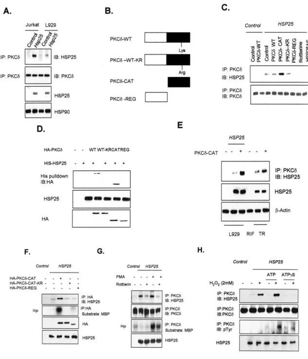
Interaction between HSP25 and the Catalytic Domain of PKC␦ and PKC␦ Kinase Activity Is Necessary for Its Interaction with HSP25—As HSP25 overexpression was found to inhibit
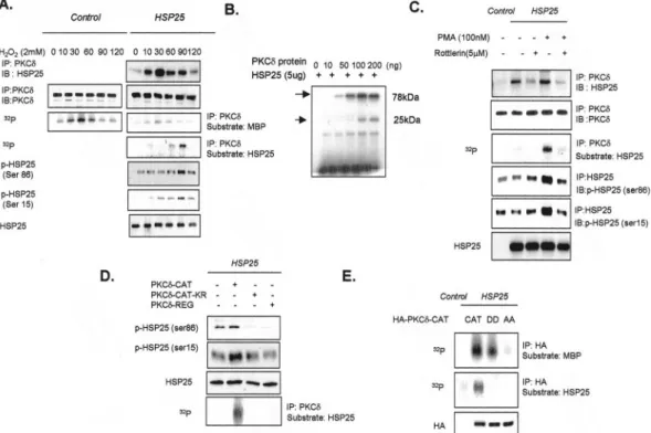
PKC␦ activity in our previous study (14), and another HSP, HSP70, was reported to bind PKC (32), we examined possible binding between HSP25 and PKC␦. HSP25 was found to coim-munoprecipitate with PKC␦ in whole cell lysates of L929 and Jurkat T cells overexpressing HSP25 (Fig. 1A). To confirm binding between PKC␦ and HSP25, several deletion constructs of PKC␦ were made (Fig. 1B). Immunoprecipitation revealed that the catalytic domain of PKC␦ (CAT), but not the regula-tory domain of PKC␦ (REG), is an HSP25 binding target. When rottlerin, a specific PKC␦ inhibitor, was pretreated, HSP25 binding disappeared, suggesting that the kinase activity of PKC␦ is important for the interaction between HSP25 and PKC␦ (Fig. 1C). A loss of binding activity after HSP25 anti-sense transfection confirmed the specific interaction between HSP25 and PKC␦ (Fig. 1C), which was further confirmed by probing histidine pull downs of HSP25 using several domain mutants of PKC␦ fused with a HA tag. Consistently, wild type (WT) PKC␦ interacted with HSP25, and PKC␦-CAT mediated this binding (Fig. 1D). TR cells, which were derived as a ther-moresistant clone of RIF and which show high expression lev-els of HSP25, were used to confirm the binding potential of endogenous HSP25 with PKC␦. Moreover, transfection of PKC␦-CAT into TR cells was found to increase the interaction between HSP25 and PKC␦-CAT (Fig. 1E). However, we did not detect any interaction between HSP70 and PKC␦ (data not shown). Because rottlerin pretreatment completely inhibited the interaction between HSP25 and PKC␦, we hypothesized that kinase-active PKC␦ is required for this interaction. Thus we transfected PKC␦-CAT-K376R (CAT-KR), which lacks PKC␦ kinase activity, and performed immunoprecipitation ex-periments. Neither PKC␦-REG (Fig. 1F, 5th lane) nor CAT-KR interacted with HSP25 (Fig. 1F, 4th lane). In contrast, PKC ␦-CAT bound to HSP25 (Fig. 1F, 3rd lane). Moreover, pretreat-ment with PKC activators such as PMA or H2O2increased the
interaction between HSP25 and PKC␦, and this was accompa-nied by an increase in the MBP kinase activity of PKC␦; how-ever, rottlerin pretreatment reduced HSP25-PKC␦ binding, even though MBP phosphorylation by PKC␦ was not com-pletely inhibited (Fig. 1G) (H2O2treatment data not shown).
Because PKC␦-CAT-KR did not interact with HSP25, we used ATP and ATP␥S to confirm the importance of the kinase activ-ity of PKC␦. An interaction was observed between HSP25 and PKC␦ after treating with H2O2, and this binding was
dramat-ically increased by cotreating H2O2 with ATP. However,
co-treatment with ATP␥S (often regarded as a nonhydrolyzable ATP analogue and is used as an inhibitor of phosphatases and ATPases (33)) blocked this interaction (Fig. 1H), suggesting that PKC␦ kinase activity (in terms of the binding and hydrol-ysis of ATP) is important for the interaction between HSP25 and PKC␦.
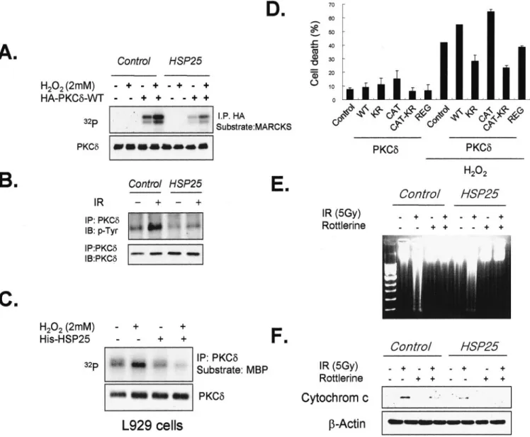
Interaction between PKC␦-CAT and HSP25 Inhibits PKC␦-mediated Cell Death—Because PKC␦ has been reported to
in-duce apoptosis (34), we examined if the interaction between HSP25 and PKC␦ affects PKC␦-mediated apoptosis. In L929 cells cotransfected with PKC␦ and HSP25, PKC␦ activation by H2O2was lower than in cells transfected with only PKC␦ (Fig. 2A). In addition, the tyrosine phosphorylation of PKC␦, which correlates with its kinase activity (35) and which induces many PKC␦-mediated effects like the induction of apoptosis (36), was also inhibited by HSP25 overexpression (Fig. 2B). In addition, we observed that H2O2treatment increased PKC␦ kinase
ac-tivity and that the addition of HSP25 protein to the cell lysates reduced PKC␦ kinase activity (Fig. 2C), which shows the inhi-bition of PKC␦ kinase activity by HSP25. PKC␦-WT increased cell death due to H2O2treatment, and PKC␦-CAT appeared to
mediate this effect, although PKC␦-REG and PKC␦-CAT-KR, which do not have PKC␦ kinase activity, reduced H2O2
-medi-ated cell death (Fig. 2D). Moreover, radiation-induced DNA fragmentation (Fig. 2E) and cytochrome c release to the cytosol were inhibited when cells were pretreated with rottlerin. The effect of HSP25 overexpression was similar to that of rottlerin in terms of cytochrome c release (Fig. 2F).
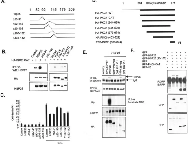
Amino Acids 90 –103 of HSP25 and V5 Region Located in the Catalytic Domain of PKC␦ Are the Binding Sites of Each Mol-ecule—To provide definitive evidence that HSP25 prevents cell
death by binding to PKC␦, several mutants of HSP25 were prepared. L929 cells were transiently transfected with an empty plasmid (control) or a plasmid containing wild type or a mutated HSP25 cDNA (refer to Fig. 3A for details of the mu-tants used). Coimmunoprecipitation studies demonstrated that deletion mutants HSP25-(⌬90–103) and HSP25-(⌬92–145) did not bind to PKC␦-CAT, whereas HSP25 mutants ⌬53–91, ⌬106 –132, and ⌬133–142 were able to bind to PKC␦-CAT as efficiently as wild type HSP25 (Fig. 3B). His pull-down analysis also confirmed that amino acids 90 –103 of HSP25 are necessary for PKC␦-CAT binding (data not shown). When H2O2-induced cell death was examined, a protective effect was observed after transfecting HSP25⌬53–91, ⌬106 –132, or ⌬133–142 mutants, which interacted with PKC␦-CAT like the wild type (Fig. 3C). However, cell death remained at the control level after transfection with HSP25-(⌬90 –103) or HSP25-(⌬92–145) mutant. To identify the PKC␦ binding locus, deletion constructs of PKC␦-CAT were made (refer to Fig. 3D) and transfected into HSP25-overexpressing L929 cells. Coimmunoprecipitation analysis revealed that the de-letion mutants⌬343– 629, ⌬344 –500, and ⌬403– 629 did not bind to HSP25, whereas mutant⌬373– 647 bound to HSP25 as efficiently as PKC␦-CAT (Fig. 3E), suggesting that the amino acid sequence 630 – 674 (the V5 region) of PKC␦ is a binding site. To determine whether the V5 region of PKC␦ binds directly to the amino acid sequence 90 –103 of HSP25, immunoprecipitation was performed using a GFP-tagged 90 – 103-amino acid sequence of HSP25 or an RFP-fused PKC␦ V5 region. HSP25 was found to bind to PKC␦-CAT and to the V5 region, and the amino acid 90 –103 sequence of HSP25 also directly bound to the PKC␦-V5 region (Fig. 3F). Confocal analysis also revealed the colocalization of the V5 region of PKC␦ and of the amino acid 90 –103 sequence of HSP25 in cytosol (data not shown). These findings demonstrate that
at Ewha Medical Library on April 25, 2019
http://www.jbc.org/
FIG. 1. Interaction between HSP25 and kinase-active PKC␦ catalytic domain. A, immunodetection of HSP25 or PKC␦ in control and in HSP25-overexpressing L929 cells or Jurkat T cells, with previous immunoprecipitation (IP) of PKC␦. B, schematic drawing of PKC␦ point or deletion mutants. Wild type PKC␦ (PKC␦-WT), Lys-Arg point mutant of wild type PKC␦ (PKC␦-WT-KR), regulatory domain deleted mutant of PKC␦ (PKC␦-CAT), catalytic domain deleted mutant (PKC␦-REG). C, after immunoprecipitation of PKC␦ in lysates from control or HSP25-overexpressed L929 cells transfected with the indicated HA-tagged PKC␦ point or deletion mutants or the HSP25 antisense construct (HSP25AS), or pretreated with rottlerin (5M), the immunodetection of HSP25 and PKC␦ was performed. D, the indicated HA-tagged PKC␦ deletion mutants were transfected to L929 cells, and cell lysates were incubated with immobilized His-HSP25 or His vector. Retained HA proteins were detected by Western blotting using an anti-HA antibody. His fusion proteins are also shown. The transfection efficiencies of HA-tagged or HSP25 vectors were confirmed by Western blotting using anti-HA or anti-HSP25 antibody. E, PKC␦ was immunoprecipitated in lysates from control or HSP25-overexpressing L929 cells, RIF, or TR (thermoresistant clone of RIF) cells with or without PKC␦-CAT transfection, and HSP25 was immunode-tected. F, immunoprecipitation of HA in lysates from control or HSP25-overexpressing L929 cells after transfecting HA-tagged PKC␦-CAT, PKC␦-CAT-KR, or PKC␦-REG vectors. HSP25 protein was immunodetected using anti-HSP25 antibody. Cellular proteins were extracted after lysing with PKC extraction buffer. HA-tagged PKC proteins from 300g of cell extracts were immunoprecipitated using an anti-HA antibody and protein G-Sepharose. Immune complex kinase reactions were performed in the presence of GST-MBP substrate and [␥-32P]ATP. The transfection
efficiencies of HA-tagged or HSP25 vectors were confirmed by Western blotting using anti-HA or anti-HSP25 antibody. G, immunoprecipitation of PKC␦ in lysates from control or HSP25-overexpressing L929 cells 30 min after adding PMA (100 nM) with or without pretreatment with rottlerin (5M) for 30 min, followed by HSP25 or PKC␦ immunodetection using anti-HSP25 or anti-PKC␦ antibodies. Cellular proteins were extracted after lysing with PKC extraction buffer. HA-tagged PKC proteins from 300g cell extracts were immunoprecipitated using an anti-HA antibody and protein G-Sepharose. Immune complex kinase reactions were performed in the presence of GST-MBP substrate and [␥-32
P]ATP. H, immunopre-cipitation of PKC␦ in lysates of control or HSP25-overexpressing L929 cells pretreated with H2O2(2 mM) with or without ATP or ATP␥S (100 M)
pretreatment. Protein extracts (500g) were immunoprecipitated with PKC␦ antibody, and immunodetection was performed using anti-phosphotyrosine (p-Tyr) antibody. IB, immunoblot.
HSP25 Binds to PKC
␦
18111
at Ewha Medical Library on April 25, 2019
http://www.jbc.org/
amino acids 90 –103 of HSP25 are a binding site for the V5 region of PKC␦.
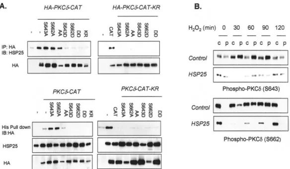
HSP25 Binds to the Unphosphorylated PKC␦-V5 Region and Inhibits PKC␦ Membrane Translocation—Binding between
HSP70 and PKC II is dependent on the phosphorylation sta-tus of PKC II (32), and several autophosphorylation sites have been identified in the V5 region of PKC␦ (37–40). Moreover, PKC activation has been reported to regulate the autophospho-rylation of these sites (37, 38). To elucidate whether autophos-phorylation status affects the interaction between HSP25 and PKC␦, we generated Ser-643 and Ser-662 phosphorylation-deficient and phosphorylation-mimicking mutants of PKC ␦-CAT. Coimmunoprecipitation revealed that the phosphoryla-tion-deficient mutants (S643A, S662A, and S643A/S662A) bound to HSP25, whereas the phosphorylation-mimicking
mu-tants (S643D, S662D, and S643D/S662D) did not, suggesting that the nonphosphorylated form of PKC␦ V5 is important for HSP25 binding (Fig. 4A). In the case of PKC␦-CAT-KR, which does not have kinase activity, all mutants failed to bind HSP25 (Fig. 4A). His pull-down analysis was used to confirm the above results. Because it has been reported that the autophosphoryl-ation of the V5 region affects the membrane translocautophosphoryl-ation of PKC (41), we examined PKC␦ phosphorylation status at Ser-643 and Ser-662. After treating the cells with H2O2for 30 min,
phosphorylated PKC␦ at Ser-643 and Ser-662 was translocated to the particulate fraction, and this was sustained 60 min later. However, in the case of HSP25-overexpressing cells, no phos-phorylated PKC␦ was translocated to the particulate fraction in accompaniment with the reduced total amount of phospho-rylated PKC␦ (Fig. 4B), which suggests that the interaction
FIG. 2. Interaction of PKC␦-CAT with HSP25 inhibits PKC␦-mediated apoptosis. A, control and HSP25-overexpressing L929 cells were transfected with PKC␦-WT and treated with or without H2O2(2 mM). Cells were then lysed, and cellular proteins were extracted. HA-tagged PKC␦
proteins were immunoprecipitated (IP) from 300g of cell extracts by using an anti-HA antibody and protein G-Sepharose. Immune complex kinase reactions were performed in the presence of GST-myristoylated alanine-rich C kinase substrate (MARCKS) and [␥-32P]ATP. B, protein
extracts (500g) from control and HSP25-overexpressing L929 cells were immunoprecipitated with anti-PKC␦ antibody and immunodetected using anti-phosphotyrosine (p-Tyr) antibody. C, cellular proteins from L929 cells pretreated with H2O2(2 mM) with or without His-tagged HSP25
protein (15g) were extracted by lysis with PKC extraction buffer. HA-tagged PKC␦ proteins were immunoprecipitated from 300 g of cell extracts using an anti-PKC␦ antibody and protein G-Sepharose. Immune complex kinase reactions were performed in the presence of GST-MBP substrate and [␥-32P]ATP. D, the cell deaths of L929 cells transfected with the indicated PKC␦ deletion mutants with or without treatment of H
2O2(2 mM)
for 4 h were followed by flow cytometry after PI staining. Results are the means⫾ S.D. of two independent experiments. DNA samples from control and HSP25-overexpressing L929 cells treated with 5-Gy gamma rays for 72 h and with or without rottlerin (5M) pretreatment were extracted from cells, subjected to agarose gel electrophoresis, stained with ethidium bromide, and visualized under UV light (E). Protein extracts were isolated, and Western blotting was performed using anti-cytochrome c antibody (F). IB, immunoblot.
at Ewha Medical Library on April 25, 2019
http://www.jbc.org/
between the V5 region of PKC␦ and HSP25, which blocks the phosphorylation of PKC␦ at Ser-643 and Ser-662, inhibits PKC␦ translocation to the particulate fraction.
Binding of Kinase-active PKC␦ with HSP25 Induces HSP25 Phosphorylation—PKC is a family of the
phospholipid-depend-ent serine/threonine protein kinases, members of which acti-vate various proteins by phosphorylation. Moreover, HSP25 is known to be phosphorylated at Ser-15 and Ser-86 by certain kinases, e.g. by MAPK-activated protein kinase 2/3 and by p38 MAPK (42, 43). Therefore, to elucidate whether PKC␦ affects HSP25 phosphorylation status after interaction, we examined the kinetics of binding between PKC␦ and HSP25, PKC␦ activ-ity, and HSP25 phosphorylation. After adding H2O2to
HSP25-overexpressing cells, binding of HSP25 and PKC␦ was observed within 10 min and peaked at 30 min, which was similar to the PKC␦ kinase activity profile (using MBP as a substrate). To determine whether PKC␦ can affect HSP25 phosphorylation, PKC␦ kinase activity was examined using HSP25 as a sub-strate in the absence or presence of [␥-32P]ATP.32P
incorpora-tion was found to increase in a time-dependent manner with maximal kinase activation 90 min after adding H2O2,
suggest-ing that PKC␦ acts as a specific kinase for HSP25 and that it activates HSP25 after interacting with HSP25. Western blot studies on the phosphorylation status of Ser-86 and Ser-15 of HSP25, using specific antibodies, showed that HSP25 phospho-rylation peaked 90 min after adding H2O2 (Fig. 5A). When
PKC␦ and HSP25 proteins were directly mixed in the presence of [␥-32P]ATP, increased HSP25 (25 kDa) phosphorylation was
accompanied by an increase in 32P-labeled PKC␦ (78 kDa)
protein, as shown Fig. 5B. Increased phosphorylation of Ser-86 and Ser-15 by PMA treatment was also inhibited by rottlerin pretreatment (Fig. 5C). Cotransfection of HSP25 with PKC ␦-CAT-KR also reduced phosphorylation at these two sites, whereas PKC␦-CAT increased the phosphorylation of HSP25 (Fig. 5D), which indicates that the interaction between HSP25 and PKC␦ induces HSP25 phosphorylation at 15 and Ser-86. Because the degree of inhibition by CAT-KR was higher at the Ser-86 site, we hypothesized that the phosphorylation of HSP25 at Ser-86 is affected more by the binding of HSP25 and PKC␦. To elucidate if the direct interaction between PKC␦ and HSP25 affects HSP25 phosphorylation, S643A/S662A (serine phosphorylation sites replaced with Ala; HSP25 binding
capac-FIG. 3. Characterization of binding sites of HSP25 and PKC␦. A, schematic drawing of the HSP27 deletion mutants used in this study. Deleted amino acids are indicated by dotted lines. B, immunodetection of HSP25 in lysates after immunoprecipitation (IP) of HA in L929 cells cotransfected with HA-tagged PKC␦-CAT and with control vector, or vectors containing either wild type HSP25 cDNA or the mutants described in A, was performed. The transfection efficiencies of HA-tagged or HSP25 mutant vectors were confirmed by Western blotting (WB) using anti-HA or anti-HSP25 antibody. C, the cell deaths of L929 cells transfected with wild type or mutant HSP25 vectors, as described in A, with or without H2O2(2 mM) treatment for 4 h were measured by flow cytometry after PI staining. Results are the means⫾ S.D. of three independent experiments.
D, schematic drawing of the PKC␦-CAT deletion mutants used in this study. E, immunoprecipitation of HA and the immunodetection of HSP25 and PKC␦ in control or HSP25-overexpressing L929 cell lysates after transfection with various HA-tagged PKC␦-CAT deletion mutants. Cellular proteins were lysed and extracted using PKC extraction buffer. HA-tagged PKC proteins from 300g of cell extracts were immunoprecipitated using anti-PKC␦ antibody and protein G-Sepharose. Immune complex kinase reactions were performed in the presence of GST-MBP substrate and [␥-32P]ATP. Transfection efficiencies were confirmed by Western blotting using anti-HA or anti-HSP25 antibody. F, after immunoprecipitation of
GFP in the lysates of cells transfected with GFP control and GFP-fused HSP25 vectors encoding wild type or truncated amino acid 92–145 of HSP25 with or without the cotransfection of RFP-tagged control and PKC␦ vectors encoding CAT or V5 region, immunodetection with anti-RFP was performed. The transfection efficiencies of GFP- or RFP-tagged vectors were confirmed by Western blotting using anti-GFP or anti-RFP antibody. IB, immunoblot.
HSP25 Binds to PKC
␦
18113
at Ewha Medical Library on April 25, 2019
http://www.jbc.org/
ity in Fig. 4A) or S643D/S662D (serine phosphorylation sites replaced with negatively charged Asp; no HSP25 binding ca-pacity in Fig. 4A) was transfected and HSP25 phosphorylation examined. PKC␦-CAT showed both MBP kinase activity and HSP25 phosphorylation activity. PKC␦ (AA) mutant showed neither MBP kinase activity nor HSP25 phosphorylation activ-ity, but PKC␦ (DD) mutant showed MBP kinase activity with-out HSP25 phosphorylation activity (Fig. 5E), suggesting that direct interaction between HSP25 and PKC␦ is necessary for HSP25 phosphorylation by PKC␦.
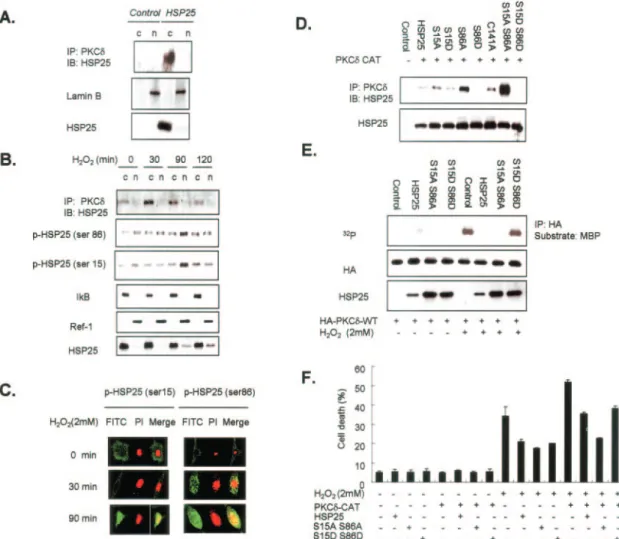
Phosphorylated HSP25 Translates into the Nucleus and Un-phosphorylated HSP25 at Ser-15 and Ser-86 Preferentially Binds to PKC␦—To determine the fate of phosphorylated
HSP25 after its interaction with PKC␦, and because phospho-rylated HSP25 has been reported to be translocated to the nucleus (44), we fractionated cell extracts and immunoprecipi-tated them with anti-PKC␦. HSP25 and PKC␦ interacted in the cytosol (Fig. 6A), and after treating H2O2this interaction in-creased, peaking at 30 min in the cytosol, but no interaction was observed in the nucleus. However, the nuclear transloca-tion of phospho-HSP25 (both Ser-15 and Ser-86) was induced by H2O2 treatment, and a translocation peak was observed after 90 min of H2O2treatment, when the interaction between
HSP25 and PKC␦ began to disappear (Fig. 6B). Confocal image analysis revealed the translocation of phospho-HSP25 to the nucleus after 90 min of H2O2treatment, and this effect was stronger for the Ser-86-phosphorylated form of HSP25 (Fig. 6C). Because interaction between HSP25 and PKC␦ occurred in the cytosol and phospho-HSP25 translocated to the nucleus, we generated phosphorylation-deficient and phosphorylation-mimicking mutants of HSP25 at Ser-15 and Ser-86 to further investigate whether PKC␦ binds to unphosphorylated or phos-phorylated HSP25. When HA-PKC␦-CAT was coexpressed with the His-HSP25 phospho-mutants in L929 cells, phosphoryla-tion-deficient mutants of HSP25 (S15A and S86A) bound to PKC␦-CAT, whereas phosphorylation-mimicking mutants (S15D and S86D) did not (Fig. 6D). Moreover, binding of the
S15A/S86A mutant of HSP25 was greater than those of the S15A or S86A mutants, implying that both sites contribute to binding. Cysteine 141 in the␣-crystalline domain of HSP25, which was reported to affect HSP27 binding to cytochrome c (7), was not found necessary for PKC␦ binding. His pull-down experiments also showed similar results, indicating that only unphosphorylated HSP25 can bind to PKC␦-CAT (data not shown). Moreover, the binding of the phosphorylation-deficient mutant S15A/S86A to PKC␦ inhibited H2O2-induced PKC␦
ac-tivation, whereas the phosphorylation-mimicking mutant (S15D/S86D), which did not bind PKC␦, did not affect H2O2
-induced PKC␦ activation (Fig. 6E), suggesting that unphospho-rylated HSP25 binds kinase-active PKC␦ and that this inter-action inhibits PKC␦ activity. Moreover, the interaction between unphosphorylated HSP25 and PKC␦-CAT inhibited the cell death triggered by H2O2(Fig. 6F), suggesting that the
interaction between unphosphorylated HSP25 (S15A/S86A) and PKC␦-CAT has a cytoprotective effect. HSP25 (S15D/ S86D) also exhibited a cytoprotective effect even though it did not bind to PKC␦-CAT, suggesting the importance of the PKC␦-independent protective activity of HSP25, which has been re-ported previously (44).
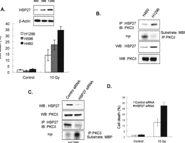
Interaction between HSP25 and PKC␦ Correlates with Ra-dioresistance in Lung Carcinoma Cell Lines—To determine the
physiological relevance of the HSP25 and PKC␦ interaction, three types of lung cancer cells with different HSP27 expres-sions and radioresistances were examined; NCI-H1299 showed highest HSP27 expression, and NCI-H460 least. HSP27 ex-pression levels were found to correlate with radiation survival (Fig. 7A). When the interaction between HSP27 and PKC␦ was checked by coimmunoprecipitating PKC␦ and HSP27, a greater interaction between PKC␦ and HSP27 and lower PKC␦ kinase activity were found for NCI-H1299 than for NCI-H460 (Fig. 7B). When NCI-H1299 cells were treated with siRNA to HSP27, the HSP27-PKC␦ interaction disappeared, and PKC␦ kinase activity was restored (Fig. 7C). Cell death also increased after treating NCI-H1299 with HSP27 siRNA (Fig. 7D),
sug-FIG. 4. Dephosphorylation-dependent binding of the PKC␦ by HSP25. A, immunodetection of HSP25 (in lysates after PKC␦ immuno-precipitation) of transfected L929 cells of HA-fused PKC␦-CAT or PKC␦-CAT-KR vectors containing the wild type and the indicated Ser-643 and Ser-662 point mutants. The transfection efficiencies of HA-tagged vectors were confirmed by Western blotting using anti-HA antibody (upper panel). Transfected L929 cells were incubated with immobilized His-HSP25 vectors. Retained HA protein was detected by Western blotting using an anti-HA antibody. His fusion proteins are also shown in the same gel. The transfection efficiencies of HA-tagged vectors were confirmed by Western blotting using anti-HA antibody. B, soluble and particulate fractions were isolated from control and HSP25-overexpressing L929 cells, and Western blotting was performed for PKC␦ phosphorylation at Ser-643 or Ser-662 using specific antibodies. IP, immunoprecipitation; IB, immunoblot.
at Ewha Medical Library on April 25, 2019
http://www.jbc.org/
gesting that cell death inhibition is correlated with the level of interaction between HSP27 and PKC␦.
DISCUSSION
The coordinated interactions of kinases, phosphatases, and other regulatory molecules with scaffolding proteins is emerg-ing as a major theme in intracellular signalemerg-ing networks (45– 47). Increasing numbers of types of PKC-binding proteins are now believed to play a role in directing the location and func-tion of individual PKC isoforms to particular subcellular loca-tions. In this study, we identified HSP25 as such a PKC ␦-binding protein.
Small heat shock proteins are pleiotropic inhibitors of cell death whose physiological protective effects are observed mainly in stressed cells (11, 48 –50). Several mechanisms have been proposed to account for this anti-apoptotic activity. HSP25 could raise defenses against oxidative stress by increas-ing glutathione content (51) or Mn-SOD enzyme activity (13). HSP27 also binds to activated Akt, a protein that generates a survival signal in response to growth factor stimulation (10, 52). Akt also inhibits cell death by phosphorylating and inac-tivating procaspase-9 (48) or by preventing the release of
cyto-chrome c from mitochondria (6). HSP25 was also found to inhibit cell growth via the inhibition of PKC␦-mediated ERK1/2 activation (12, 14). Moreover, the present study identifies other mechanisms by which HSP25 interferes with cell death path-ways. This cytoprotective activity is potentiated in the follow-ing two ways: by interaction between HSP25 and the V5 region of the catalytic domain of PKC␦, thereby preventing PKC␦-mediated cell death; and by interaction between kinase-active PKC␦ and HSP25, which induces HSP25 phosphorylation at Ser-15 and Ser-86 and potentiates HSP25 cytoprotection.
In a previous study, we found that HSP25 inhibits PKC␦ translocation to the membrane, the kinase activity, and tyro-sine phosphorylation of PKC␦ (14). In the present study, im-munoprecipitation experiments were performed using HSP25-overexpressing L929 and Jurkat T cells, which do not express endogenous HSP25 or HSP27 protein. Most interestingly, HSP25 directly bound PKC␦ in HSP25-overexpressing cells. HSP25/27 shares several properties with HSP70, another in-ducible HSP. When overexpressed, both stress proteins inhibit apoptosis in vitro and in vivo, induce resistance to most che-motherapeutic agents, and enhance tumorigenesis in rodents
FIG. 5. Interaction of PKC␦ with HSP25 induces HSP25 phosphorylation. A, immunoprecipitation (IP) of PKC␦ in lysates from control or HSP25-overexpressing L929 cells after H2O2(2 mM) pretreatment at the indicated time points. HSP25 or PKC␦ protein was detected using
anti-HSP25 or anti-PKC␦ antibodies. Cellular proteins were extracted after lysis with PKC extraction buffer. PKC proteins from 300 g of cell extracts were immunoprecipitated using anti-PKC␦ antibody and protein G-Sepharose. Immune complex kinase reactions were performed in the presence of GST-MBP or His-HSP25 substrate and [␥-32P]ATP. Western blotting was performed using anti-HSP25, anti-phospho-HSP25 (Ser-15),
and anti-phospho-HSP25 (Ser-86) antibodies. B, the indicated amounts of recombinant PKC␦ protein were immunoprecipitated using an anti-PKC␦ antibody and protein G-Sepharose. Immune complex kinase reactions were performed in the presence of His-HSP25 substrate and [␥-32P] with or without PKC lipid activator. C, immunoprecipitation of PKC␦ in lysates from control or HSP25-overexpressing L929 cells treated
with PMA (100 nM) and with or without rottlerin (5M). HSP25 or PKC␦ proteins were detected using anti-HSP25 or PKC␦ antibody. Cellular proteins were extracted after lysis using PKC extraction buffer. PKC proteins from 300g of cell extracts were immunoprecipitated using an anti-PKC␦ antibody and protein G-Sepharose. Immune complex kinase reactions were performed in the presence of His-HSP25 substrate and [␥-32
P]ATP. Western blotting was performed using anti-HSP25, anti-phospho-HSP25 (Ser-15), and anti-phospho-HSP25 (Ser-86) antibodies after immunoprecipitating HSP25 with anti-HSP25 antibody. The light chain band of immunoglobulin G overlapped with the phospho-HSP25 band. D, Western blotting of lysates from PKC␦-CAT, PKC␦-CAT-KR, or PKC␦-REG in HSP25-overexpressing L929 cells was performed using anti-HP25, anti-phospho-HSP25 (Ser-15), or anti-phospho-HSP25 (Ser-86) antibodies after immunoprecipitating HSP25 with anti-HSP25 antibody. The light chain band of immunoglobulin G overlapped the phospho-HSP25 band. Cellular proteins were lysed and extracted using PKC extraction buffer. PKC proteins from 300g of cell extracts were immunoprecipitated using an anti-PKC␦ antibody and protein G-Sepharose. Immune complex kinase reactions were performed in the presence of His-HSP25 substrate and [␥-32P]ATP. E, cellular proteins were lysed and extracted using PKC
extraction buffer from control or HSP25-overexpressing L929 cells after cotransfecting cells with HA-tagged PKC␦-CAT (CAT) or point mutants of PKC␦-CAT on S643D/S662D (DD) or S643A/S662A (AA). PKC proteins from 300 g of cell extracts were immunoprecipitated using an anti-HA antibody and protein G-Sepharose. Immune complex kinase reactions were performed in the presence of GST-MBP or His-HSP25 substrate and [␥-32P]ATP. The transfection efficiency of HA-tagged vectors was confirmed by Western blotting using anti-HA antibody. IB, immunoblot.
HSP25 Binds to PKC
␦
18115
at Ewha Medical Library on April 25, 2019
http://www.jbc.org/
(49). However, several differences between these two chaper-ones have been identified. The first concerns ATP hydrolysis dependence. The second is that HSP70 as an early response gene and HSP25/27 is a late response gene. The third is that different molecular mechanisms are required for their anti-apoptotic effects (48). The present study also suggests that they differ in terms of their interaction with the PKC␦ protein, although it should be noted that HSP70 did not bind PKC␦ (data not shown). The PKC␦-binding site for HSP25 was PKC␦-CAT, and PKC␦ kinase activity was important for the PKC␦-HSP25 interaction, because treatment with the PKC␦ kinase inhibitor rottlerin, PKC␦-CAT-KR, or ATP␥S inhibited the in-teraction between PKC␦ and HSP25. An in vitro translation assay also confirmed the interaction between PKC␦-CAT and HSP25.
Because PKC␦ has been reported to induce cell death with the concomitant activation of PKC␦ or PKC␦ tyrosine phospho-rylation (36, 53, 54) and HSP25 was found to inhibit cell death
(Fig. 2), we concluded that the interaction between kinase-active PKC␦-CAT and HSP25 inhibits PKC␦ activity and PKC␦-mediated cell death. When the binding sites of HSP25 and PKC␦-CAT were investigated using deletion mutants of PKC␦ or HSP25, we found that amino acids 90–103 of HSP25 and the V5 region (amino acids 630 – 674 of PKC␦) of PKC␦ (Figs. 4 and 5) are essential for the interaction between HSP25 and PKC␦. Amino acids 92–145 of HSP25 are overlapping with the cytochrome c-binding site (7) and with the 26 S proteosomal PA700-binding site (8), which suggests that the-sheets of the ␣-crystalline domain of HSP25/27 (indispensable for the HSP25/27 chaperone function in vitro (55)) might also be im-portant for the interaction between HSP25 and PKC␦. More-over, the deletion of amino acids 90 –103 in the HSP25 se-quence inhibited HSP25-mediated cytoprotection, suggesting that binding between HSP25 and PKC␦ is required for the HSP25-mediated cytoprotection (Fig. 3). Because the 90 –103-amino acid region of HSP25 overlaps the cytochrome c-binding
FIG. 6. Unphosphorylated HSP25 preferentially binds to PKC␦ and phosphorylated HSP25 produced by the HSP25-PKC␦ interaction is translocated to the nucleus. A, cytosolic (c) and nuclear (n) fractions of control and HSP25-overexpressing L929 cells were isolated; PKC␦ was immunoprecipitated (IP) in lysates, and HSP25 protein was immunodetected using anti-HSP25 antibody. Nucleus specific protein lamin B was detected by Western blotting. B, cytosolic and nuclear fractions of HSP25-overexpressing L929 cells were isolated; PKC␦ was immunoprecipitated from lysates, and HSP25 protein was immunodetected using anti-HSP25 antibody. Nucleus specific protein Ref-1, and cytosol-specific proteins IB-␣, HSP25 (Ser-15), and HSP25 (Ser-86) were detected by Western blotting. C, localization changes of HSP25 (Ser-15) and phospho-HSP25 (Ser-86) in L929 cells which stably overexpress phospho-HSP25 after treatment of H2O2(2 mM). Cells were fixed with formaldehyde and immunostained
with either anti-phospho-HSP25 (Ser-15) or anti-phospho-HSP25 (Ser-86) antibodies. The results shown are representative of two independent experiments. D, PKC␦ was immunoprecipitated from the lysates of transfected L929 cells of control or vector containing either wild type HSP25 cDNA or the indicated point mutants with or without PKC␦-CAT cotransfection, and HSP25 protein was immunodetected using anti-HSP25 antibodies. E, cellular proteins were obtained using PKC extraction buffer from control or vector containing wild type HSP25 cDNA or the indicated point mutants cotransfected with HA-tagged PKC␦-WT in the presence or absence of H2O2(2 mM). PKC proteins from 300g of cell extracts were immunoprecipitated
using an anti-PKC␦ antibody and protein G-Sepharose. Immune complex kinase reactions were performed in the presence of GST-MBP or His-HSP25 substrate and [␥-32P]ATP. F, the cell deaths of L929 cells cotransfected with vector containing either wild type HSP25 cDNA or the indicated point
mutants and PKC␦-CAT in the presence or absence of H2O2(2 mM) were measured by flow cytometry after PI staining. Results are the means and
standard deviations of three independent experiments. IB, immunoblot.
at Ewha Medical Library on April 25, 2019
http://www.jbc.org/
site (amino acids 51–141 of HSP27) and the PA700-binding site (amino acids 88 –141 of HSP27), this cytoprotective effect rep-resents more than an inhibition of PKC␦-mediated activity. Moreover, it appears reasonable to conclude that the inhibition of PKC␦-mediated cell death by HSP25 interaction with PKC␦ is involved in the inhibition of cell death by HSP25.
Oxidative stress or ionizing radiation permits PKC to trans-locate to the membrane in an open conformation, allowing its pseudosubstrate region to be released from its bind-ing cavity and enablbind-ing C-terminal access to the substrate-binding site and autophosphorylation at Ser-643 and Ser-662 by intramolecular mechanisms, which activates downstream signaling (56). Because HSP70 has been reported to interact with the dephosphorylated turn motif of PKC II, we examined whether PKC␦ interacts with HSP25 in a similar manner. Phosphorylation-deficient mutants but not phosphorylation-mimicking mutants of the V5 region of PKC␦ were found to bind to HSP25. Moreover, HSP25 inhibited PKC␦ translocation to the particulate fraction (Fig. 4), suggesting that the primed activation of PKC␦ permits V5 regions to be exposed, autophos-phorylated, and fully activated. As soon as the V5 region is exposed and before autophosphorylation at 643 and Ser-662, HSP25 may bind to the V5 region of PKC␦ to stabilize PKC␦ and thus inhibit re-phosphorylation and re-activation. Thus, PKC␦ activity and its translocation to the particulate
fraction are inhibited by the PKC␦-HSP25 interaction. HSP25 is regulated by post-translational modifications like phosphorylation, deamination, and acylation (42). Moreover, the phosphorylation of HSP25 is catalyzed by MAPK-activated protein kinase 2/3 (a serine protein kinase), which is phospho-rylated and activated by p38 MAPK or EKR1/2 (44, 57), in a stress-dependent manner. In addition, the inhibition of HSP25 phosphorylation resulted in the destruction of actin filaments and the blocking of the protective effect mediated by HSP25 under heat shock conditions, suggesting that the phosphoryla-tion of HSP25 is important for the stability of actin filament and for thermoresistance (44). In the present study, we found for the first time, that PKC␦ induces HSP25 phosphorylation at Ser-15 and Ser-86 and that the direct interaction between PKC␦ and HSP25 induces HSP25 phosphorylation (Fig. 5). However, it remains to be determined why PKC␦ phosphoryl-ated MBP before HSP25. One possibility is that the phospho-rylation of HSP25 by PKC␦, unlike the phosphorylation of MBP by PKC␦, requires structural changes in the PKC␦-HSP25 com-plex. Indeed, PKC␦ activation may expose the V5 region of PKC␦, and HSP25 then binds directly to this region to induce some conformational change. Another possibility is that PKC␦ has a different substrate binding affinity, and thus the kinetics of the phosphorylations of MBP or HSP25 by PKC␦ may differ. The interaction between PKC␦ and HSP25 usually occurred
FIG. 7. The interaction between HSP25 and PKC␦ correlates with radioresistance. A, the cell deaths of the human lung carcinoma cell lines, NCI-H460, -H596, and -H1299 after 10 Gy of␥-radiation exposure was measured by flow cytometric analysis after PI staining. Results are the means ⫾ S.D. of three independent experiments. Western blotting (WB) was performed using anti-HSP27 antibody. B, HSP27 was immunoprecipitated (IP) in lysates from NCI-H460, and-H1299 with anti-PKC␦ antibodies. Cellular proteins were extracted by lysis using PKC extraction buffer, and PKC proteins were immunoprecipitated from 300g of cell extracts using anti-PKC␦ antibody and protein G-Sepharose. Immune complex kinase reactions were performed in the presence of GST-MBP substrate and [␥-32P]ATP. C, immunoprecipitation of HSP27 from
lysates of NCI-H1299 after transfection with control siRNA or HSP27 siRNA; PKC␦ protein was immunodetected using anti-PKC␦ antibodies. Cellular proteins were obtained by lysing and then extracting with PKC extraction buffer. PKC proteins from 300g of cell extracts were immunoprecipitated using anti-PKC␦ antibody and protein G-Sepharose. Immune complex kinase reactions were performed in the presence of GST-MBP substrate and [␥-32P]ATP. D, NCI-H1299 cell death after transfecting with control siRNA or HSP27 siRNA with or without exposure
to 10 Gy of gamma radiation was measured by flow cytometry after PI staining. Results are the means⫾ S.D. of three independent experiments. IB, immunoblot.
HSP25 Binds to PKC
␦
18117
at Ewha Medical Library on April 25, 2019
http://www.jbc.org/
in cytosol (Fig. 6), and phosphorylated HSP25 was translocated to the nucleus. The results of our kinetic experiments involving H2O2 additions and phosphorylation-deficient mutants of
HSP25 at Ser-15 and Ser-86 suggest that exposed V5 regions of PKC␦ after activation interact with unphosphorylated HSP25 in the cytosol and that after phosphorylation by PKC␦, phos-phorylated HSP25 translocates to the nucleus.
The physiological importance of the correlation between PKC␦-HSP25 binding and radioresistance in lung carcinoma cell lines implies that HSP27 overexpression, which is related to radioresistance (Fig. 7), is in part determined by the HSP25/ 27-PKC␦ interaction. Because HSP27 expression in lung carci-noma cells is well correlated with radioresistance, the V5 re-gion of PKC␦ might be therapeutically useful for inhibiting radioresistance by HSP27.
PKC␦ activity plays an essential role in the apoptosis of cells, and small HSP is constitutively expressed in many cancer cells to negatively regulate apoptotic induction. The small HSP-PKC␦ interaction could thus indicate the physiologic impor-tance of this small HSP (Fig. 8). Moreover, this property might account for the observed protective effect of this protein when induced in response to radiation or oxidative stress.
REFERENCES
1. Wagstaff, M. J., CollacoMoraes, Y., Smith, J., de Belleroche, J. S., Coffin, R. S., and Latchman, D. S. (1999) J. Biol. Chem. 274, 5061–5069
2. Guay, J., Lambert, H., Gingras-Breton, G., Lavoie, J. N., Huot, J., and Landry, J. (1997) J. Cell Sci. 110, 357–368
3. Huot, J., Houle, F., Marceau, F., and Landry, J. (1997) Circ. Res. 80, 383–392 4. Landry, J., and Hout, J. (1999) Biochem. Soc. Symp. 64, 79 – 80
5. Mehlen, P., Coronas, V., Ljubic-Thibal, V., Ducasse, C., Granger, L., Jourdan, F., and Arrigo, A. P. (1999) Cell Death Differ. 6, 227–233
6. Paul, C., Manero, F., Gonin, S., Kretz-Remy, C., Virot, S., and Arrigo, A. P. (2002) Mol. Cell. Biol. 22, 816 – 834
7. Bruey, J. M., Ducasse, C., Bonniaud, P., Ravagnan, L., Susin, S. A., Diaz-Latoud, C., Gurbuxani, S., Arrigo, A. P., Kroemer, G., Solary, E., and Garrido, C. (2000) Nat. Cell Biol. 2, 645– 652
8. Parcellier, A., Schmitt, E., Gurbuxani, S., Seigneurin-Berny, D., Pance, A.,
Chantome, A., Plenchette, S., Khochbin, S., Solary, E., and Garrido, C. (2003) Mol. Cell. Biol. 23, 5790 –5802
9. Charette, S. J., and Landry, J. (2000) Ann. N. Y. Acad. Sci. 926, 126 –131 10. Rane, M. J., Pan, Y., Singh, S., Powell, D. W., Wu, R., Cummins, T., Chen, Q.,
Leish, K. R., and Klein, J. B. (2003) J. Biol. Chem. 278, 27828 –27835 11. Park, S. H., Cho, H. N., Lee, S. J., Kim, T. H., Lee, Y., Park, Y. M., Lee, Y. J.,
Cho, C. K., Yoo, S. Y., and Lee, Y. S. (2000) Radiat. Res. 154, 421– 428 12. Cho, H. N., Lee, S. J., Park, S. H., Lee, Y. J., Cho, C. K., and Lee, Y. S. (2001)
Int. J. Radiat. Biol. 77, 225–233
13. Yi, M. J., Park, S. H., Cho, H. N., Chung, H. Y., Kim, J. I., Cho, C. K., Lee, S. J., and Lee, Y. S. (2002) Radiat. Res. 158, 641– 649
14. Lee, Y. J., Cho, H. N., Jeoung, D. I., Soh, J. W., Cho, C. K., Bae, S., Chung, H. Y., Lee, S. J., and Lee, Y. S. (2004) Free Radic. Biol. Med. 36, 429 – 444 15. Lee, Y. J., Lee, D. H., Cho, C. K., Chung, H. Y., Bae, S., Jhun, G. J., Soh, J. W.,
Jeoung, D. I., Lee, S. J., and Lee, Y. S. (2005) Oncogene, in press 16. Cho, H. N., Lee, Y. J., Cho, C. K., Lee, S. J., and Lee, Y. S. (2002) Cell Death
Differ. 9, 448 – 456
17. Watanabe, T., Ono, Y., Taniyam, Y., Hazama, K., Igarashi, K., Ogita, K., Kikkawa, U., and Nishizuka, Y. (1992) Proc. Natl. Acad. Sci. U. S. A. 89, 10159 –10163
18. Lu, Z., Hornia, A., Jiang, Y. W., Zang, Q., Ohno, S., and Foster, D. A. (1997)
Mol. Cell. Biol. 17, 3418 –3428
19. Cross, T., Griffiths, G., Deacon, E., Sallis, R., Gough, M., Watters, D., and Lord, M. (2000) Oncogene 19, 2331–2337
20. Basu, A., Woolard, M. D., and Johnson, C. L. (2001) Cell Death Differ. 8, 899 –908
21. Denning, M. F., Wang, Y., Tibudan, S., Alkan, S., Nickoloff, B. J., and Qin, J. Z. (2002) Cell Death Differ. 9, 40 –52
22. Yuan, Z., Ishiko, T., Nakada, S., Huang, Y., Kharbanda, S., Weischselbaum, R., and Kufe, D. (1998) Oncogene 16, 1643–1648
23. Reyland, M. E., Anderson, S. M., Matassa, A. A., Barzen, K. A., and Quissell, D. O. (1999) J. Biol. Chem. 274, 19115–19123
24. Godbout, J. P., Pesavento, J., Hartman, M. E., Manson, S. R., and Freund, G. C. (2002) J. Biol. Chem. 277, 2554 –2561
25. Ghayur, T., Hugunin, M., Talanian, R. V., Ratnofsky, S., Quinlan, C., Emoto, Y., Pandey, P., Datta, R., Huang, Y., Kharbanda, S., Allen, H., Kamen, R., Wong, W., and Kufe, D. (1996) J. Exp. Med. 184, 2399 –2404
26. Mizuno, K., Noda, K., Araki, T., Imaoka, T., Kobayashi, Y., Akita, Y., Shi-monaka, M., Kishi, S., and Ohno, S. (1997) Eur. J. Biochem. 250, 7–18 27. Matassa, A. A., Carpenter, L., Biden, T. J., Humphries, M. J., and Reyland,
M. E. (2001) J. Biol. Chem. 276, 29719 –29728
28. Leverrier, S., Vallentin, A., and Joubert, D. (2002) Biochem. J. 368, 905–913 29. Ho, S. N., Hunt, H. D., Horton, R. M., Pullen, J. K., and Pease, L. R. (1989)
Gene (Amst.) 77, 51–59
30. Soh, J. W., Lee, E. H., Prywes, R., and Weinstein, I. B. (1999) Mol. Cell. Biol. 19, 1313–1324
31. Ory, D. S., Neugeboren, B. A., and Mulligan, R. C. (1996) Proc. Natl. Acad. Sci.
FIG. 8. Hypothetical scheme for the role of HSP25 in the inhibition of PKC␦-mediated apoptosis. In response to a stimulus, such as radiation (IR) or oxidative stress, PKC␦ is primed for activation by PDK-1 and tyrosine kinase, and then the V5 region is exposed. Primed PKC␦ becomes fully active by the autophosphorylation of Ser-643 and Ser-662 in the V5 region and by membrane translocation. HSP25 directly binds to the exposed V5 region of PKC␦, which inhibits PKC␦ activity by blocking the autophosphorylation of PKC␦ at Ser-662 and Ser-643 and its subsequent translocation to the membrane fraction, which is correlated with the induction of apoptosis. The interaction between PKC␦ and HSP25 also induces HSP25 phosphorylation at Ser-15 and Ser-86, and this phosphorylation facilitates the dissociation of phosphorylated HSP25 from PKC␦. Phosphorylated HSP25 translocates to the nucleus, although some remains in the cytoplasm. When HSP25 is overexpressed, apoptotic induction by radiation or oxidative stress is inhibited because PKC␦ remains bound to HSP25. at Ewha Medical Library on April 25, 2019
http://www.jbc.org/
U. S. A. 93, 11400 –11406
32. Gao, T., and Newton, A. C. (2002) J. Biol. Chem. 277, 31585–31592 33. Shimabukuro, K., Yasuda, R., Muneyuki, E., Hara, K. Y., Kinosita, K., Jr., and
Yoshida, M. (2003) Proc. Natl. Acad. Sci. U. S. A. 100, 14731–14736 34. Lee, Y. J., Soh, J. W., Dean, N. M., Cho, C. K., Kim, T. H., Lee, S. J., and Lee,
Y. S. (2002) Cell Growth & Differ. 13, 237–246
35. Li, W., Zhang, J., Bottaro, D. P., and Pierce, J. H. (1997) J. Biol. Chem. 272, 24550 –24555
36. Blass, M., Kronfeld, I., Kazimirsky, G., Blumberg, P. M., and Brodie, C. (2002)
Mol. Cell. Biol. 22, 182–195
37. Li, W., Mischak, H., Yu, J. C., Wang, L. M., Mushinski, J. F., Heidaran, M. A., and Pierce, J. H. (1994) J. Biol. Chem. 269, 2349 –2352
38. Stempka, L., Schnolzer, M., Radke, S., Rincke, G., Marks, F., and Gschwendt, M. (1999) J. Biol. Chem. 274, 8886 – 8892
39. Parekh, D., Ziegler, W., Yonezawa, K., Hara, K., and Parker, P. J. (1999)
J. Biol. Chem. 274, 34758 –34764
40. Dutil, E. M., Keranen, L. M., DePaoli-Roach, A. A., and Newton, A. C. (1994)
J. Biol. Chem. 269, 29359 –29362
41. Wang, Q. J., Lu, G., Schlapkohl, W. A., Goerke, A., Larsson, C., Mischak, H., Blumberg, P. M., and Mushinski, J. F. (2004) Mol. Cancer Res. 2, 129 –140 42. Welsh, M. J., and Gaestel, M. (1998) Ann. N. Y. Acad. Sci. 851, 28 –35 43. Knauf, U., Jakob, U., Engel, K., Buchner, J., and Gaestel, M. (1994) EMBO J.
13, 54 – 60
44. Geum, D., Son, G. H., and Kim, K. (2002) J. Biol. Chem. 277, 19913–19921
45. Dutil, E. M., Toker, A., and Newton, A. C. (1998) Curr. Biol. 6, 806 – 809 46. Pawson, T., and Scott, J. D. (1997) Science 278, 2075–2080
47. Jaken, S., and Parker, P. J. (2000) BioEssays 22, 245–254
48. Garrido, C., Bruey, J. M., Fromentin, A., Hammann, A., Arrigo, A. P., and Solary, E. (1999) FASEB J. 13, 2061–2070
49. Garrido, C., Fromentin, A., Bonnotte, B., Favre, N., Moutet, M., Arrigo, A. P., Mehlen, P., and Solary, E. (1998) Cancer Res. 58, 5495–5499
50. Garrido, C., Ottavi, P., Fromentin, A., Hammann, A., Arrigo, A. P., Chauffert, B., and Mehlen, P. (1997) Cancer Res. 57, 2661–2667
51. Baek, S. H., Min, J. N., Park, E. M., Han, M. Y., Lee, Y. S., Lee, Y. J., and Park, Y. M. (2002) J. Cell. Physiol. 183, 100 –107
52. Konishi, H., Matsuzaki, H., Tanaka, M., Takemura, Y., Kuroda, S., Ono, Y., and Kikkawa, U. (1997) FEBS Lett. 410, 493– 498
53. Fujii, T., Garcia-Bermejo, M. L., Bernabo, J. L., Caamano, J., Ohba, M., Kuroki, T., Li, L., Yuspa, S. H., and Kazanietz, M. G. (2000) J. Biol. Chem. 275, 7574 –7582
54. Pongracz, J., Webb, P., Wang, K., Deacon, E., Lunn, O. J., and Lord, J. M. (1999) J. Biol. Chem. 274, 37329 –37334
55. Guo, Z., and Cooper, L. F. (2000) Biochem. Biophys. Res. Commun. 270, 183–189
56. Newton, A. C. (2003) Biochem. J. 370, 361–371
57. Maizels, E. T., Peters, C. A., Kline, M., Cutler, R. E., Shanmugam, M., and Hunzicker-Dunn, M. (1998) Biochem. J. 332, 703–712
HSP25 Binds to PKC
␦
18119
at Ewha Medical Library on April 25, 2019
http://www.jbc.org/
Jae-Won Soh and Yun-Sil Lee
Yoon-Jin Lee, Dae-Hoon Lee, Chul-Koo Cho, Sangwoo Bae, Gil-Ja Jhon, Su-Jae Lee,
-mediated Cell Death through Direct Interaction
δ
HSP25 Inhibits Protein Kinase C
doi: 10.1074/jbc.M501131200 originally published online February 24, 2005
2005, 280:18108-18119.
J. Biol. Chem.
10.1074/jbc.M501131200
Access the most updated version of this article at doi:
Alerts:
When a correction for this article is posted
•
When this article is cited
•
to choose from all of JBC's e-mail alerts
Click here
http://www.jbc.org/content/280/18/18108.full.html#ref-list-1
This article cites 56 references, 31 of which can be accessed free at
at Ewha Medical Library on April 25, 2019
http://www.jbc.org/