Impact of simulation-based
anesthesiology training using
an anesthetized porcine
model for ultrasound-guided
transversus abdominis plane
block
Sang Jun Park
1,2, Hyun Joo Kim
1,2,
Hun-Mu Yang
3, Kyung Bong Yoon
1,2,
Ki-Young Lee
1,2, Taehoon Ha
1, Hun Jang
1and Shin Hyung Kim
1,2Abstract
Objective: This study was performed to assess the impact of simulation-based training for ultrasound-guided transversus abdominis plane (TAP) block using anesthetized pigs.
Methods: In this prospective study, 23 participating residents (10 in their second year, 13 in their third year) underwent simulation-based training for ultrasound-guided TAP block. The residents completed standard questionnaires comprising 10 multiple-choice questions regarding essential general knowledge of abdominal ultrasound and TAP block before and after the training session. On a 5-point Likert scale, they reported their levels of comfort with the use of ultrasound and block equipment, subject/operator positioning, proper block technique, image documentation, needle handling, anxiety, and their overall confidence with the procedure.
Results: Compared with those before training, the comfort levels of the residents significantly improved for all measures except needle handling. The participants also indicated significantly reduced anxiety regarding performance of the TAP block technique.
Conclusion: The use of anesthetized pigs in simulation-based training for ultrasound-guided TAP block improves procedural knowledge and confidence while reducing the associated anxiety in anesthesiology trainees.
1
Department of Anesthesiology and Pain Medicine, Yonsei University College of Medicine, Seoul, Korea
2
Anesthesia and Pain Research Institute, Yonsei University College of Medicine, Seoul, Korea
3
Department of Anatomy, Yonsei University College of Medicine, Seoul, Korea
Corresponding author:
Shin Hyung Kim, Department of Anesthesiology and Pain Medicine, Anesthesia and Pain Research Institute, Yonsei University College of Medicine, 50 Yonsei-ro, Seodaemun-gu, Seoul 03722, Korea.
Email: [email protected]
Journal of International Medical Research 48(3) 1–7 ! The Author(s) 2019 Article reuse guidelines: sagepub.com/journals-permissions DOI: 10.1177/0300060519896909 journals.sagepub.com/home/imr
Creative Commons Non Commercial CC BY-NC: This article is distributed under the terms of the Creative Commons Attribution-NonCommercial 4.0 License (https://creativecommons.org/licenses/by-nc/4.0/) which permits non-commercial use, reproduction and distribution of the work without further permission provided the original work is attributed as specified on the SAGE and Open Access pages (https://us.sagepub.com/en-us/nam/open-access-at-sage).
Keywords
Abdominal ultrasound, anesthesiology residents, porcine, questionnaire, simulation-based train-ing, transversus abdominis plane block
Date received: 9 June 2019; accepted: 4 December 2019
Introduction
Abdominal interfascial plane block has become more common than previously with the introduction of simple and effective techniques, such as the transversus abdomi-nis plane (TAP) block, and the increased use of ultrasound,1–3which enables direct visu-alization of the abdominal wall layers, needle placement, and dispersion of local anesthetics. However, block procedures can be risky and result in ineffective analgesia for patients when performed by inadequately trained anesthesiologists.4
The traditional “see one, do one, teach one” training model for health professio-nals is expensive and time-consuming and produces inconsistent results for complex procedures.5Alternatively, hands-on simu-lation-based training enables trainees to gain experience in a safe and controlled environment. Practicing regional anesthesia via simulation training may improve needle handling and facilitate the localization of targets.6–8 Simulation training can entail virtual reality, bench model simulators, human cadavers, and animal carcasses.8
The ultrasound appearance of the abdominal muscles and visceral organs of pigs is almost identical to that of humans, and the respiration-induced positional changes of organs and muscles are also sim-ilar. Therefore, we hypothesized that an anesthetized porcine model is ideal for simulation-based training for the TAP block. In the present study, we investigated the experiences of anesthesiology residents in simulation-based ultrasound-guided TAP block training with anesthetized pigs.
Methods
Study participants
This study was approved by the institutional review board of Severance Hospital, Korea (no. 4-2018-0208) and registered at Clinical Trials.gov (NCT 03516058). Ten second-year and 13 third-year anesthesiology residents (n¼ 23) without experience in ultrasound-guided regional block were recruited from a single academic institution to participate in a TAP block training session from June to July 2018. Participation in the study was optional, and informed consent was obtained from each trainee. This study was approved by our institutional review board.
Animal preparation
Three swine weighing 20 to 30 kg were anes-thetized via inhalation of isoflurane (5%) in oxygen at 6 L/min with a standard animal mask. After intubation, anesthesia was main-tained with 2% isoflurane and oxygen (2 L/ min) via mechanical ventilation. The ventilator was adjusted to maintain the end-tidal carbon dioxide at 35 to 45 mmHg throughout the study. All procedures were approved by and performed in accordance with the guidelines of the local Institutional Animal Care and Use Committee and Animal and Plant Quarantine Agency of the Korean government.
Ultrasound-guided TAP block procedure
and training
The 1-hour-long simulation-based TAP block training session in anesthetized pigs
was led by a single fellowship-trained pain specialist (Figure 1). After a brief didactic session and demonstration, which covered needle safety, patient positioning for the block, and proper TAP block technique, the participating residents familiarized themselves with the functions of the
ultrasound machine and practiced the TAP block on the swine placed in a lateral or prone position. The ultrasound probe (high-frequency linear probe, 5–10 MHz) was placed transverse to the lateral abdom-inal wall between the lower costal margin and iliac crest, revealing the skin (from skin to peritoneum), subcutaneous tissue, fat, external and internal obliques, and transversus abdominis. The deeper peritone-um and bowel loops were also often visual-ized. Needles were introduced directly under the ultrasound probe at the same plane and advanced until they reached the plane between the internal oblique and transversus abdominis muscles. Next, 2 mL of saline was injected to confirm the correct needle posi-tion (hydrolocaposi-tion), and 10 mL of saline was then injected to visualize the expansion of the TAP (Figure 2). The residents per-formed at least three blocks and were allowed to ask questions during the training session. Feedback was provided throughout the session by the anesthesiology staff.
Procedure questionnaire
Each resident’s training level was recorded, and they were given questionnaires to
Figure 1. Ultrasound-guided transversus abdomi-nis block simulation training in the anesthetized porcine model
Figure 2. Ultrasound images demonstrating needle placement and injectate spread of TAP block. (a) Lateral and (b) posterior approaches in an anesthetized porcine model. EO, external oblique muscle; IO, internal oblique muscle; TA, transversus abdominis muscle; TAP, transversus abdominis plane; P, peritoneum
anonymously assess their overall knowledge and confidence of the procedure before and after the training session. The participants rated each of the following questions according to a 5-point Likert scale (1¼ very low, 2¼ low, 3 ¼ moderate, 4 ¼ high, and 5¼ very high):
1. I know the anatomical basis, clinical indications, and procedure of the TAP block (basic knowledge).
2. I can handle the ultrasound machine, including selection of the probe type and control of image depth, gain, and focus (ultrasound machine functionality). 3. I know the correct positions of the sub-ject and operator to facilitate the proce-dure (subject/operator positioning). 4. I can prepare the devices and materials
for TAP block (block equipment preparation).
5. I can distinguish the three muscle layers of the abdominal wall and peritoneum in an ultrasound image (image documentation).
6. I can manipulate the needle safely and skillfully during TAP block (needle manipulation).
7. I know the estimated trajectory of the needle and the optimal needle tip posi-tion for successful TAP block (optimal needle tip position).
8. I can confirm the correct injectate spread within the TAP (hydrolocation) in an ultrasound image (confirmation of injectate spread).
9. I am confident in my ability to perform TAP block for patients in the future (overall confidence level).
10. I am worried about performing TAP block (overall anxiety level).
Statistical analysis
The mean scores from the questionnaires were compared before and after the training session via the Wilcoxon signed rank test.
McNemar’s test was used to compare the scores for each question. SPSS software, version 23.0 (IBM Corp., Armonk, NY, USA), was used for all analyses. Results with a P value of <0.05 were considered statistically significant.
Results
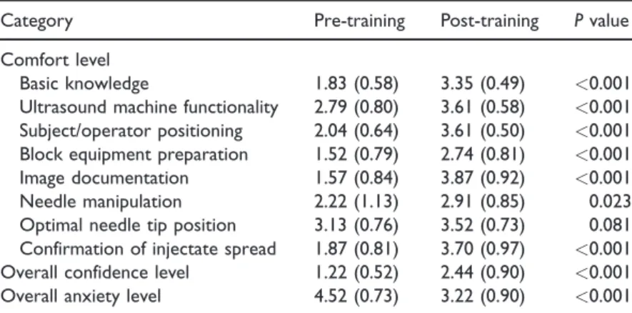
Compared with the scores obtained before the training session, those reported after the training were significantly higher (P< 0.05) for all questions concerning the level of comfort with the procedure except for that pertaining to the optimal needle tip position, for which the mean Likert score indicated moderate knowledge of the appro-priate trajectory before training (Table 1). Before the training session, most residents (22/23 [96%]) cited low to very low confi-dence in their ability (scores of 1 or 2 on the Likert scale) and high to very high anx-iety (scores of 4 or 5) with regard to per-forming ultrasound-guided TAP block.
Unfamiliarity with nerve blocks because of lack of practice (21 of 23 residents) and lack of foundational knowledge (15 of 23 resi-dents) were the two main factors that contrib-uted to trainee anxiety in performing ultrasound-guided TAP block. Moreover, 22 of the 23 residents (96%) chose simulation-based training over apprenticeship methods such as demonstration videos, lectures, and observing faculty members to learn procedur-al skills. In addition, 20 of the 23 residents (87%) marked the educational value of the nerve block training session as high or very high (score of 4 or 5, respectively), and 100% added that a simulation-based training ses-sion on performing TAP block should be included in the residency curriculum.
Discussion
The results of this study indicate that simulation-based training for ultrasound-guided TAP block using an anesthetized
porcine model reduced anesthesiology trainees’ anxiety and improved their knowl-edge and confidence in performing the pro-cedure. The three basic requirements for ultrasound-guided regional block training are (i) pattern recognition, (ii) probe han-dling and scanning, and (iii) manual dexter-ity to align the needle with the ultrasound beam.9 The latter two requirements are challenging for residents with no experience in regional block using ultrasound.10 Correct identification of the needle tip on ultrasound during regional block, particu-larly as the tip proceeds to abdominal muscle layers without a bony landmark, is important for patient safety and determines block success.
For effective analgesia, the needle should be placed between the internal oblique and transversus abdominis muscles. TAP blocks were historically performed in a blind manner, but current clinical trends show a reliance on ultrasound to check the needle tip and thus prevent possible damage to internal organs.4Although needle handling and optimal positioning may still be diffi-cult after a single training session, residents gain realistic practice and confidence with this hydrodissection technique. To strength-en resident training, simulation-based
training programs should be further incor-porated for both skill retention and famil-iarity for trainees.
Water, gelatin, meat, cadavers, and animal carcasses have been used for ultra-sound training.11–13 However, water and gelatin are more suitable for spine models, and fascial plane blocks are difficult to reproduce with these materials. A meat phantom provides tactile feedback from the needle, with echogenicity resembling that of human tissue.14 Cadaver phantoms are similar in this regard. However, these materials are expensive and not readily available. Additionally, these models are fixed and do not incorporate movement, such as that of the abdominal wall caused by respiration of the patient, which can interfere with the TAP block. For this reason and because of their similar anato-my, live pigs are superior for practicing the TAP block. Indeed, most (87%) of the res-idents surveyed for this study reported that the experience was closer to that with real patients.
In our institution, the anesthetized por-cine model has been incorporated for educat-ing residents about other regional anesthesia techniques and anatomy. Notably, this model provides vivid sonoanatomies of the
Table 1. Likert scale scores for trainee confidence before and after simulation-based training
Category Pre-training Post-training P value
Comfort level
Basic knowledge 1.83 (0.58) 3.35 (0.49) <0.001
Ultrasound machine functionality 2.79 (0.80) 3.61 (0.58) <0.001
Subject/operator positioning 2.04 (0.64) 3.61 (0.50) <0.001
Block equipment preparation 1.52 (0.79) 2.74 (0.81) <0.001
Image documentation 1.57 (0.84) 3.87 (0.92) <0.001
Needle manipulation 2.22 (1.13) 2.91 (0.85) 0.023
Optimal needle tip position 3.13 (0.76) 3.52 (0.73) 0.081
Confirmation of injectate spread 1.87 (0.81) 3.70 (0.97) <0.001
Overall confidence level 1.22 (0.52) 2.44 (0.90) <0.001
Overall anxiety level 4.52 (0.73) 3.22 (0.90) <0.001
ribs, intercostal muscle layers, and most importantly, the sliding pleura sign, all of which are helpful for performing intercostal nerve blocks. Because the sonoanatomy of the porcine lumbar spine is similar to that of humans, this model can be used to prac-tice block techniques, including facet joint injection, medial branch block, and psoas compartment block. Blocks in the thoracic region, such as paravertebral blocks, have a risk of pneumothorax, and a living porcine model with breathing patterns can provide an opportunity to practice avoiding such complications. Nevertheless, the use of live pigs for nerve blocks involves general anes-thesia requiring veterinarian assistance and equipment. In addition, training can only occur while the pigs are anesthetized, which limits training time and space. Regardless of the extra effort, a live porcine model provides a clinical and anatomical environment simi-lar to that of real patients as a desirable option for training in various ultrasound-guided regional block techniques.
This study has some limitations. First, we surveyed a relatively small sample of residents from a single institution. Second, the residents had no opportunity to per-form the TAP block on actual patients; thus, we could not directly assess the out-come of the nerve block training. Third, the efficacy of the training performed in this study was not compared with that of other simulation-based training models. Finally, no objective parameters were eval-uated, such as inter-evaluator reliability, internal consistency, convergent validity, or error reduction. Although a typical process validation of such a technical simulation-based scenario should include data derived from both subjective and objective parameters, we only addressed subjective parameters in this study.
In conclusion, the results of the present study suggest that simulation-based ultra-sound-guided TAP block training using an anesthetized porcine model positively
impacts ultrasound knowledge and block technique education. After the training ses-sion, residents expressed higher levels of comfort and confidence with the technical aspects of the TAP block as well as reduced anxiety regarding performance of the procedure. Our findings are in line with those of prior studies that demonstrated improved confidence levels in performing ultrasound-guided regional blocks after simulation-based training.
Future direction
Practicing on actual patients will become more difficult over time with patients’ increasing concerns regarding medicolegal issues. Anesthetized pigs represent an appropriate model for training in various interfascial plane blocks, such as the erector spinae plane block, quadratus lumborum block, serratus plane block, and many others, and should be considered an effec-tive tool in regional anesthesia education. Anesthetized pigs are also expected to be used for training in advanced pain interven-tions such as introduction of spinal cord stimulation or intrathecal drug delivery sys-tems for pain specialist.
Author contributions
KYL and SHK designed the study. All authors contributed to the literature search. TH, HJ, and SHK collected the data. SJP, H-MY, KBY, HJK, KYL, HJ, and SHK analyzed the data. SJP, H-MY, KBY, KYL, TH, HJ, and SHK prepared the manuscript. SJP, H-MY, KBY, HJK, KYL, TH, and SHK reviewed the manuscript.
Availability of data and materials
The datasets generated and analyzed during the present study are available from the correspond-ing author on reasonable request.
Consent for publication
Declaration of conflicting interest
The authors declare that there is no conflict of interest.
Ethics approval and consent to participate
This study was approved by the institutional review board of Severance Hospital, Korea
(no. 4-2018-0208). Each patient read and
signed a consent form before enrolling in the study. All procedures using swine were approved by the local Institutional Animal Care and Use Committee (no. MIC-IACUC 201801) and Animal and Plant Quarantine Agency of the Korean government.
Funding
This work was supported by a National Research Foundation (NRF) of Korea grant funded by the Korean government (MSIT) (grant no. 2017R1C1B5074007) and a faculty research grant of Yonsei University College of Medicine (6-2018-0088).
ORCID iDs
Sang Jun Park
https://orcid.org/0000-0002-2496-7764
Hyun Joo Kim
https://orcid.org/0000-0003-1963-8955
Taehoon Ha
https://orcid.org/0000-0001-9035-8003
References
1. Rafi AN. Abdominal field block: a new
approach via the lumbar triangle.
Anaesthesia2001; 56: 1024–1026.
2. Hebbard P. Subcostal transversus abdomi-nis plane block under ultrasound guidance.
Anesth Analg 2008; 106: 674–675; author
reply 5.
3. Hebbard P, Fujiwara Y, Shibata Y, et al. Ultrasound-guided transversus abdominis plane (TAP) block. Anaesth Intensive Care 2007; 35: 616–617.
4. Farooq M and Carey M. A case of liver trauma with a blunt regional anesthesia needle while performing transversus abdom-inis plane block. Reg Anesth Pain Med 2008; 33: 274–275.
5. Long DM. Competency-based residency training: the next advance in graduate medical education. Acad Med 2000; 75: 1178–1183.
6. Sites BD, Gallagher JD, Cravero J, et al. The learning curve associated with a simu-lated ultrasound-guided interventional task by inexperienced anesthesia residents. Reg
Anesth Pain Med2004; 29: 544–548.
7. Niazi AU, Haldipur N, Prasad AG, et al. Ultrasound-guided regional anesthesia per-formance in the early learning period: effect of simulation training. Reg Anesth Pain Med2012; 37: 51–54.
8. Chen XX, Trivedi V, AlSaflan AA, et al. Ultrasound-guided regional anesthesia sim-ulation training: a systematic review. Reg
Anesth Pain Med2017; 42: 741–750.
9. Awad IT and Chan V. Ultrasound imaging of peripheral nerves: a need for a new trend.
Reg Anesth Pain Med2005; 30: 321–323.
10. Sites BD, Spence BC, Gallagher JD, et al. Characterizing novice behavior associated with learning ultrasound-guided peripheral regional anesthesia. Reg Anesth Pain Med 2007; 32: 107–115.
11. Bellingham GA and Peng PW. A low-cost ultrasound phantom of the lumbosacral spine. Reg Anesth Pain Med 2010; 35: 290–293.
12. Hocking G, Hebard S and Mitchell CH. A review of the benefits and pitfalls of phan-toms in ultrasound-guided regional anesthe-sia. Reg Anesth Pain Med 2011; 36: 162–170. 13. Kim YH. Ultrasound phantoms to protect patients from novices. Korean J Pain 2016; 29: 73–77.
14. Xu D, Abbas S and Chan VW. Ultrasound phantom for hands-on practice. Reg Anesth