저작자표시-비영리-변경금지 2.0 대한민국 이용자는 아래의 조건을 따르는 경우에 한하여 자유롭게 l 이 저작물을 복제, 배포, 전송, 전시, 공연 및 방송할 수 있습니다. 다음과 같은 조건을 따라야 합니다: l 귀하는, 이 저작물의 재이용이나 배포의 경우, 이 저작물에 적용된 이용허락조건 을 명확하게 나타내어야 합니다. l 저작권자로부터 별도의 허가를 받으면 이러한 조건들은 적용되지 않습니다. 저작권법에 따른 이용자의 권리는 위의 내용에 의하여 영향을 받지 않습니다. 이것은 이용허락규약(Legal Code)을 이해하기 쉽게 요약한 것입니다. Disclaimer 저작자표시. 귀하는 원저작자를 표시하여야 합니다. 비영리. 귀하는 이 저작물을 영리 목적으로 이용할 수 없습니다. 변경금지. 귀하는 이 저작물을 개작, 변형 또는 가공할 수 없습니다.
Investigation of the optimal
combination of external beam
radiotherapy and high-dose-rate
intracavitary brachytherapy in
definitive radiotherapy for uterine
cervical cancer patients
Kyung Hwan Kim
Department of Medicine
Investigation of the optimal
combination of external beam
radiotherapy and high-dose-rate
intracavitary brachytherapy in
definitive radiotherapy for uterine
cervical cancer patients
Directed by Professor Yong Bae Kim
The Master's Thesis
submitted to the Department of Medicine,
the Graduate School of Yonsei University
in partial fulfillment of the requirements for the degree
of Master of Medical Science
Kyung Hwan Kim
This certifies that the Master's Thesis
of Kyung Hwan Kim is approved.
---Thesis Supervisor : Yong Bae Kim
---Thesis Committee Member#1 : Sunghoon Kim
---Thesis Committee Member#2 : Woong Sub Koom
The Graduate School
Yonsei University
ACKNOWLEDGEMENTS
First of all, I would like to thank my supervisor,
Professor Yong Bae Kim, for giving me such inspiring advice
and support for preparing this thesis and setting a good
example for me by devoting himself to patients suffering from
cancer. Also I would like to express my gratefulness to
professor Sunghoon Kim and Woong Sub Koom for giving me
a great amount of advice essential for completing this article. I
would like to appreciate to professor Gwi Eon Kim, Chang-Ok
Suh, Jinsil Seong, Chang Geol Lee, Ki Chang Keum, Jaeho
Cho, Ik Jae Lee, and Jun Won Kim, for always offering the
great instructions with careful concern.
<TABLE OF CONTENTS>
ABSTRACT ···1
I. INTRODUCTION ···4
II. MATERIALS AND METHODS···7
1. Patients ···7
2. Radiotherapy···7
3. Chemotherapy ···10
4. Follow-up and toxicity evaluation···10
5. Statistical analysis···11
III. RESULTS ···13
1. Patient and treatment characteristics ···13
2. Late toxicity rate with or without midline block···15
3. Association between the cumulative ICRU point dose (EQD2) and
late toxicity rate···17
4. Treatment outcome according to midline block ···20
IV. DISCUSSION ···23
V. CONCLUSION ···29
REFERENCES ···30
LIST OF FIGURES
Figure 1. The grade ≥2 late complication rates of rectum,
genitourinary (GU), and small bowel ··· 16
Figure 2. Probability of grade ≥2 late rectal toxicity according to
biologically equivalent dose in 2-Gy fractions (EQD2) of rectal
point (A) and probability of grade ≥2 late bladder toxicity
according to EQD2 of bladder point (B) ··· 19
Figure 3. Overall survival (A), progression-free survival (B),
local recurrence (C), and regional recurrence (D) rates of
Midline block (MLB) group and non-MLB group ··· 21
LIST OF TABLES
Table 1. Patient and Treatment Characteristics··· 13
Table 2. Late Toxicity Rates in the Midline Block (MLB) Group
and non-MLB Group ··· 15
Table 3. Patient and Treatment Characteristics after Propensity
ABSTRACT
Investigation of the optimal combination of external beam radiotherapy
and high-dose-rate intracavitary brachytherapy in definitive radiotherapy
for uterine cervical cancer patients
Kyung Hwan Kim
Department of Medicine
The Graduate School, Yonsei University
(Directed by Professor Yong Bae Kim)
Purpose: Intracavitary brachytherapy (ICBT) and external beam radiotherapy (EBRT) are both essential components of definitive radiotherapy for patients with uterine cervical cancer. From previous phase II trials, reduced cumulative central dose using midline block (MLB) did not compromise the treatment outcome while reducing late toxicity rate compared to other studies. However, no randomized evidence is available and long term results are needed to confirm the efficacy and safety of this treatment approach. We aimed to assess the efficacy and toxicity of low cumulative central dose using MLB during EBRT.
Patients and Methods: Between January 1988 and December 2010, a total of 1559 patients with uterine cervical cancer (FIGO stage IB 410, stage IIA 133, stage IIB 1016) who received definitive radiotherapy (n = 1054, 67.6%) or platinum-based
chemoradiotherapy (n = 504, 32.4%) consisting of EBRT and high-dose-rate intracavitary brachytherapy (HDR-ICBT) were retrospectively analyzed. The median EBRT dose was 45.0 Gy (range, 30.6‒60.0 Gy) in 1.8 Gy per fraction and median HDR-ICBT dose prescribed at point A was 30 Gy (range, 12‒63 Gy) in median 5 Gy (3.0‒6.0 Gy) per fraction. During EBRT, tumor response was checked every week and when sufficient response was achieved to place the ICBT applicator, MLB of 4 cm width and 8‒10 cm in height was placed (n = 1195, MLB group). For patients with slow tumor response during EBRT, full dose was applied without MLB (n = 364, non-MLB group). non-MLB was performed after ≤ 27 Gy (n = 229), > 27 Gy and ≤ 36 Gy (n = 847), or > 36 Gy (n = 119) of EBRT. The rectal and bladder doses were estimated using doses at the International Commission on Radiation Units and Measurements points. To calculate the cumulative dose from EBRT and ICBT, the biologically equivalent dose in 2-Gy fractions (EQD2) using the linear quadratic model was used (α/β value of 3 for normal tissue and 10 for tumor). Propensity score matching was also performed to balance the characteristics between MLB and non-MLB group. Results: Median follow-up period was 89.0 months (range, 2.4-320.2 months). The 10-year overall survival (OS), progression-free survival (PFS), regional recurrence (RR), and local recurrence (LR) rates were 82.3%, 74.7%, 2.6%, and 9.5%, respectively. The 10-year OS, PFS, RR, and LR (all Ps <0.05) were significantly superior in the MLB group compared to the non-MLB group. The MLB group was older in age, had smaller tumor size, lower FIGO stage, higher pelvic and para-aortic lymph node metastases rate than the non-MLB group. EQD2point A(72.9 Gy vs. 86.4
Gy), EQD2rectal(64.5 Gy vs. 74.8 Gy), and EQD2bladder(67.4 Gy vs. 75.9 Gy) were all
significantly lower in the MLB group (all Ps <0.05). Grade ≥2 late rectal toxicity was significantly lower in MLB group (8.1% vs. 11.5%, P = 0.045). There was no significant difference in late genitourinary and small bowel toxicity. After all patient and tumor characteristics were well balanced using propensity score matching, the 10-year OS, PFS, RR, and LR were similar between the MLB and non-MLB group (all Ps >0.05) despite the lower EQD2point A(72.9 Gy vs. 86.4 Gy; P <0.001).
Conclusion: Lowering the cumulative central dose using EBRT with MLB according to tumor response may reduce rectal toxicity without compromising treatment outcome. The efficacy of MLB should further be evaluated in prospective trials.
---Key words: uterine cervical cancer, midline block, radiotherapy, high-dose-rate, intracavitary brachytherapy
Investigation of the optimal combination of external beam radiotherapy
and high-dose-rate intracavitary brachytherapy in definitive radiotherapy
for uterine cervical cancer patients
Kyung Hwan Kim
Department of Medicine
The Graduate School, Yonsei University
(Directed by Professor Yong Bae Kim)
I. INTRODUCTION
Uterine cervical cancer is the seventh most common cancer worldwide and the tenth leading cause of cancer mortality.1 However, it is the second most common
and third leading cause of cancer mortality in developing countries.2 Definitive
radiotherapy (RT) and concurrent chemoradiotherapy (CRT) has been the standard treatments for early stage and locally advanced uterine cervical cancer.3,4Standard RT
for uterine cervical cancer consists of external beam RT (EBRT) and intracavitary brachytherapy (ICBT). Recently high-dose-rate ICBT (HDR-ICBT) is taking over the place of low-dose-rate ICBT5-7 and the dose schedules of EBRT and HDR-ICBT
is yet to be established.
ICBT plays a major role in definitive treatment of patients with uterine cervical cancer. The impact of ICBT on survival was reported from two population based studies, which showed significantly higher overall survival in patients who received ICBT compared to those who did not.9,10 One of the concerns in utilizing
HDR-ICBT would be its late toxicity. The late toxicity rate of rectum and bladder would increases in correlation to the increasing radiation dose delivered to both organs. From a previous report from our institution, keeping the biological effective dose of point A not higher than 90 Gy was associated with lesser late rectal and bladder complications.11More recent studies showed that the dose volume parameters
of the rectum and bladder were associated with the rate of late toxicity.12,13Therefore
to enhance the therapeutic ratio, the establishment of an optimal dose schedule of HDR-ICBT and EBRT is necessary.
In an attempt of reducing the central dose, using the midline block (MLB) during EBRT was tested in a prospective multi-institutional study from Japan delivering biologically equivalent dose in 2-Gy fractions (EQD2) of 52‒65 Gy to point A.14,15They reported a comparable outcome and lower incidence of late toxicity
compared to other studies that used higher point A doses. However, the median follow-up period was relatively short in both studies. In our institution, we have adopted MLB during EBRT since 1988 for the last three decades. The initial results were reported previously and showed that the results were comparable to other reports that used global dose schedules not using MLB.16 Here we present the long-term
follow-up data on treatment outcome and toxicity of using MLB during EBRT in conjunction with HDR-ICBT.
II. MATERIALS AND METHODS
1. Patients
The study included patients with histologically confirmed FIGO stage IB‒ IIB uterine cervical carcinoma treated with definitive RT or CRT using EBRT and HDR-ICBT between January 1988 and December 2010. Sixty-five patients were excluded due to double primary cancer (n = 15), small cell histology (n = 5), ICBT refusal (n = 13), previous hysterectomy (n = 2), and distant metastases (n = 30). Eventually, data of 1559 patients were retrospectively analyzed.
The staging evaluation routinely included physical examination, complete blood count, liver function test, sigmoidoscopy, cystoscopy, intravenous pyelogram, and chest radiographs. Either computed tomograghy (CT) scans (n = 1234) or magnetic resonance imaging (MRI) (n = 409) were performed to evaluate lymph node involvement for all patients. Lymph nodes larger than 1 cm in the short-axis dimension or with central necrosis were considered metastatic. Since 2004, 18F-fluorodeoxyglucose positron emission tomography (FDG-PET) was commonly performed and 158 patients underwent FDG-PET.
2. Radiotherapy
The treatment protocol at Yonsei Cancer Center has been described previously.16-18 RT consisted of a combination of EBRT and HDR-ICBT. EBRT
included the whole pelvis and was delivered by a 10-MV linear accelerator using the four-field box technique. The upper border of the pelvic field was L4/5 and lower
border was below the obturator foramen or 2 cm inferior to the caudal margin of tumor. The lateral borders were 1.5 cm beyond the lateral margin of bony pelvis for the anteroposterior (AP)-posteroanterior (PA) portals. The anterior border was placed at the anterior margin of symphysis pubis and the posterior border was placed at the posterior surface of the second sacrum. For patients with paraaortic lymph node involvement, extended field RT to T11/12 or T12/L1 was performed.19 In patients
with pelvic lymph node involvement near the aortic bifurcation, a semi-extended field was used which the superior border had extended to the upper margin of L2. EBRT was delivered 5 days per week to a total dose of 45‒50.4 Gy in 1.8 Gy per fraction. MLB was placed after a sufficient tumor regression was achieved for ICBT applicator insertion which was checked by weekly pelvic examination (MLB group, n = 1195). The MLB usually were 4-cm wide and covered the inserted tandem. However, patients with insufficient response received full dose EBRT without placing MLB (non-MLB group, n = 364). The portal arrangement was changed to the AP-PA technique after the MLB was inserted. Usually the MLB was placed after 27‒36 Gy of EBRT and HDR-ICBT was immediately initiated. After completion of HDR-ICBT, EBRT with MLB was delivered. In cases without sufficient response after 45 Gy of EBRT, an additional boost to the primary tumor mass was attempted to a total dose of 60 Gy. Since 1979, the Ralstron 303 (Shimadzu, Kyoto, Japan), utilizing Co-60 sources, had been used three times per week at 3 Gy per fraction to a total dose of 39 Gy. Later, GammaMed II (Sauerwein, Haan, Germany) with Ir-192 was applied at 5 Gy per fraction to a total dose of 30 Gy since 1989. Applicator insertion could be
done easily on an outpatient basis without anesthesia, and the time required for each treatment was approximately 10 to 15 minutes. For HDR-ICBT planning, both orthogonal AP-PA and lateral X-ray films were taken and the position of point A, bladder point, and rectal point were defined according to the International Commission on Radiation Units and Measurements (ICRU) 38 recommendation. Lymph node boost of 5.4‒14.4 Gy in 1.8 Gy per fraction was performed in cases with lymph node metastases. Parametrial boost with MLB was used for patients with suspected residual parametrial disease after the planned course of EBRT and HDR-ICBT.
EQD2 using the linear quadratic model was used (α/β value of 3 for normal tissue [Gy3] and 10 for tumor [Gy10]) to calculate the cumulative dose from EBRT and
ICBT at point A (EQD2pointA), bladder point (EQD2bladder), and rectal point
(EQD2rectum). The equation used to calculate the EQD2 was as follows20:
EQD2total= EQD2EBRT+ EQD2ICBT = NEBRTdEBRT
dEBRT+ α/β
2 + α/β + NICBTdICBT
dICBT + α/β 2 + α/β
where NEBRT is the number of fractions delivered before placing MLB, dEBRT is the
fraction dose of EBRT, NICBTis the number of delivered fractions of ICBT, and dICBT
3. Chemotherapy
Concurrent chemotherapy was applied in 505 patients and most of them received platinum-based chemotherapy (n = 484). The commonly applied regimens were combination of carboplatin and fluorouracil or combination of cisplatin and 5-fluorouracil performed at first, fourth, and seventh weeks of RT or weekly administration of cisplatin during RT. For patients with adenocarcinoma a combination of cisplatin, cyclophosphamide, and adriamycin was used.18
4. Follow-up and toxicity evaluation
Treatment response was evaluated 3 months after completion of treatment. Complete remission (CR) was defined as 100% decrease, partial response (PR) as ≥50% decrease, and progressive disease (PD) as >25% increase in size of gross tumor on clinical evaluation or radiologic images. Follow-up examinations were performed every 3 months for the first two years, every 6 months for the next 3 years, and then once per year every year afterwards. Patients were evaluated for disease status and treatment related toxicity, which included complete physical examinations in addition to laboratory, radiologic tests, and biopsies, when clinically indicated. Recurrences involving the cervix, vagina, or parametrial tissue were classified as local and lymph node failures within the RT field were defined as regional recurrences. Lymph node or hematogeneous metastasis outside the RT field were defined as distant recurrences. Late toxicities were defined as those occurring 3 months after treatment and graded according to the Radiation Therapy Oncology Group and European Organization for
Research and Treatment of Cancer late radiation morbidity scoring scheme.21 Late
distal ureteral strictures due to RT were counted as late genitourinary (GU) toxicity and graded according to the National Cancer Institute Common Terminology Criteria for Adverse Events, version 4.0.
5. Statistical analysis
Categorical data were analyzed by using Fisher’s exact test or χ2 analyses and continuous data were compared using Mann-Whitney U test between the two groups. The Kaplan-Meier method and log-rank test were used to estimate and compare rates of overall survival (OS) and progression-free survival (PFS). Rates of local recurrence (LR), regional recurrence (RR), and toxicity were estimated by means of the cumulative incidence method and were compared between the two groups with the use of the Gray’s test.22 OS, PFS, PC, and LC rates were measured
from the date of treatment start to the date of death from any cause, date of recurrence or death, date of regional recurrence, and date of local recurrence, respectively. Only death was considered a competing risk for local recurrence, regional recurrence, or toxicity. To determine the optimal cut-off values for EQD2rectum and EQD2bladder in
predicting late rectal and bladder toxicity, we applied receiver operating characteristic (ROC) curve analyses. The optimal cutoff values were established by determining the values with maximum Youden index (sensitivity + specificity –1). Logistic regression analyses was performed to analyze the dose-response relationship between the late toxicity rate and ICRU dose. To balance the patient and tumor characteristics (age,
histology, tumor size, FIGO stage, lymph node involvement, use of concurrent chemotherapy) between MLB and non-MLB group, propensity score matching was performed using the Match-it package for R software version 3.1.0.23 P <0.05 was
considered statistically significant. All analyses were performed using IBM SPSS version 20.0 (SPSS, Chicago, IL).
III. RESULTS
1. Patient and treatment characteristics
Patient and treatment characteristics are summarized in Table 1. MLB was performed after ≤ 27 Gy (n = 229), > 27 Gy and ≤ 36 Gy (n = 847), or > 36 Gy (n = 119) of EBRT. The MLB group presented with older age, lower FIGO stage, smaller tumor size, lower pelvic and para-aortic lymph node metastases rate, and higher complete response rate than the non-MLB group. More patients in the non-MLB group received a whole pelvis dose of >45 Gy. There was no significant difference in use of concurrent chemotherapy. The EQD2rectum, EQD2bladder, and EQD2pointA were
significantly lower in the MLB group compared to the non-MLB group.
Table 1. Patient and Treatment Characteristics
non-MLB group MLB group
N = 364 N = 1195
Characteristic N (%) N (%) P
Age, median (range), years 53 (21‒87) 57 (24‒86) <0.001
Histologic subtype 0.198 Squamous cell 330 (90.7) 1108 (92.7) Non-squamous cell 34 (9.3) 87 (7.3 ) FIGO stage <0.001 IB 44 (12.1) 366 (30.6) IIA 28 (7.7) 105 (8.8) IIB 292 (80.2) 724 (60.6) Tumor size, cm <0.001 <4.0 105 (28.8) 682 (57.1)
non-MLB group MLB group
N = 364 N = 1195
Characteristic N (%) N (%) P
≥4.0 259 (71.2) 513 (42.9)
Pelvic lymph node <0.001
Negative 274 (75.3) 998 (83.5)
Positive 90 (24.7) 197 (16.5)
Paraaortic lymph node 0.005
Negative 347 (95.3) 1171 (98.0)
Positive 17 (4.7) 24 (2.0)
Chemotherapy regimen 0.230
Platinum based regimen 121 (23.2) 363 (30.5)
Others 5 (1.4) 16 (1.3) Radiotherapy alone 238 (65.4) 816 (68.3) Radiotherapy field 0.001 Whole pelvis 320 (87.9) 1116 (93.4) Semi-extended field 25 (6.9) 55 (4.6) Extended field 19 (5.2) 24 (2.0) Complete response <0.001 No 25 (6.9) 11 (0.9) Yes 339 (93.1) 1184 (99.1) EBRT dose <0.001 ≤45 Gy 324 (89.0) 1154 (96.6) >45 Gy 40 (11.0) 41 (3.4)
EQD2ICBT, median (range), Gy
Point A dose, Gy10 42.3 (16.3‒77.9) 37.5 (26.0‒62.0) 0.293
Bladder point dose, Gy3 32.3 (8.1‒100.9) 35.0 (7.1‒102.2) 0.001
Rectal point dose, Gy3 30.9 (4.1‒103.3) 31.3 (1.9‒160.0) 0.073
EQD2EBRT + ICBT, median (range), Gy
Point A dose, Gy10 86.4 (54.9‒122.2) 72.9 (51.7‒98.0) <0.001
Bladder point dose, Gy3 75.9 (49.0‒144.1) 67.4 (33.9‒136.8) <0.001
Abbreviations: FIGO = International Federation of Gynecology and Obstetrics; MLB = midline block; EQD2 = biologically equivalent dose in 2-Gy fractions; EBRT = external beam radiation therapy; ICBT = intracavitary brachytherapy
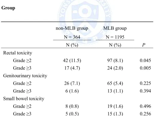
2. Late toxicity rate with or without midline block
Table 2 summarizes the crude rate of grade ≥2 and of grade ≥3 late toxicity after RT according to MLB application.
Table 2. Late Toxicity Rates in the Midline Block (MLB) Group and non-MLB Group
non-MLB group MLB group
N = 364 N = 1195 N (%) N (%) P Rectal toxicity Grade ≥2 42 (11.5) 97 (8.1) 0.045 Grade ≥3 17 (4.7) 24 (2.0) 0.005 Genitourinary toxicity Grade ≥2 26 (7.1) 65 (5.4) 0.225 Grade ≥3 6 (1.6) 13 (1.1) 0.394
Small bowel toxicity
Grade ≥2 8 (0.8) 19 (1.6) 0.496
Grade ≥3 5 (0.5) 15 (1.3) 0.256
Abbreviations: MLB = midline block
toxicity are shown in Figure 1.
Fig. 1. Grade ≥2 late complication rates of rectum, genitourinary (GU), and small bowel.
The 10-year rate of grade ≥2 late rectal toxicities, late GU toxicities and late small bowel toxicities were 10.4% (95% confidence interval [CI], 8.8‒12.3%), 7.2% (95% CI, 5.8‒9.0%), and 1.7% (95% CI, 1.1‒2.7%), respectively. Most of the rectal toxicity occurred within 2 years after treatment but the incidence of GU toxicity consistently increased throughout the follow-up period for more than 20 years (Fig 1). There was no difference in 10-year grade ≥2 late small bowel toxicity between the non-MLB and MLB groups (1.4% versus 1.8%, P = 0.565). The 10-year grade ≥2 late rectal toxicity rates were 13.5% (95% CI, 10.2‒18.2%) and 9.4% (95% CI, 7.7‒11.6%) for non-MLB and MLB groups, respectively (P = 0.012), and the difference was
consistent until 20 years (16.0% and 10.5%). The 10-year rates of grade ≥2 late GU toxicities were 8.5% (95% CI, 5.4‒13.1%) and 6.8% (95% CI, 5.3‒8.9%) for non-MLB and non-MLB groups, respectively (P = 0.158). Although the difference in grade ≥2 late GU toxicity rates between the two groups was not significant, it continued to increase after 10 years. At 20 years, the grade ≥2 late GU toxicity rate was 16.6% (95% CI, 9.8‒28.1%) and 9.5% (7.0‒13.0%), in the non-MLB and MLB groups, respectively. When GU toxicity was subdivided to bladder and distal ureteral strictures the 20-year rates of grade ≥2 late bladder toxicity and distal ureteral stricture rates were 10.4% (95% CI, 9.0 ‒12.1%) and 1.3% (95% CI, 0.04‒4.4%). Although the difference in the 20-year rate of distal ureter stricture was not statistically different between the two groups (0.5% versus 1.5%, P = 0.339), the difference in 20-year rate of grade ≥2 late bladder toxicities was borderline significant (16.6% versus 8.3%, P = 0.070).
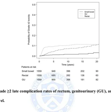
3. Association between the cumulative ICRU point dose (EQD2) and late toxicity rate
The median EQD2rectum was median 74.2 Gy3 (range, 35.4‒160.3 Gy) for
patients who experienced grade ≥2 rectal toxicity and median 66.0 Gy3(range, 29.3‒
198.0 Gy3) for those who did not (P <0.001). The median EQD2bladderof patients who
experienced grade ≥2 GU toxicity was also significantly higher compared to the patients who did not (75.6 Gy3[range, 40.7‒127.9 Gy3] versus 69.5 Gy3[range, 33.9‒
significantly higher area under curve (AUC) of 0.65 than the line of no discrimination (defined as AUC = 0.50) (P <0.001). For grade ≥2 late GU toxicity, EQD2bladder
exhibited an AUC of 0.57 which was also significantly higher than the line of no discrimination (P = 0.019). The optimal cutoff value of EQD2rectum was 63.3 Gy3for
grade ≥2 late rectal toxicity and the optimal cutoff value of EQD2bladder was 74.3 Gy3
for grade ≥2 late GU toxicity. The 10-year grade ≥2 late rectal toxicity rate for patients with EQD2rectum of ≤63.3 Gy3and >63.3 Gy3 were 4.7% (95% CI, 3.2‒6.9%)
and 14.4% (95% CI, 12.0‒17.3%), respectively (P <0.001). The 10-year grade ≥2 late GU toxicity rate for patients with EQD2bladder of ≤74.3 Gy3and >74.3 Gy3 were 5.3%
(95% CI, 4.0‒7.1%) and 10.0% (95% CI, 7.4‒13.5%), respectively (P = 0.001). The median EQD2pointAwas significantly higher in patients who experienced grade ≥2 late
rectal toxicity (77.0 Gy10 [64.1‒99.6 Gy10] versus 72.9 Gy10 [51.7‒122.2 Gy10], P
<0.001) but no difference was observed between the patients who experienced grade ≥2 late GU toxicity and those who did not. ROC curve analysis revealed that EQD2pointA could predict grade ≥2 late rectal toxicity (AUC 0.61, P = 0.022). Using
the optimal cutoff value of 74.0 Gy10, the incidence of grade ≥2 late rectal toxicity
was 12.9% (95% CI, 9.2‒18.2%) in patients with EQD2pointA>74.0 Gy10and 6.7% (95%
CI, 4.6‒9.6%) in patients with EQD2pointA ≤74.0 Gy10 (P <0.001). No significant
differences in EQD2rectum, EQD2bladder, and EQD2pointA were observed between the
patients who experienced grade ≥2 late small bowel toxicity and those who did not. ROC curve analyses revealed that EQD2rectum(P = 0.631), EQD2bladder(P = 0.490), and
A significant dose-response relationship between the ICRU dose and occurrence of grade ≥2 late rectal and GU toxicity was shown (Fig 2). The 5% and 10% probability EQD2rectum on grade ≥2 late rectal toxicity was 47.0 Gy3 (95% CI, 34.4‒74.3 Gy3) and 75.1 Gy3 (95% CI, 54.9‒118.7 Gy3), respectively and the 5% and 10% probability EQD2bladder on grade ≥2 late GU toxicity was 79.1 Gy3 (95% CI, 44.9‒ 329.2 Gy3) and 126.3 Gy3 (95% CI, 71.8‒526.2 Gy3).
Fig. 2. Probability of grade ≥2 late rectal toxicity according to biologically equivalent dose in 2-Gy fractions (EQD2) of rectal point (A) and probability of grade ≥2 late genitourinary (GU) toxicity according to EQD2 of bladder point (B).
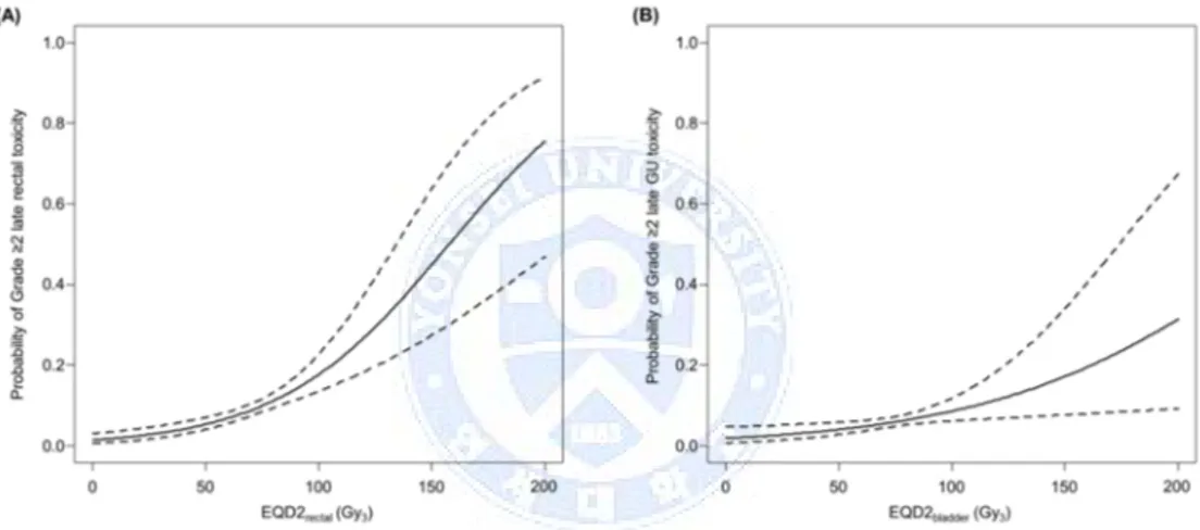
4. Treatment outcome according to midline block
The median follow-up period was 89.0 months (range, 2.4‒320.2 months). The 10-year OS, PFS, LR, and RR rates were 82.3% (95% CI, 80.2‒84.5%), 74.7% (95% CI, 72.2‒77.2%), 9.5% (95% CI, 8.0‒11.2%), and 2.6% (95% CI, 1.8‒3.7%), respectively. The 10-year OS, PFS, LR, and RR rates of the MLB group were 83.4% (95% CI, 81.1‒85.9%), 76.6% (95% CI, 73.9‒79.4%), 8.3% (95% CI, 6.7‒10.2%), and 2.3% (95% CI, 1.8‒3.5%), respectively, and 78.5% (95% CI, 73.8‒83.5%), 68.2% (95% CI, 62.6‒74.2%), 13.5% (95% CI, 10.1‒17.9%), and 3.4% (95% CI, 1.6‒7.3%), respectively, for the non-MLB group (Fig. 3). All endpoints were significantly superior in the MLB group.
After propensity score matching, the patient and tumor characteristics were well balanced (Table 3). The 10-year OS, PFS, LR, and RR rates of the MLB group were 78.9% (95% CI, 74.4‒83.8%), 70.3% (95% CI, 65.2‒75.8%), 13.0% (95% CI, 9.6‒17.5%), and 2.9% (95% CI, 1.5‒5.4%), respectively. Despite the significantly lower EQD2pointAin the MLB group, all endpoints were similar compared to the
Fig. 3. Overall survival (A), progression-free survival (B), local recurrence (C), and regional recurrence (D) rates of the midline block (MLB) group and non-MLB group.
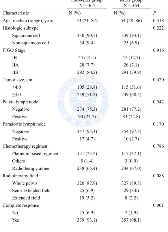
Table 3. Patient and treatment characteristics after propensity score matching non-MLB group N = 364 MLB group N = 364 Characteristic N (%) N (%) P
Age, median (range), years 53 (21‒87) 54 (28‒86) 0.418
Histologic subtype 0.222 Squamous cell 330 (90.7) 339 (93.1) Non-squamous cell 34 (9.4) 25 (6.9) FIGO Stage 0.916 IB 44 (12.1) 47 (12.7) IIA 28 (7.7) 26 (7.1) IIB 292 (80.2) 291 (79.9) Tumor size, cm 0.420 <4.0 105 (28.8) 115 (31.6) ≥4.0 259 (71.2) 249 (68.4)
Pelvic lymph node 0.542
Negative 274 (75.3) 281 (77.2)
Positive 90 (24.7) 83 (22.8)
Paraaortic lymph node 0.170
Negative 347 (95.3) 354 (97.3)
Positive 17 (4.7) 10 (2.7)
Chemotherapy regimen 0.786
Platinum based regimen 121 (23.2) 117 (32.1)
Others 5 (1.4) 3 (0.9) Radiotherapy alone 238 (65.4) 244 (67.0) Radiotherapy field 0.088 Whole pelvis 320 (87.9) 327 (89.8) Semi-extended field 25 (6.9) 29 (8.0) Extended field 19 (5.2) 8 (2.2) Complete response 0.001 No 25 (6.9) 7 (1.9) Yes 339 (93.1) 357 (98.1)
non-MLB group N = 364 MLB group N = 364 Characteristic N (%) N (%) P EBRT dose, Gy <0.001 ≤45 324 (89.0) 346 (95.1) >45 40 (11.0) 18 (4.9)
EQD2EBRT + ICBT, median (range), Gy
Point A dose, Gy10 86.4 (54.9‒122.2) 72.9 (59.1‒98.0) <0.001
Bladder point dose, Gy3 75.9 (49.0‒144.1) 68.3 (33.9‒136.8) <0.001
Rectal point dose, Gy3 74.8 (47.3‒160.3) 66.1 (34.0‒117.5) <0.001
Abbreviations: FIGO = International Federation of Gynecology and Obstetrics; MLB = midline block; 5-FU = 5-fluorouracil; EQD2 = biologically equivalent dose in 2-Gy fractions; EBRT = external beam radiation therapy; ICBT = intracavitary brachytherapy
IV. DISCUSSION
This retrospective study is one of the largest series to date investigating the benefit and feasibility of using MLB for patients with cervix cancer who received definitive RT or CRT. The cumulative dose prescribed to point A in the MLB group was lower than the dose recommended in current guidelines, which recommends more than 80 Gy to EQD2pointA.24 Although the central dose was reduced, our results
suggest that MLB based on response to EBRT may not compromise local control or survival and rather reduce the incidence of late rectal toxicity.
with improved survival.9,10 The role of EBRT, which is more limited compared to
HDR-ICBT, is to 1) improve the geometry of the tumor for optimal ICBT by reducing the size of tumor and bringing the tumor volume within a higher dose portion of HDR-ICBT dose distribution, 2) sterilize disease in the parametrium and lymph nodes that cannot receive adequate dose by HDR-ICBT. Therefore in our institution we have blocked the central region as soon as the tumor regressed and the geometry of the tumor was appropriate for ICBT. The attempt to find the optimal fractionation schedule of EBRT and HDR-ICBT has been made but no clear conclusions were drawn.25 The efficacy of reducing the cumulative central dose using MLB in FIGO
stages IB to IIB, non-bulky tumors was investigated in a small prospective trial from Japan.14 MLB was performed after 20 Gy of pelvic EBRT, and the cumulative dose
prescribed to point A was EQD2 52 Gy10. The results were promising with a 3-yr
pelvic control rate of 96% and no grade ≥3 toxicity. Several retrospective studies with long-term follow-up using a low cumulative central dose protocols have also shown comparable results with other studies using higher dose protocols.16,26In our study, the
MLB group showed better local control and survival compared to the non-MLB group. Since the MLB group presented with more favorable characteristics we performed propensity score matching. After matching the characteristics were well balanced and the local control and survival rate were similar between the two groups even though the median EQD2pointAwas lower by 13.5 Gy10in the MLB group.
However, the fore-mentioned studies including our study do not provide information on dose-volume parameters of tumor volume, which is also a critical
factor in addition to the prescribed dose. Recently Dyk et al. reported the results of using IMRT and HDR-ICBT.27 During EBRT the cervix region received 20 Gy and
the nodal region received 50 Gy in 28 fractions. MRI guided HDR-ICBT was performed in 6 weekly fractions of 6.5 Gy prescribed to point A. The minimum dose delivered to 100 and 90% of gross tumor volume (D100, D90) was significantly lower in the patients who experienced local failure compared to the patients who achieved local control. They estimated that the D100, D90 for ≥90% local control was 69 and 98 Gy10. In a similar manner, the Vienna group has compared the spatial dose
distribution within the high-risk clinical target volume (HR-CTV) for patients who failed locally and patients who achieved continuous complete remission.28 The mean
minimum point dose within the HR-CTV was 72 Gy10 for patients with local failure
and 99 Gy10in patients who achieved local control. It implies that patients with poor
target coverage may more likely result in local recurrence. In our study the median dose prescribed to point A in the MLB group was 72.9 Gy10. Although we cannot
evaluate the dose-volume parameters, some possibilities can be brought up why the lower point A dose did not result in poorer local control. After significant tumor reduction during EBRT, the central dose from HDR-ICBT would be remarkably higher than the dose prescribed to point A. In addition, small tumors or bulky tumors that show a major response may not need as much radiation as large tumors without response after EBRT. This was observed from the data from the Vienna group. They showed that D90 and D100 of HR-CTV were correlated with local control.29However,
significant dependence on dose for local control. Recently, Mazeron et al. proposed a dose-response relationship for local control in patients treated with image guided pulsed-dose rate brachytherapy.30 In this model, the HR-CTV volume had significant
impact on the dose thresholds and D90 required to achieve a 90% local control was ≥92.0 Gy10in cases of HR-CTV volume ≥30 cm3and ≥73.9 Gy10in cases of HR-CTV
volume <30 cm3.
The 10-year actuarial rate of developing grade ≥2 late rectal, GU, small bowel toxicities were 10.4%, 7.2%, and, 1.7%, respectively. Although, it may not be appropriate to compare the toxicity rates among different studies due to the difference in patient population and the retrospective nature of this study, our results were comparable or lower compared to other studies.14,31Previous studies have shown that
the ICRU rectal dose has a relatively better correlation with late rectal toxicity compared to ICRU bladder dose with late bladder toxicity.13In the era of 3D
image-based ICBT, the dose-volume parameters such as D2cc have been introduced in
substitution to ICRU point doses in reporting dose delivered to organs at risk.20
Considering the inhomogeneous dose distribution for organs near the source, dose-volume parameters seem more appropriate compared to single point dose assessment and several studies have reported on the predictability of dose-volume parameters on late rectal or bladder complications.12,13 The Vienna group reported the predictive
value of dose-volume parameters, such as the minimum EQD2 to the most exposed 2 cm3 (D2cc) that patients. Patients with rectal D2cc>75 Gy had a higher incidence of grade ≥2 rectal morbidity (12% versus 4%). In addition, patients with bladder
D2cc>100 Gy had a higher incidence of grade ≥2 bladder toxicity (13% versus 9%).13
However, still a large portion of patients are planned in a conventional fashion7,32and
dose-volume parameters may not be available. Previous reports showed that the ICRU rectal dose provided a good estimate of D2cc for the rectum while the ICRU bladder
dose did not.33,34In our study both ICRU rectal and bladder dose showed significant
correlation with late rectal and bladder toxicities, respectively, though the correlation was weaker between ICRU bladder dose and late bladder toxicity. We have analyzed the optimal cutoff value for grade ≥2 late rectal (EQD2rectum = 63.3 Gy3) and bladder
toxicities (EQD2bladder = 74.3 Gy3) and the dose of 10% probability in developing
grade ≥2 late rectal (EQD2rectum = 75.1 Gy3) and GU toxicities (EQD2bladder= 126.3
Gy3).
The MLB is aimed to shield the centrally located rectum and bladder and eventually lower the dose delivered to these organs. Therefore the MLB group could be anticipated to have lesser late rectal and bladder toxicities. However, in our study, the MLB group and non-MLB group showed significant difference in the rate of late rectal toxicity but not in GU toxicity. The minimal difference in GU toxicity may be due to the higher ICRU bladder point dose delivered during HDR-ICBT in the MLB group (35.0 Gy3 versus 32.3 Gy3). The 2.7 Gy3difference seems small but it has been
reported that ICRU bladder dose usually underestimates the D2cc of bladder33,34and
the ICRU dose difference may have been underestimated. Although not statistically significant, the difference of 7% in the 20-year rate of grade ≥2 late GU toxicity rate may need some attention. With a longer median follow-up time the data may not only
show clinical but also statistical significance. We have also observed the fact that the bladder toxicity, which includes hematuria or vesicovaginal fistula, mainly contributed to the difference in GU toxicity and the difference in rate of distal ureteral stricture was minimal. Since the MLB covers 4 cm of the central region, the shielding effect to the distal ureter would have been minimal.
Some limitations of our study should be noted. First, this was an institution-based retrospective study for a long study period. However, a large volume of patients were treated with a consistent policy and a long-term follow-up was performed which was sufficient enough to evaluate late complications. Second, the baseline patient and tumor characteristics were different between the MLB group and non-MLB group. In attempt to reduce the difference in baseline characteristics we have performed propensity score matching. However, tumor response itself can be prognostic for further treatment outcome30,35 and the different response to RT may represent the
different tumor biology which is out of the scope of this study. Third, MLB has innate limitations that should be considered. The rectangular shape of MLB does not match the pear shaped HDR- ICBT dose line. In addition, until now, we have no way to precisely calculate the cumulative dose of HDR-ICBT and EBRT using MLB on 3-dimensional basis. Even in patients who underwent CT simulation, there are uncertainties in evaluating the cumulative dose delivered to tumor and organs at risk.36 Fourth, we did not perform 3D image-based brachytherapy during the study
period. The reduction of complications and improvement of treatment outcome by using based brachytherapy has been reported and we have also started
image-based brachytherapy since 2011.37,38To further evaluate the feasibility of our approach,
patients who receive 3D image-based brachytherapy in addition to EBRT with MLB should be analyzed for their dose-volume parameters.
V. CONCLUSION
This single-institution retrospective study with a large patient volume and a long-term follow-up period investigated the safety and benefit of selectively applying MLB for uterine cervical cancer patients who showed response to EBRT. We found that the rectal toxicity was significantly lower in patients who received MLB and the positive dose-response relationship between cumulative ICRU doses and late rectal and GU toxicity. Additionally, the treatment outcome does not seem to be compromised in the MLB group even though the cumulative central dose was lower. These data should be confirmed in further prospective randomized trials comparing the treatment outcome and toxicity rates according to dose response relationship.
REFERENCES
1. Jung KW, Won YJ, Kong HJ, Oh CM, Cho H, Lee DH, et al. Cancer statistics in Korea: incidence, mortality, survival, and prevalence in 2012. Cancer Res Treat 2015;47:127-41.
2. Torre LA, Bray F, Siegel RL, Ferlay J, Lortet-Tieulent J, Jemal A. Global cancer statistics, 2012. CA Cancer J Clin 2015;65:87-108.
3. Landoni F, Maneo A, Colombo A, Placa F, Milani R, Perego P, et al. Randomised study of radical surgery versus radiotherapy for stage Ib-IIa cervical cancer. Lancet 1997;350:535-40.
4. Chemoradiotherapy for Cervical Cancer Meta-Analysis C. Reducing uncertainties about the effects of chemoradiotherapy for cervical cancer: a systematic review and meta-analysis of individual patient data from 18 randomized trials. J Clin Oncol 2008;26:5802-12.
5. Tomita N, Toita T, Kodaira T, Shinoda A, Uno T, Numasaki H, et al. Patterns of radiotherapy practice for patients with cervical cancer in Japan, 2003-2005: changing trends in the pattern of care process. Int J Radiat Oncol Biol Phys 2012;83:1506-13.
6. Pearce A, Craighead P, Kay I, Traptow L, Doll C. Brachytherapy for carcinoma of the cervix: a Canadian survey of practice patterns in a changing era. Radiother Oncol 2009;91:194-6.
7. Viswanathan AN, Erickson BA. Three-dimensional imaging in gynecologic brachytherapy: a survey of the American Brachytherapy Society. Int J Radiat
Oncol Biol Phys 2010;76:104-9.
8. Potter R, Georg P, Dimopoulos JC, Grimm M, Berger D, Nesvacil N, et al. Clinical outcome of protocol based image (MRI) guided adaptive brachytherapy combined with 3D conformal radiotherapy with or without chemotherapy in patients with locally advanced cervical cancer. Radiother Oncol 2011;100:116-23.
9. Han K, Milosevic M, Fyles A, Pintilie M, Viswanathan AN. Trends in the utilization of brachytherapy in cervical cancer in the United States. Int J Radiat Oncol Biol Phys 2013;87:111-9.
10. Gill BS, Lin JF, Krivak TC, Sukumvanich P, Laskey RA, Ross MS, et al. National Cancer Data Base analysis of radiation therapy consolidation modality for cervical cancer: the impact of new technological advancements. Int J Radiat Oncol Biol Phys 2014;90:1083-90.
11. Lee SW, Suh CO, Chung EJ, Kim GE. Dose optimization of fractionated external radiation and high-dose-rate intracavitary brachytherapy for FIGO stage IB uterine cervical carcinoma. Int J Radiat Oncol Biol Phys 2002;52:1338-44.
12. Koom WS, Sohn DK, Kim JY, Kim JW, Shin KH, Yoon SM, et al. Computed tomography-based high-dose-rate intracavitary brachytherapy for uterine cervical cancer: preliminary demonstration of correlation between dose-volume parameters and rectal mucosal changes observed by flexible sigmoidoscopy. Int J Radiat Oncol Biol Phys 2007;68:1446-54.
13. Georg P, Lang S, Dimopoulos JC, Dorr W, Sturdza AE, Berger D, et al. Dose-volume histogram parameters and late side effects in magnetic resonance image-guided adaptive cervical cancer brachytherapy. Int J Radiat Oncol Biol Phys 2011;79:356-62.
14. Toita T, Kato S, Niibe Y, Ohno T, Kazumoto T, Kodaira T, et al. Prospective multi-institutional study of definitive radiotherapy with high-dose-rate intracavitary brachytherapy in patients with nonbulky (<4-cm) stage I and II uterine cervical cancer (JAROG0401/JROSG04-2). Int J Radiat Oncol Biol Phys 2012;82:e49-56.
15. Toita T, Kitagawa R, Hamano T, Umayahara K, Hirashima Y, Aoki Y, et al. Phase II study of concurrent chemoradiotherapy with high-dose-rate intracavitary brachytherapy in patients with locally advanced uterine cervical cancer: efficacy and toxicity of a low cumulative radiation dose schedule. Gynecol Oncol 2012;126:211-6.
16. Park HC, Suh CO, Kim GE. Fractionated high-dose-rate brachytherapy in the management of uterine cervical cancer. Yonsei Med J 2002;43:737-48.
17. Kim YB, Lee IJ, Kim SY, Kim JW, Yoon HI, Kim SW, et al. Tumor heterogeneity of FIGO stage III carcinoma of the uterine cervix. Int J Radiat Oncol Biol Phys 2009;75:1323-8.
18. Kim YB, Cho JH, Keum KC, Lee CG, Seong J, Suh CO, et al. Concurrent chemoradiotherapy followed by adjuvant chemotherapy in uterine cervical cancer patients with high-risk factors. Gynecol Oncol 2007;104:58-63.
19. Yoon H, Cha J, Keum K, Lee H, Nam E, Kim S, et al. Treatment outcomes of extended-field radiation therapy and the effect of concurrent chemotherapy on uterine cervical cancer with para-aortic lymph node metastasis. Radiat Oncol 2015;10:18.
20. Potter R, Haie-Meder C, Van Limbergen E, Barillot I, De Brabandere M, Dimopoulos J, et al. Recommendations from gynaecological (GYN) GEC ESTRO working group (II): concepts and terms in 3D image-based treatment planning in cervix cancer brachytherapy-3D dose volume parameters and aspects of 3D image-based anatomy, radiation physics, radiobiology. Radiother Oncol 2006;78:67-77.
21. Cox JD, Stetz J, Pajak TF. Toxicity criteria of the Radiation Therapy Oncology Group (RTOG) and the European Organization for Research and Treatment of Cancer (EORTC). Int J Radiat Oncol Biol Phys 1995;31:1341-6. 22. Gray RJ. A class of K-sample tests for comparing the cumulative incidence of
a competing risk. Ann Stat 1998;16:1141-54.
23. Ho DE, Imai K, King G, Stuart EA. MatchIt: Nonparametric Preprocessing for Parametric Causal Inference. Journal of Statistical Software 2011;42. 24. Viswanathan AN, Beriwal S, De Los Santos JF, Demanes DJ, Gaffney D,
Hansen J, et al. American Brachytherapy Society consensus guidelines for locally advanced carcinoma of the cervix. Part II: high-dose-rate brachytherapy. Brachytherapy 2012;11:47-52.
fractionation schedules in the treatment of cervical cancer: is there an optimal fractionation schedule? Int J Radiat Oncol Biol Phys 1999;43:359-66.
26. Nakano T, Kato S, Ohno T, Tsujii H, Sato S, Fukuhisa K, et al. Long-term results of high-dose rate intracavitary brachytherapy for squamous cell carcinoma of the uterine cervix. Cancer 2005;103:92-101.
27. Dyk P, Jiang N, Sun B, DeWees TA, Fowler KJ, Narra V, et al. Cervical gross tumor volume dose predicts local control using magnetic resonance imaging/diffusion-weighted imaging-guided high-dose-rate and positron emission tomography/computed tomography-guided intensity modulated radiation therapy. Int J Radiat Oncol Biol Phys 2014;90:794-801.
28. Schmid MP, Kirisits C, Nesvacil N, Dimopoulos JC, Berger D, Potter R. Local recurrences in cervical cancer patients in the setting of image-guided brachytherapy: a comparison of spatial dose distribution within a matched-pair analysis. Radiother Oncol 2011;100:468-72.
29. Dimopoulos JC, Potter R, Lang S, Fidarova E, Georg P, Dorr W, et al. Dose-effect relationship for local control of cervical cancer by magnetic resonance image-guided brachytherapy. Radiother Oncol 2009;93:311-5.
30. Mazeron R, Castelnau-Marchand P, Dumas I, Del Campo ER, Kom LK, Martinetti F, et al. Impact of treatment time and dose escalation on local control in locally advanced cervical cancer treated by chemoradiation and image-guided pulsed-dose rate adaptive brachytherapy. Radiother Oncol 2015;114:257-63.
31. Kato S, Ohno T, Thephamongkhol K, Chansilpa Y, Cao J, Xu X, et al. Long-term follow-up results of a multi-institutional phase 2 study of concurrent chemoradiation therapy for locally advanced cervical cancer in east and southeast Asia. Int J Radiat Oncol Biol Phys 2013;87:100-5.
32. Viswanathan AN, Creutzberg CL, Craighead P, McCormack M, Toita T, Narayan K, et al. International brachytherapy practice patterns: a survey of the Gynecologic Cancer Intergroup (GCIG). Int J Radiat Oncol Biol Phys 2012;82:250-5.
33. Wachter-Gerstner N, Wachter S, Reinstadler E, Fellner C, Knocke TH, Wambersie A, et al. Bladder and rectum dose defined from MRI based treatment planning for cervix cancer brachytherapy: comparison of dose-volume histograms for organ contours and organ wall, comparison with ICRU rectum and bladder reference point. Radiother Oncol 2003;68:269-76.
34. Pelloski CE, Palmer M, Chronowski GM, Jhingran A, Horton J, Eifel PJ. Comparison between CT-based volumetric calculations and ICRU reference-point estimates of radiation doses delivered to bladder and rectum during intracavitary radiotherapy for cervical cancer. Int J Radiat Oncol Biol Phys 2005;62:131-7.
35. Oh D, Lee JE, Huh SJ, Park W, Nam H, Choi JY, et al. Prognostic significance of tumor response as assessed by sequential 18F-fluorodeoxyglucose-positron emission tomography/computed tomography during concurrent chemoradiation therapy for cervical cancer. Int J Radiat
Oncol Biol Phys 2013;87:549-54.
36. Fenkell L, Assenholt M, Nielsen SK, Haie-Meder C, Potter R, Lindegaard J, et al. Parametrial boost using midline shielding results in an unpredictable dose to tumor and organs at risk in combined external beam radiotherapy and brachytherapy for locally advanced cervical cancer. Int J Radiat Oncol Biol Phys 2011;79:1572-9.
37. Rijkmans EC, Nout RA, Rutten IH, Ketelaars M, Neelis KJ, Laman MS, et al. Improved survival of patients with cervical cancer treated with image-guided brachytherapy compared with conventional brachytherapy. Gynecol Oncol 2014;135:231-8.
38. Kang HC, Shin KH, Park SY, Kim JY. 3D CT-based high-dose-rate brachytherapy for cervical cancer: clinical impact on late rectal bleeding and local control. Radiother Oncol 2010;97:507-13.