J Echocardiogr Vol.5, No.2, 55-57 (2007)
J Echocardiogr Vol.5, No.2, 2007 55
Aortic regurgitation (AR) may be congenital or acquired, caused by either abnormalities of the aortic root or the valve itself. Surgical management is recom-mended in patients with significant AR enough to reduce left ventricular function. Aortic valve (AV) replacement is a common surgical strategy for signifi-cant AR, but in some patients who have annuloaortic ectasia (AAE) without leaflets abnormalities, valve-pre-serving aortic replacement can be considered[1]. Therefore, the knowledge of aortic annulus and leaflets geometry is very important in considering the surgical strategy.
Recently developed real-time 3-dimensional (3-D) echocardiography can be used to provide fast and non-invasive estimates with high image resolution that is more accurate and physiologically realistic than those measured by conventional imaging techniques[2]. We have already demonstrated the geometry of the mitral leaflets and annulus in patients with ischemic function-al mitrfunction-al regurgitation using this system[3-5]. In the
present study we evaluated the 3-D geometry of the aortic annulus and leaflets in normal subjects and in patients who had different etiologies of significant AR.
Methods
We performed real-time 3-D echocardiographic examination in 3 normal subjects and 5 patients who had different etiologies of significant AR: AR with AAE (n=2), AR with degenerative valve (n=1), AR with leaflet prolapse (n=1), and AR with bicuspid AV (n=1). Left ventricular (LV) systolic function and degree of AR were quantified by using 2-dimensional echocardio-graphy. Transthoracic volumetric images were acquired by the real-time 3-D echocardiography sys-tem in apical views. By utilizing our original software system for 3-D aortic valve analysis, we radially cropped the 3-D volumetric data into 18 planes to mark and trace the aortic annulus and leaflets. Then, 3-D images of the aortic leaflets and annulus were recon-structed in early diastole for the 3-D quantitative mea-surements. Aortic annular size was calculated by using those 3-D data sets (area, circumference). The aortic leaflets’ surface area on LV side was calculated from the 3-D data as well. All subjects provided written informed consent to the study protocol, which was approved by the Committee for the Protection of Human Subjects in Research at Kawasaki Medical School.
Received February 20, 2007; revision received May 1, 2007; accept-ed June 13, 2007
Address for correspondence: Nozomi Watanabe, MD Department of Cardiology, Kawasaki Medical School, 577 Matsushima, Kurashiki, 701-0192, Japan. Telephone: +81-86-462-1111
Fax: +81-86-464-4060
E-mail: [email protected]
2007 Japanese Society of Echocardiography
Three-Dimensional Geometry of Aortic Valve: A new Trial of
Visualization With Real-Time Three-Dimensional Echocardiography
Chi Young Shim, MD*, Nozomi Watanabe, MD**, Miwako Tsukiji, MD**,Yasuko Yamaura, MD**, Yasuo Ogasawara, PhD**** Jong-Won Ha, MD, PhD*, Se-Joong Rim, MD, PhD*, Kazuo Tanemoto, MD***, Namsik Chung, MD, PhD*, and Kiyoshi Yoshida, MD**
*Division of Cardiology, Yonsei Cardiovascular Center, Yonsei University College of Medicine, Seoul, Korea
**Department of Cardiology,
***Thoracic and Cardiovascular Surgery,
****Medical Engineering and Systems of Cardiology, Kawasaki Medical School, Kurashiki, Japan
Results
We could visualize and measure the AV annular and leaflets geometry and size by using novel software sys-tem for AV with 3-D transthoracic echocardiography
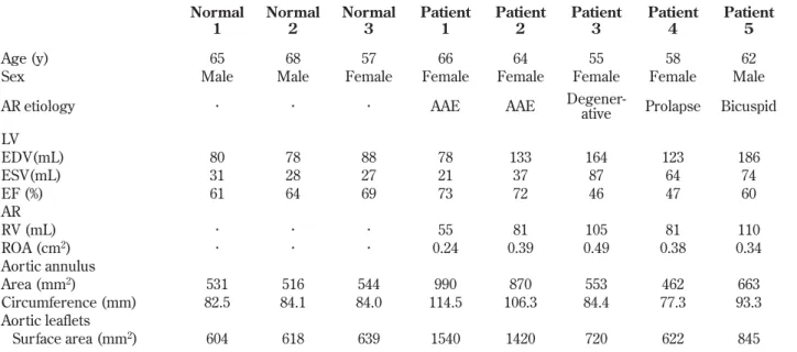
not only in 3 normal subjects but also in 5 patients with significant AR. Table 1 shows baseline characteristics, measured 2-D parameters, and calculated 3-D parame-ters in each subject.
In normal subjects, we could see the three-curved,
J Echocardiogr Vol.5, No.2, 2007 Shim et al. 56
Table 1. Clinical characteristics and 3-D measurements of aortic annulus and leaflets
Fig. 1. Three-dimensional images of the aortic leaflets and annulus in early diastole obtained from subjects: A, Anatomic 3-D images. The anatomic 3-D images shows the actual configuration of the annulus and leaflets with surface colorations. B, 3-D surface images in the vertical view from the LV. The aortic leaflets' config-uration was represented in contour. R, right coronary cusp; L, left coronary cusp; N, non coronary cusp; LV, left ventricle
semilunar and non-planar geometry of aortic annulus and define three normal leaflets. In patients with AAE, the area and circumference of aortic annulus and the aortic leaflets’ surface area were apparently large. We could find that the non-planar curvature of the annulus seemed to be blunted in a patient with degenerative AV. In a patient with bicuspid AV, the size of the aortic annulus was larger than that of normal subjects, and we could define only two leaflets on the aortic leaflets’ configuration (Table 1, Figure 1).
Discussion
Although there have been many studies of the sad-dle-shaped mitral annulus, the geometry of aortic annulus and leaflets has not been studied with real-time 3-D echocardiography. Like the mitral annulus, the annular configuration of aortic valve also appeared curved-shaped and non-planar because aortic leaflets were attached in a semilunar fashion rather than a ring-like planar fashion[6,7]. Therefore, precise and comprehensive understanding of the 3-D geometric changes of the aortic annulus and leaflets is needed in various clinical settings, especially for preoperative evaluation before AV surgery. In this study, we suc-cessfully demonstrated the three-curved, non-planar configuration of aortic annulus and quantified their cir-cumferences and areas in normal subjects and in patients with significant AR who had different etiolo-gies using our novel software system with real-time 3-D echocardiography.
Even though this study was performed with a small number of subjects, it was enough to show a new pos-sibility of the AV geometric evaluation. Further inves-tigations are needed to clarify the geometric differ-ences of various AV diseases.
In conclusion, this 3-D technique would be helpful to understand the 3-D geometry of AV, especially before
AV surgery, and to make a proper decision for surgical strategy for each individual.
References
1. David TE, Ivanov J, Armstrong S, Feindel CM, Webb GD. Aortic valve-sparing operations in patients with aneurysms of the aortic root or ascending aorta. Ann Thorac Surg. 2002; 74(suppl): S1758-61; discussion S1792-9.
2. Yamaura Y, Watanabe N, Ogasawara Y, Yamamoto K, Kawamoto T, Toyota E, et al. Geometric demonstration and three-dimensional quantitative analysis of the mitral valve with real-time three-dimensional echocardiography: novel anatomical image creation system. J Echocardiogr. 2004; 2: 99-104.
3. Watanabe N, Ogasawara Y, Yamaura Y, Kawamoto T, Toyota E, Akasaka T, et al. Quantification of mitral valve tenting in ischemic mitral regurgitation by thransthoracic real-time three-dimensional echocardiography. J Am Coll Cardiol. 2005; 45: 763-9.
4. Watanabe N, Ogasawara Y, Yamaura Y, Kawamoto T, Toyota E, Akasaka T. et al. Mitral annulus flattens in ischemic mitral regurgitation: geometric differences between inferior and anterior myocardial infarction: a real-time 3-dimensional echocardiographic study. Circulation 2005; 112: 1458-62.
5. Yamaura Y, Watanabe N, Ogasawara Y, Wada N, Kawamoto T, Toyota E, et al. Geometric change of mitral valve leaflets and annulus after reconstructive surgery for ischemic mitral regurgitation: Real-time 3-dimensional echocardiographic study. J Thorac and Cardiovasc Surg. 2005; 5: 1 459-61.
6. Anderson RH, Devine WA, Ho SY, Smith A, McKay R. The myth of the aortic annulus: the anatomy of the subaortic outflow tract. Ann Thorac Surg. 1991; 52: 640-6. 7. Sutton III JP, Ho SY, Anderson RH. The forgotten
inter-leaflet triangles: a review the surgical anatomy of the aor-tic valve. Ann Thorac Surg. 1995; 59: 419-27.
Aortic valve geometry by 3D echocardiography 57