Transcriptional regulation of
adrenomedullin by oncostatin M in
human astroglioma cells: Implications for
tumor invasion and migration
Seul Ye Lim
1,2, So-Hee Ahn
1,2, Hyunju Park
1,2, Jungsul Lee
3, Kyungsun Choi
3, Chulhee Choi
3, Ji Ha Choi
2,4,
Eun-Mi Park
2,4& Youn-Hee Choi
1,21Department of Physiology, Global Top5 Research Program, School of Medicine, Ewha Womans University, Seoul, Korea,2Tissue Injury Defense Research Center, School of Medicine, Ewha Womans University, Seoul, Korea,3Department of Bio and Brain Engineering, KAIST, Daejeon, Korea,4Department of Pharmacology, School of Medicine, Ewha Womans University, Seoul, Korea.
Adrenomedullin (ADM), a secretory peptide with multiple functions in physiological to pathological
conditions, is upregulated in several human cancers, including brain, breast, colon, prostate, and lung
cancer. However, the molecular mechanisms underlying the regulation of ADM expression in cancerous
cells are not fully understood. Here, we report that oncostatin M (OSM), a cytokine belonging to the
interleukin-6 family, induces ADM expression in astroglioma cells through induction of signal transducer
and activator of transcription-3 (STAT-3) phosphorylation, nuclear translocation, and subsequent DNA
binding to the ADM promoter. STAT-3 knockdown decreased OSM-mediated expression of ADM,
indicating that ADM expression is regulated by STAT-3 in astroglioma cells. Lastly, scratch wound healing
assay showed that astroglioma cell migration was significantly enhanced by ADM peptides. These data
suggest that aberrant activation of STAT-3, which is observed in malignant brain tumors, may function as
one of the key regulators for ADM expression and glioma invasion.
M
alignant gliomas are the most common subtype of primary brain tumors. They are characterized by
cellular pleomorphism, microvascular proliferation, areas of necrosis, and extensive invasion into the
surrounding brain tissues, which leads to poor prognosis for patients
1,2. Because of the extraordinary
ability to invade the surrounding healthy brain tissue, complete elimination of malignant gliomas by surgical
resection is almost impossible
3. Thus, the identification of molecular mechanisms involved in invasion is an
important objective in glioma research, to develop an effective therapeutic modality for this particular tumor.
In glioma, a large number of microglia/macrophages are found within the tumor mass, and they are known to
be involved in the tumor microenvironment which favors glioma growth and invasion, through releasing several
microglia/macrophages-derived molecules
4. Oncostatin M (OSM), one of interleukin-6 (IL-6) family cytokines, is
secreted by activated macrophages and microglia
5,6. Increased OSM expression has been reported in a variety of
cancers, including malignant glioma
7. OSM mainly activates signal transducer and activator of transcription
(STAT)-3, which is involved in glioma development and progression
8–11. Constitutive activation and
phosphor-ylation of STAT-3 is frequently detected in glioma, and this activation is believed to promote tumor formation
and progression via transcriptional activation of downstream genes
12,13.
Adrenomedullin (ADM), a 52-amino acid ring-structure peptide originally isolated from a human
pheochro-mocytoma, is expressed in human cancer cell lines, including brain, breast, colon, prostate, and lung cancer cells
14.
In physiologic conditions, ADM performs important roles as a vasodilator, bronchodilator, regulator of hormone
secretion, neurotransmitter, antimicrobial agent, and controller of renal functions
15. In brain tumors, the extent of
ADM mRNA expression is related to the tumor type and grade
16. However, the stimuli involved in the increased
expression of ADM and the molecular mechanisms regulating ADM expression in brain tumors are not fully
understood.
In the present study, we showed that OSM induces ADM upregulation through the activation of STAT-3 and
that ADM contributes to increased invasion activity in human astroglioma cell lines. Our data support the notion
that ADM expression level is influenced by OSM, which is secreted by activated microglia/macrophages,
pro-SUBJECT AREAS:
CNS CANCER CANCER MICROENVIRONMENTReceived
28 April 2014
Accepted
28 August 2014
Published
23 September 2014
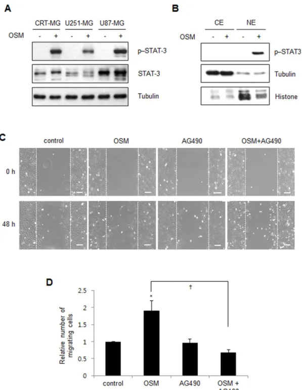
Correspondence and requests for materials should be addressed to Y.-H.C. ([email protected]. kr)OSM induces STAT-3 activation and migration in astroglioma
cells.
OSM is known to predominantly activate the STAT-3
signal-ing pathway
17,18. To examine whether OSM induces STAT-3
phos-phorylation in human astroglioma cells, CRT-MG, U251-MG, and
U87-MG cells were incubated in the absence or presence of hOSM
(10 ng/mL) for 30 min and then analyzed by immunoblotting.
STAT-3 phosphorylation at residue Tyr705 was significantly enhanced by
OSM treatment in all cell lines (Fig. 2A). Next, to examine whether
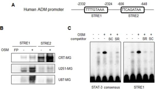
STAT-3 binds to the ADM promoter in the presence of OSM.
The
promoter region of human ADM contains two putative STAT-3
response elements (STRE1 and STRE2; TTN
5AA) (Fig. 3A). To
test whether activated STAT-3 binds to the ADM promoter, three
human astroglioma cell lines were treated with hOSM for 30 min.
The nuclear extracts (NEs) from each cell line were then subjected to
an electrophoretic mobility shift assay (EMSA) (Fig. 3B). In
CRT-MG cells, DNA binding of STAT-3 to STRE2 in human ADM
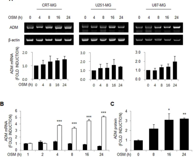
Figure 1
|
OSM induces ADM expression in astroglioma cells. (A) CRT-MG, U251-MG and U87-MG astroglioma cells were either untreated controls (time zero) or treated with hOSM (10 ng/mL) as indicated, and RNA were extracted and analyzed by RT-PCR for ADM mRNA. Data are presented as the fold induction compared with each untreated control cells. Data shown are representative of three independent experiments. (B) qRT-PCR analysis was performed for CRT-MG cells incubated in the absence or presence of hOSM (10 ng/mL) for 0–24 h. ***P , 0.001 vs. each untreated control cells. (C) ADM protein from the culture supernatants was analyzed by ELISA. Data are presented as the fold induction in relative ADM protein levels compared with the untreated cells at the time point 0 h. Data shown are representative of at least three experiments. *P , 0.05, **P , 0.01 vs. untreated control. OSM, oncostatin M.promoter was strongly enhanced by OSM treatment, while STAT-3
binding to STRE1 was slightly increased. In U251-MG and U87-MG
cells, STAT-3 binding to STRE1 was strongly enhanced by OSM and
a modest increase in STRE2 binding was observed. The same NEs
obtained from CRT-MG cells were then exposed to the STAT-3
consensus oligonucleotide (Fig. 3C). STAT-3 binding to the ADM
promoter STRE1 and STAT-3 consensus oligonucleotide was
enhanced by OSM treatment. Competition with an excess of
Figure 2
|
OSM induces STAT-3 activation and migration in astroglioma cells. (A) Whole cell lysates from the CRT-MG, U251-MG and U87-MG cells treated with OSM (10 ng/mL) for 30 min were analyzed by immunoblotting against total STAT-3 and p-Y705-STAT-3. Tubulin was used as the loading control. Cropped blots are used. Full-length blots are shown in supplementary Figure 1. (B) Nuclear and cytoplasmic extracts from CRT-MG cells treated with hOSM (10 ng/ml) for 30 min and analyzed by immunoblotting against total STAT-3 and p-Y705-STAT-3. Tubulin and Histone were used as loading and purity controls of each cellular fraction. Data shown are representative of at least three experiments. P, phospho; CE, cytoplasmic extracts; NE, nuclear extracts. Full-length blots are shown in supplementary Figure 1. (C) Representative micrographs of the scratch wound healing assay in CRT-MG cells in the absence or presence of either OSM or AG490 at 48 h, as indicated. Bar 5 100 mm. (D) Confluent CRT-CRT-MG cells were assayed for migration into the wound at 48 h after scratching, in the absence or presence of OSM and AG490, as indicated. Migrating cells were measured by counting the cell numbers. The results are expressed as the mean 6 SD values (compared with untreated controls at 48 h time point); normalized values are shown. The data shown are representative of three independent experiments. (*P , 0.05 vs. untreated control;{P , 0.05 between OSM-treated cells and OSM/ AG490-treated cells.).
unlabeled STRE1 or STAT-3 oligonucleotide abrogated the complex
formation.
STAT-3 knockdown reduces OSM-induced ADM expression.
To
determine whether the enhanced ADM expression level in the
presence of OSM is mediated by STAT-3, siRNA-mediated
downre-gulation of STAT-3 was performed. CRT-MG cells were transiently
transfected with a STAT-3 siRNA or a green fluorescence protein
(GFP) siRNA (negative control) and incubated with or without
hOSM. As shown in Figure 4B, ADM mRNA expression level in
the STAT-3 siRNA-transfected cells was decreased compared to
that in the GFP siRNA-transfected cells. The reduction in the
expression of STAT-3 protein in the STAT-3 siRNA-transfected
cells was confirmed by immunoblotting (Fig. 4A). These results
demonstrate that STAT-3 knockdown reduces OSM-mediated
transcription of the ADM gene, indicating that ADM expression is
regulated by STAT-3 in astroglioma cells.
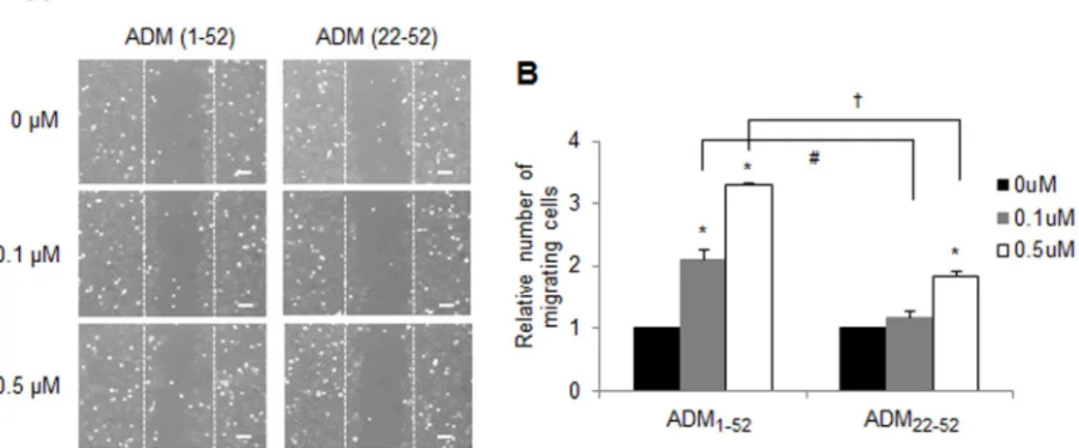
ADM enhances astroglioma cell invasion.
A previous study
demonstrated that the extent of ADM mRNA expression is related
Figure 3
|
STAT-3 binds to the ADM promoter in the presence of OSM. (A) The human ADM promoter. The putative STAT-3 response elements are indicated as STRE1 and STRE2. (B) The NEs from CRT-MG, U251-MG and U87-MG astroglioma cells treated with hOSM (10 ng/mL) for 30 min were incubated in the presence of a radiolabeled DNA probe for the human ADM promoter and subjected to an electrophoretic mobility shift assay. (C) NEs from CRT-MG cells were analyzed with a radiolabeled DNA probe of either STAT-3 consensus oligonucleotide or human ADM promoter (STRE1). The competition assay was performed by adding a 100-fold molar excess of cold STRE1 and the STAT-3 probe. Data shown is representative of at the least three experiments. STRE, STAT-3 response elements; FP, free probe; SC, STAT-3 consensus; SR, STRE1. Lane 1: FP; lane 2: CRT-MG nuclear extract; lane 3: OSM-treated CRT-MG nuclear extract; lanes 4–5: OSM-treated CRT-MG nuclear extract with competing unlabeled probe as indicated.Figure 4
|
STAT-3 knockdown reduces OSM-induced ADM expression in astroglioma cell lines. (A) CRT-MG cells were transiently transfected with STAT-3 siRNA or GFP siRNA, as negative control. Two days after transfection, cells were incubated in the absence or presence of hOSM (10 ng/mL) for 24 h. Effective siRNA-mediated suppression of STAT-3 protein expression was verified for each assay by immunoblotting. Cropped blots are used. Full-length blots are shown in supplementary Figure 1. (B) ADM mRNA expression was analyzed by RT-PCR and normalized by actin. The relative expression of ADM was calculated as the normalized amount divided by the normalized amount of CRT-MG cells which were transiently transfected with GFP siRNA in the absence of OSM, and the expression level of parental cells was arbitrarily set at 1. Data shown are representative of three independent experiments. *P , 0.05 between STAT-3 siRNA-transfected cells and GFP siRNA-transfected cells in the presence of OSM.to the brain tumor type and grade
16. In two independent data sets,
ADM mRNA was found to be consistently overexpressed in brain
tumor samples when compared to non-cancerous samples (Fig. 5,
brain samples from patients with epilepsy in GSE4290 and normal
adult brain samples in GSE16011). To explore the effect of enhanced
ADM on astroglioma cells, CRT-MG cells were incubated in the
absence or presence of full-length ADM peptide (ADM
1-52) or the
truncated ADM peptide (ADM
22-52) for 48 h in a medium
contain-ing 1% FBS, and wound-healcontain-ing assay was performed. As shown in
Figure 6, the number of cells migrating to the wound region
significantly increased in the ADM
1-52-treated cells, in a
dose-dependent manner, compared to the untreated cells. Compared to
the ADM
1-52-treated cells, ADM
22-52treatment of CRT-MG cells
displayed a modest enhancement of cell migration.
Discussion
In this study, we demonstrated, for the first time, that OSM enhances
ADM expression levels in astroglioma cells. OSM induced STAT-3
phosphorylation and nuclear translocation, leading to enhanced
DNA binding of STAT-3 to the ADM promoter, increased ADM
mRNA expression, and increased secretion of ADM. Moreover, we
showed that increased ADM expression contributes to astroglioma
cell migration.
Several reports have demonstrated that STAT-3 or ADM is
upre-gulated in a variety of cancers and contribute to tumor
progres-sion
14,16,21,22. Hsieh et al. reported that ADM level correlates with
elevation of STAT-3 phosphorylation in breast cancer, suggesting
that ADM is one of the potential downstream genes regulated by
STAT-3
23. However, the molecular mechanisms involved in ADM
upregulation and the direct relationship between STAT-3 and ADM
are yet to be fully understood. Our results provide initial evidence
that ADM is induced by OSM, a multifunctional cytokine, through a
STAT-3-dependent mechanism.
STAT-3 is a cytoplasmic transcription factor that is activated in
response to a variety of cytokines, chemokines, and growth factors.
The IL-6 family of cytokines, including OSM, preferentially activates
STAT-3, leading to dimerization, nuclear translocation, and binding
Figure 5
|
ADM mRNA expression is increased in human astroglioma. Relative ADM mRNA expression levels were compared in non-tumorous and tumorous brain samples from two independent microarray data-sets in the GEO database (GSE4290 and GSE16011). Patient astrocytoma samples represent the brain tumor group. Epilepsy (GSE4290) or normal adult brain (GSE16011) samples were used as the non-brain tumor group. ***P , 0.001 between non-tumor and tumor group.Figure 6
|
ADM enhances the invasion activity of human astroglioma cells. (A) Representative micrographs of the scratch wound healing assay in CRT-MG cells in the absence or presence of either ADM1-52or ADM22-52at 48 h. Bar 5 100 mm. (B) Confluent CRT-MG cells were assayed for migration into the wound at 48 h after scratching, in the absence or presence of either ADM1-52or ADM22-52, as indicated. Migrating cells were measured by counting the cell numbers that advanced into the cell-free space. The results are expressed as the mean 6 SD values (compared with untreated controls at 48 h time point); normalized values are shown. The data shown are representative of three independent experiments. The error bars represent SD. (*P , 0.05 vs. each untreated control;#P , 0.05 and{P , 0.05 between ADM(Fig. 2D).
It has been reported that increased ADM expression levels are
found in glioblastoma, and that ADM functions as a potent inducer
of glioblastoma cell growth
16. In addition, ADM is known to affect
cell proliferation and angiogenesis in cancer
15,22,25. ADM
22-52
is a
truncated peptide, derived from the full-length ADM. In contrast
to the full-length ADM (ADM
1-52) peptide, the truncated ADM
(ADM
22-52) peptide is reported to act as an agonist or an antagonist,
depending on the cell type
26. In our system, ADM seemed to be an
inducer of tumor invasion and migration. Although there was a
difference in the extent of migration, both ADM
1-52and ADM
22-52enhanced astroglioma cell migration (Fig. 6B).
Cancer cells synthesize and release many secretory molecules such
as growth factors and inflammatory chemokines
27. These factors
promote and aggravate tumor progression, neoangiogenesis, and
metastasis by remodeling both tumor cells and their
microenviron-ment. Consistent with these findings, our results demonstrate that
astroglioma cells express and secrete ADM in response to OSM,
indicating that astroglioma cells provide a substantial source of
ADM. Inflammatory cytokines, such as TNF-a, IL-1b, and oxidative
stress, are known to stimulate ADM synthesis and secretion
28,29,
although ADM constitutive secretion was reported in
keratino-cytes
30. With regard to oxidative stress, hypoxia is also known to
be a potent inducer of ADM in various cancers
31,32. Once a tumor
develops, numerous inflammatory cells infiltrate into the tumor
region and the tumor cells are exposed to these inflammatory cells
and mediators as well as to hypoxic conditions. Thus, it would be
interesting to compare ADM expression patterns in response to
inflammatory conditions and/or hypoxic conditions and to identify
the major transcription factors that regulate ADM expression in
certain cellular contexts.
Methods
Cells.The CRT-MG, U87-MG and U251-MG human astroglioma cell lines were maintained at 37uC and 5% CO2in Dulbecco’s Modified Eagle’s Medium (DMEM) supplemented with L-glutamine, 100 U/ml penicillin, 10 mg/ml streptomycin, and 10% FBS.
Reagents and antibodies.Recombinant human OSM (hOSM) was purchased from Stem cell technologies Inc (Grapevine, TX, USA). Antibody to p-Y705-STAT-3 was purchased from Cell Signaling Technology (Beverly, MA, USA). Antibody to STAT-3 was purchased from Santa Cruz Biotechnology (Santa Cruz, CA, USA), and anti-tubulin antibody was purchased from Sigma-Aldrich, Co. (St. Louis, MO, USA). Human ADM peptide 1-52 and ADM fragment 22-52 were purchased from Sigma-Aldrich, Co. (St. Louis, MO, USA).
RNA isolation, reverse transcription polymerase chain reaction (RT-PCR).Total cellular RNA was isolated from confluent monolayers of cells using RNA extract kit (Qiagen, Hilden, Germany) according to the manufacturer’s protocol. cDNA was synthesized from 5 mg of total RNA with reverse transcription and PCR was performed using 3 ml cDNA with a thermal cycler (Bio-Rad Laboratories, Hercules, CA, USA). The primer sequences were as follows: ADM, forward 59-AAGAAGTGG-AATAAGTGGGCT-39, reverse 59-TGTGAACTGGTAGATCTGGT-39.
washing, plates were incubated with TMB substrate solution for 1 h at room temperature. Absorbance was measured spectrophotometrically at 450 nm using a microplate reader. The concentration of ADM in each sample was determined by reference to a standard curve generated using known amounts of ADM.
Nuclear and cytoplasmic fractionation.Cells (2 3 106) were plated and incubated
with medium or hOSM (10 ng/ml) for 30 min and then collected. Nuclear and cytoplasmic fractionations were purified using the NE-PER kit (Rockford, IL, USA). Cell pellet was resuspended in 200 ml CER I for 30 min on ice and then 11 ml of ice-cold CER II was added to and incubated for 1 min on ice. After centrifugation (12,000 3g) at 4uC for 5 min, the supernant (cytoplasmic extract) was transferred to clean pre-chilled tube. The pellet was resuspended in 100 ml of ice-cold NER for 40 min on ice with continued vortexing for 15 sec every 10 min. The tube was centrifuged (12,000 3 g) at 4uC for 20 min and the supernant (nuclear extract) was transferring to clean pre-chilled tube. 15 mg of nuclear extracts and 40 mg of cytoplasmic extracts were analyzed by immunoblotting.
Immunoblotting.Cells were plated and incubated with or without hOSM (10 ng/ml) for 30 min, followed by lysis in cold radioimmunoprecipitation assay (RIPA) buffer with protease inhibitors (1% Nonidet-P40, 0.5% sodium deoxycholate, 10 mM disodium hydrogen phosphate, 150 mM sodium chloride, 1 mM EDTA, 0.1% SDS, 1 mM sodium orthovanadate, 10 mg/ml aprotinin, 10 mg/ml leupeptin and 1 mM PMSF) for 30 min on ice. Lysates were centrifuged (12,000 3 g) at 4uC for 30 min and 20 mg of total protein was subjected to 10% SDS/PAGE, and then blots were probed with STAT-3, p-Y705-STAT-3 and and tubulin antibodies. After incubation with secondary antibody, blots were developed using ECL chemiluminescence system (Amersham, Buckinghamshire, UK).
Nuclear extracts and electrophoretic mobility shift assays (EMSA).EMSA was performed with 8–10 mg of nuclear extracts, as described34. Cells were plated and
incubated with or without hOSM (10 ng/ml) for 30 min. Nuclear extracts were incubated with either the ADM sequence including putative STAT-3 Response Element (hSTRE1, forward 59- TCACGAGCTTTTGTAAAGGGCAGCG-39; hSTRE2, forward 59-TCTGAAATTTCAGATAATTCCCCCC-39) or consensus STAT-3 binding sequence (SC-2573, Santa Cruz Biotechnology) which was end-labeled with [32P]ATP for 30 min. For competition experiments, a 100-molar excess
of unlabeled oligonucleotide was added to the nuclear extracts for 30 min before addition of the labeled probe. Bound and free DNA were then resolved by electrophoresis through a 5% polyacrylamide gel and exposed for autoradiography. siRNA transfection.Cells (4.5 3 105) were transiently transfected using
LipofectamineTMRNAiMAX (Invitrogen, Carlsbad, CA, USA) with either 100 nmol
of SMART pools of siRNA against STAT-3 (Lafayette, CO, USA) or negative control siRNA against GFP, according to the manufacturer’s instructions. Cells were allowed to recover for 48 h before treatment with or without 10 ng/ml of hOSM for 24 h, and then analyzed by RT-PCR.
ADM mRNA expression profiles in public database.Two microarray data-sets of brain tumor were downloaded from Gene Expression Omnibus (GEO). In GSE429035
samples annotated as astrocytoma or glioblastoma patients (N 5 103; astrocytoma grade II: 7, astrocytoma grade III: 19, and astrocytoma grade IV: 77) were used as brain tumor samples. Samples of epilepsy patients (N 5 23) were used as non-tumor samples. In GSE1601136samples annotated as glioma (N 5 188; astrocytoma grade II:
13, astrocytoma grade III: 16, and astrocytoma grade IV: 159) were used as brain tumor samples and samples of normal adult brain (N 5 8) were used as non-tumor samples. For GSE4290, probes with ABS_CALL value P were used. All probes in GSE16011 were used because there is no detection p-value in this data-set. For each data-set, probe intensities were quantile-quantile normalized within data-set. Probes were converted to gene ID in HPRD37by averaging probe intensities for the same gene
ID.
Wound-healing assay.Adherent cells were scraped off the bottom of a culture plate using a pipette tip to create a cell-free area. The cell culture was washed with PBS to
remove cell debris and then incubated with OSM, full length of ADM peptide (ADM1-52), or truncated ADM peptide (ADM22-52) for 48 h in 1% FBS DMEM. The wound area was photographed after scratching for control. The number of cells migrating into the initial wound area was counted at 48 h after scratching. Statistical analyses.Statistical analyses were performed using the Student’s t-test to compare between sample groups, and one-way ANOVA with Tukey’s post hoc test was used to determine differences among multiple groups (SPSS 12.0 K for Windows, SPSS Inc., Chicago, IL). Statistical significance of the data was set at P , 0.05.
1. Brandes, A. A., Franceschi, E., Tosoni, A., Hegi, M. E. & Stupp, R. Epidermal growth factor receptor inhibitors in neuro-oncology: hopes and disappointments. Clin cancer Res 14, 957–960 (2008).
2. Maher, E. A. et al. Malignant glioma: genetics and biology of a grave matter. Genes Dev 15, 1311–1333 (2001).
3. Merzak, A. & Pilkington, G. J. Molecular and cellular pathology of intrinsic brain tumours. Cancer Metastasis Rev 16, 155–177 (1997).
4. Li, W. & Graeber, M. B. The molecular profile of microglia under the influence of glioma. Neuro Oncol 14, 958–978 (2012).
5. Morikawa, Y. et al. Essential function of oncostatin m in nociceptive neurons of dorsal root ganglia. J Neurosci 24, 1941–1947 (2004).
6. Repovic, P. & Benveniste, E. N. Prostaglandin E2 is a novel inducer of oncostatin-M expression in macrophages and microglia. J Neurosci 22, 5334–5343 (2002). 7. Repovic, P., Fears, C. Y., Gladson, C. L. & Benveniste, E. N. Oncostatin-M
induction of vascular endothelial growth factor expression in astroglioma cells. Oncogene 22, 8117–8124 (2003).
8. Mizoguchi, M. et al. Activation of STAT3, MAPK, and AKT in malignant astrocytic gliomas: correlation with EGFR status, tumor grade, and survival. J Neuropathol Exp Neurol 65, 1181–1188 (2006).
9. Wang, H., Zhang, W., Huang, H. J., Liao, W. S. & Fuller, G. N. Analysis of the activation status of Akt, NFkappaB, and Stat3 in human diffuse gliomas. Lab Invest 84, 941–951 (2004).
10. Brantley, E. C. & Benveniste, E. N. Signal transducer and activator of transcription-3: a molecular hub for signaling pathways in gliomas. Mol Cancer Res 6, 675–684 (2008).
11. Heinrich, P. C. et al. Principles of interleukin (IL)-6-type cytokine signalling and its regulation. Biochem J 374, 1–20 (2003).
12. Bromberg, J. F. et al. Stat3 as an oncogene. Cell 98, 295–303 (1999).
13. Buettner, R., Mora, L. B. & Jove, R. Activated STAT signaling in human tumors provides novel molecular targets for therapeutic intervention. Clin Cancer Res 8, 945–954 (2002).
14. Miller, M. J. et al. Adrenomedullin expression in human tumor cell lines. Its potential role as an autocrine growth factor. J Biol Chem 271, 23345–23351 (1996).
15. Martinez, A. et al. The effects of adrenomedullin overexpression in breast tumor cells. J Natl Cancer Inst 94, 1226–1237 (2002).
16. Ouafik, L. et al. Neutralization of adrenomedullin inhibits the growth of human glioblastoma cell lines in vitro and suppresses tumor xenograft growth in vivo. Am J Pathol 160, 1279–1292 (2002).
17. Dey, G. et al. Signaling network of Oncostatin M pathway. J Cell Commun Signal 7, 103–108 (2013).
18. Fossey, S. L., Bear, M. D., Kisseberth, W. C., Pennell, M. & London, C. A. Oncostatin M promotes STAT3 activation, VEGF production, and invasion in osteosarcoma cell lines. BMC cancer 11, 125 (2011).
19. Caffarel, M. M. & Coleman, N. Oncostatin M receptor is a novel therapeutic target in cervical squamous cell carcinoma. J Pathol 232, 386–390 (2014).
20. Bolin, C. et al. Oncostatin M promotes mammary tumor metastasis to bone and osteolytic bone degradation. Genes Cancer 3, 117–130 (2012).
21. Yu, H., Pardoll, D. & Jove, R. STATs in cancer inflammation and immunity: a leading role for STAT3. Nat Rev Cancer 9, 798–809 (2009).
22. Ramachandran, V. et al. Adrenomedullin is expressed in pancreatic cancer and stimulates cell proliferation and invasion in an autocrine manner via the adrenomedullin receptor, ADMR. Cancer Res 67, 2666–2675 (2007).
23. Hsieh, F. C., Cheng, G. & Lin, J. Evaluation of potential Stat3-regulated genes in human breast cancer. Biochem Biophys Res Commun 335, 292–299 (2005). 24. Shuai, K. & Liu, B. Regulation of JAK-STAT signalling in the immune system. Nat
Rev Immunol 3, 900–911 (2003).
25. Fernandez-Sauze, S. et al. Effects of adrenomedullin on endothelial cells in the multistep process of angiogenesis: involvement of CRLR/RAMP2 and CRLR/ RAMP3 receptors. Int J Cancer 108, 797–804 (2004).
26. Zudaire, E. et al. Adrenomedullin is a cross-talk molecule that regulates tumor and mast cell function during human carcinogenesis. Am J Pathol 168, 280–291 (2006).
27. Karagiannis, G. S., Pavlou, M. P. & Diamandis, E. P. Cancer secretomics reveal pathophysiological pathways in cancer molecular oncology. Mol Oncol 4, 496–510 (2010).
28. Sugo, S. et al. Interleukin-1, tumor necrosis factor and lipopolysaccharide additively stimulate production of adrenomedullin in vascular smooth muscle cells. Biochem Biophys Res Commun 207, 25–32 (1995).
29. Ando, K., Ito, Y., Kumada, M. & Fujita, T. Oxidative stress increases adrenomedullin mRNA levels in cultured rat vascular smooth muscle cells. Hypertens Res 21, 187–191 (1998).
30. Kapas, S., Tenchini, M. L. & Farthing, P. M. Regulation of adrenomedullin secretion in cultured human skin and oral keratinocytes. J invest dermatol 117, 353–359 (2001).
31. Fujita, Y. et al. Involvement of adrenomedullin induced by hypoxia in angiogenesis in human renal cell carcinoma. Int J Urol 9, 285–295 (2002). 32. Drimal, J., Drimal, J., Jr. & Drimal, D. Hypoxic stress-enhanced expression and
release of adrenomedullin (AM) and up-regulated AM receptors, while glucose starvation reduced AM expression and release and down-regulated AM receptors in monkey renal cells. Physiol Res 55, 535–542 (2006).
33. Livak, K. J. & Schmittgen, T. D. Analysis of relative gene expression data using real-time quantitative PCR and the 2(-Delta Delta C(T)) Method. Methods 25, 402–408 (2001).
34. Choi, Y. H., Bernardi, R., Pandolfi, P. P. & Benveniste, E. N. The promyelocytic leukemia protein functions as a negative regulator of IFN-gamma signaling. Proc Natl Acad Sci U S A 103, 18715–18720 (2006).
35. Sun, L. et al. Neuronal and glioma-derived stem cell factor induces angiogenesis within the brain. Cancer Cell 9, 287–300 (2006).
36. Gravendeel, L. A. et al. Intrinsic gene expression profiles of gliomas are a better predictor of survival than histology. Cancer Res 69, 9065–9072 (2009). 37. Keshava Prasad, T. S. et al. Human Protein Reference Database--2009 update.
Nucleic Acids Res 37, D767–772 (2009).
Acknowledgments
This work was supported by a National Research Foundation of Korea (NRF) grant funded by the Korean government (MSIP) Grant 2012R1A5A2A32671866 and by
NRF-2013R1A1A3009978.
Author contributions
S.Y.L. and Y.H.C. designed research; S.Y.L., S.H.A., H.P., J.L., K.C. and Y.H.C. conducted. Research; S.Y.L., C.C., J.H.C., E.M.P. and Y.H.C. analysed data; S.Y.L. and Y.H.C. wrote the paper; S.Y.L. and Y.H.C. had primary responsibility for final content. All authors read and approved the final manuscript.
Additional information
Supplementary informationaccompanies this paper at http://www.nature.com/ scientificreports
Competing financial interests:The authors declare no competing financial interests. How to cite this article:Lim, S.Y. et al. Transcriptional regulation of adrenomedullin by oncostatin M in human astroglioma cells: Implications for tumor invasion and migration. Sci. Rep. 4, 6444; DOI:10.1038/srep06444 (2014).
This work is licensed under a Creative Commons Attribution-NonCommercial-NoDerivs 4.0 International License. The images or other third party material in this article are included in the article’s Creative Commons license, unless indicated otherwise in the credit line; if the material is not included under the Creative Commons license, users will need to obtain permission from the license holder in order to reproduce the material. To view a copy of this license, visit http:// creativecommons.org/licenses/by-nc-nd/4.0/