INTRODUCTION
Gaucher disease (OMIM 230800, 230900, 231000), the most common lysosomal storage disorder, is caused by a defi-ciency in the activity of the enzyme glucocerebrosidase (D-glucosyl-N-acylsphingosine glucohydrolase, EC 3.2.1.45; GC). Glucocerebroside or glucosylsphingosine, two substrates of this enzyme, accumulate in the lysosomes of cells of the monocyte/macrophage lineage, particularly in the spleen and liver (1, 2). Clinically, patients with Gaucher disease are divided into three major phenotypes: chronic nonneuronopathic (type I); acute neuronopathic (type II); and chronic neuronopathic (type III), based on symptoms of the nervous system, the severi-ty of these symptoms, and the age of disease onset (3). The characteristics of patients with neuronopathic Gaucher dis-ease include oculomotor abnormalities, hypertonia of the neck muscles with extreme arching of the neck, bulbar signs, limb rigidity, seizures and occasional choreoathetoid move-ments, and neuronal loss (1). However, the mechanisms lead-ing to the neurological symptoms of this disorder remain unknown.
In a previous study, we analyzed the mRNA expression pro-file in the brain of a mouse model for Gaucher disease using
DNA microarrays and found that apoptosis was associated with neuronal loss (4). Moreover, proinflammatory cytokines were upregulated in the Gaucher mouse brain. TNF- and IL-6 mRNA expression levels in the brains of Gaucher mice were 2.5-fold and 8.5-fold higher than that of wild-type mice, respectively. This implies that inflammation is associated with neuronal degeneration in Gaucher disease. There are some reports of elevated proinflammatory cytokines and sustained inflammatory reactions in the liver of Gaucher patients and the Gaucher mouse (5-7). Inflammation has also been reported in the brains of mouse models for lysosomal storage diseases. In mouse models of Niemann-Pick type C disease, Tay-Sachs disease, and Sandhoff disease, activation of microglia and as-trocytes, and secretion of proinflammatory cytokines, such as TNF- and interleukin 1 (IL-1 ), are increased (8, 9). More-over, deletion of the gene for macrophage-inflammatory pro-tein 1 retards neurodegeneration and extends the lifespan in a mouse model for Sandhoff disease (10).
In this study, we investigated the levels of proinflamma-tory cytokines and their possible role in neurodegeneration in Gaucher mice.
Young Bin Hong, Eun Young Kim, Sung-Chul Jung*
Department of Biomedical Sciences, National Institute of Health; Department of Biochemistry*, College of Medicine, Ewha Womans University, Seoul, Korea
Address for correspondence
Sung-Chul Jung, M.D.
Department of Biochemistry, College of Medicine, Ewha Womans University, 911-1 Mok-dong, Yangcheon-gu, Seoul 158-710, Korea Tel : +82.2-2650-5725, Fax : +82.2-6008-7850 E-mail : [email protected]
*This work was supported by a grant from Ministry of Health and Welfare, Republic of Korea (A01-0384-A70604-05A5-00050B).
733
Upregulation of Proinflammatory Cytokines in the Fetal Brain of the
Gaucher Mouse
Gaucher disease is caused by a deficiency of glucocerebrosidase. Patients with Gaucher disease are divided into three major phenotypes: chronic nonneurono-pathic, acute neuronononneurono-pathic, and chronic neuronononneurono-pathic, based on symptoms of the nervous system, the severity of symptoms, and the age of disease onset. The characteristics of patients with acute and chronic neuronopathic-type Gaucher disease include oculomotor abnormalities, bulbar signs, limb rigidity, seizures and occasional choreoathetoid movements, and neuronal loss. However, the mechanisms leading to the neurodegeneration of this disorder remain unknown. To investigate brain dysfunction in Gaucher disease, we studied the possible role of inflammation in neurodegeneration during development of Gaucher disease in a mouse model. Elevated levels of the proinflammatory cytokines, IL-1 , IL-1 , IL-6, and TNF- , were detected in the fetal brains of Gaucher mice. Moreover, the levels of secreted nitric oxide and reactive oxygen species in the brains of Gaucher mice were higher than in wild-type mice. Thus, accumulated glucocerebroside or gluco-sylsphingosine, caused by glucocerebrosidase deficiency, may mediate brain inflam-mation in the Gaucher mouse via the elevation of proinflammatory cytokines, nitric oxide, and reactive oxygen species.
Key Words : Gaucher Disease; Glucosylceramidase; Glucocerebrosidase; Models, Animal; Mice; Brain; Cytokines; Nitric Oxide
Received : 9 November 2005 Accepted : 27 December 2005
MATERIALS AND METHODS Mouse care
Glucocerebrosidase-deficient mice (C57BL/6J-Gbatm1Nsb, ‘Gaucher mice’) were obtained from the Jackson Laboratory (Bar Harbor, ME, U.S.A.). This mouse, which is homozygous for the GC gene-knockout, expresses <4% of normal GC activity, stores glucocerebroside in the lysosomes of cells of the reticuloendothelial system, and dies within a day of birth (11). All the animals were housed in the Korean Food and Drug Administration, which is accredited by the American Association for the Accreditation of Laboratory Animal Care, and were treated according to the Korean Food and Drug Administration and National Institutes of Health guidelines for animal care.
Cell culture
Microglia were prepared from the cerebral cortices of post-natal ICR-strain mice, and from the brains of wild-type mice and Gaucher mice (12). Brain tissues, devoid of meninges and blood vessels, were dissociated by a mild mechanical tritu-ration. The isolated cells (1×10⁶) were seeded in 75 cm2T flasks in MEM medium (Life Technologies, Grand Island, NY, U.S.A.) containing 10% heat-inactivated fetal bovine serum, for two weeks. Microglia were then detached from the flasks by mild shaking and seeded in plates with or with-out 200 M conduritol B epoxide (CBE; Sigma, St Louis, MO, U.S.A.), a specific GC inhibitor (13), at 37℃in a 5% CO2incubator for 8 days.
Semiquantitative reverse transcriptase-polymerase chain reaction (RT-PCR)
Fetal brains of postnatal wild-type mice and Gaucher mice were separated into cerebral cortex, brainstem, and cerebel-lar regions. Total RNA was isolated using an RNeasy Mini kit (Qiagen GmbH, Germany). Reverse transcriptase (RT) reactions were performed with 2 g of total RNA using 50 g/mL of oligo (dT) primer at 42℃for 1 hr in the presence of 20 units of MMLV RT (Promega, Madison, WI, U.S.A.). Aliquots of the RT product were amplified by polymerase chain reaction (PCR) using 20 cycles of 94℃for 45 sec, 60℃ for 30 sec, and 72℃for 30 sec. The PCR cycles were performed in a reaction volume of 25 L, including 2 L RT products as a template, 1×reaction buffer, 1 unit of Ex Taq DNA poly-merase (TaKaRa, Japan), and 20 pmol of each primer. Primer sequences were as follows: -actin, forward 5′ -CCCACAC-TGTGCCCATCTAC-3′, reverse 5′ -AGTACTTGCGCTC-AGGAGGA-3′; IL 1 , forward 5′ -CGTCAGGCAGAAG-TTTGTCA-3′, reverse 5′ -GTGCACCCGACTTTGTTC-TT-3′; IL 1 , forward 5′ -CAGGCAGGCAGTATCACTC-A-3′, reverse 5′-AGGCCACAGGTATTTTGTCG-3′; IL
6, forward 5′-GTTCTCTGGGAAATCGTGGA-3′, reverse 5′-GGAAATTGGGGTAGGAAGGA-3′; and TNF- , for-ward 5′-ACGGCATGGATCTCAAAGAC-3′, reverse 5′
-CGGACTCCGCAAAGTCTAAG-3′. The PCR products
were analyzed on 1% agarose gels.
Detection of cytokines
To measure cytokines secreted from microglial cells, we used the Beadlyte Mouse Multi-Cytokine Detection System 2 (Upstate, Lake Placid, NY, U.S.A.) following the manufac-turer’s instructions. Fifty microliter aliquots of media from cultured microglia were loaded into 96-well microplates with 25 L of Luminex bead-conjugated anti-mouse multi-cytokine antibody. Two hours later, 25 L of biotin-conjugated anti-mouse multi-cytokine antibody was added to each well. After incubation for 1.5 hr, 25 L of streptavidin-phycoerythrin solution was added. After 30 min, 25 L of stop solution was added. Samples were analyzed using a Luminex 100 lumi-nometer (Luminex, Austin, TX, U.S.A.).
Determination of nitric oxide and reactive oxygen species
The amount of nitric oxide (NO) produced from microglia of wild-type mice and Gaucher mice was determined using the Griess method (14). Using 96-well cell culture plates, each well was filled with 100 L of MEM medium mixed with an equal volume of Griess reagent (0.1% naphthylethy-lenediamine, 1% sulfanylamide, 2.5% H3PO4). After 10 min at room temperature, the optical density of samples was mea-sured using a microplate reader at 550 nm. To measure intra-cellular reactive oxygen species (ROS) production, cells iso-lated from wild-type and Gaucher mouse brains were load-ed with 10 M 5,6-chloromethyl-2′, 7′ -dichlorodihydroflu-orescein diacetate acetyl ester (CM-H2DCFDA, Molecular Probes, Eugene, OR, U.S.A.) for 30 min at room tempera-ture in the dark. The cells were then washed, resuspended in phosphate buffered saline and analyzed on a flow cytome-ter (FACS Vantage, BD, Franklin Lakes, NJ, U.S.A.) equipped with a 488 nm argon-ion laser and a 525 nm bandpass emis-sion filter.
Statistical analysis
The statistical significance of differences between groups was determined using Student’s t-test. The data are present-ed as mean±SD.
RESULTS
Genes for the cytokines IL-1 , IL-1 , IL-6, and TNF-were upregulated in the cerebral cortex, brainstem, and cere-bellum of Gaucher mice compared with those of wild-type
mice by RT-PCR (Fig. 1A). We induced Gaucher disease in a cell model using primary cell cultures of microglia from ICR-strain mice. After incubation with 200 M CBE for 8 days, IL-6 and TNF- mRNAs were upregulated in CBE-treated microglia (Fig. 1B). However, there was no increase in IL-1 or IL-1 mRNA levels (data not shown). Using the Beadlyte Mouse Multi-Cytokine Detection System 2, which can detect 10 cytokines (MG-CSF, IFN- , IL-1 , IL-2, IL-4, IL-5, IL-6, IL-10, IL-12 and TNF- ), we analyzed cytokine levels in CBE-treated microglia. Three of them were detectable and secreted at high levels from CBE-treated microglia (Fig. 1C). The mean level of secreted IFN- was 2.2-fold greater in CBE-treated microglia (35.3±5.3 pg/mL) compared with control microglia (16.2±5.9 pg/mL). The amount of secret-ed IL-6 was 1.8-fold higher in CBE-treatsecret-ed microglia (219.5 ±49 pg/mL) compared with control microglia (125.8±9.9 pg/mL), and the level of TNF- secreted from CBE-treated microglia (183.8±25.2 pg/mL) was 2.8-fold higher
com-pared with control microglia (65.3±4.1 pg/mL).
Cells from the cerebral cortex, brainstem, and cerebellum of Gaucher mice produced 4.04±0.19 M NO, 4.31±0.18 M NO, and 3.78±0.20 M NO, respectively. The same tissues from wild-type mice produced 2.57±0.09 M NO, 2.58±0.15 M NO, and 3.03±0.17 M NO, respective-ly (Fig. 2). Thus, the level of production of NO in cells of Gaucher mice was 1.25-fold higher in the cerebellum (p< 0.01) and 1.67-fold higher in the brainstem (p<0.001) com-pared with NO production in the corresponding cells of wild-type mice.
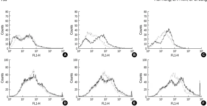
ROS generation in microglial cells of the mouse brain was analyzed using flow cytometry (Fig. 3). Microglial cells from the brainstem and cerebellum of Gaucher mice had higher levels of ROS compared with microglial cells from the brain-stem and cerebellum wild-type mice at embryonic day 19.5 (E19.5) and postnatal day 1 (P1). However, there was no dif-ference in ROS levels in cells from the cerebral cortex of wild-type mice and Gaucher mice.
DISCUSSION
Gaucher disease is mainly caused by the accumulation of glucocerebroside, a metabolic intermediate in the synthesis and degradation of complex glycosphingolipids. This causes the release of calcium from intracellular stores, leading to enhanced sensitivity to agents that induce neuronal death (13, 15). The deacylated analog of glucocerebroside, gluco-sylsphingosine, accumulates in the cerebral and cerebellar cortices of patients with type II and type III Gaucher disease (16), and in the brains of Gaucher mice (17), and has been suggested to be a candidate neurotoxin (18, 19). However, no explanation has yet been proposed to link these experi-mental results to the known neurodegenerative symptoms of Gaucher disease (20). IL 1 IL 1 IL 6 TNF -actin IL 6 200 M CBE - + TNF -actin W-Cx G-Cx W-Bs G-Bs W-Cbll G-Cbll A B C Secreted cytokine (pg/mL) 300 250 200 150 100 50 0 IFN- IL 6
TNF-Fig. 1.RT-PCR analysis of IL-1 , IL-1 , IL-6 and TNF- mRNA expression in mouse brains (A) and of IL-6 and TNF- mRNA expression in cultured microglia with or without 200 M of CBE (conduritol B epoxide; see Materials and Methods) (B). Cytokines secreted from cul-tured microglia were analyzed (C): Open bar, untreated microglia; closed bar, microglia treated with 200 M of CBE for 8 days. W-Cx, cerebral cortex of wild type mouse; G-Cx, cerebral cortex of Gaucher mouse; W-Bs, brainstem of wild type; G-Bs, brainstem of Gaucher mouse; W-Cbll, cerebellum of wild type; G-Cbll cerebellum of Gaucher mouse..
Nitric oxide ( M ) 5 4 3 2 1 0
Cerebral cortex Brainstem Cerebellum
Fig. 2.NO release from cultured brain cells of cerebral cortex, bra-instem and cerebellum. Open bars, NO levels from cells of wild type mice; closed bars, NO levels from cells of Gaucher mice. *p< 0.01, �
p<0.001 vs. wild type mice (by paired t tests).
�
�
Gaucher disease is characterized by the accumulation of glucosylceramide in macrophages and macrophage-derived cells in various tissues (3). Among the earliest and more pro-vocative suggestions to explain phenotypic variability in Gau-cher disease was the suggestion that it is an immune or auto-immune reaction to a chronic storage of glucocerebroside (21). Systemically, an excessive immune response involves the inhi-bition of proinflammatory cytokines, such as TNF- , which is produced by macrophages and is necessary for adhesion molecule expression and leukocyte recruitment to inflamma-tory sites (22).
Inflammation is an important contributor to neuronal da-mage in other neurodegenerative conditions, such as Parkin-son’s disease, Alzheimer’s disease, multiple sclerosis, and amyo-trophic lateral sclerosis (23-25). One of the best-known fea-tures of inflammation in the central nervous system (CNS) is the production of NO, which is a product of macrophages that have been activated by cytokines and a microbial com-pound found in the inflammatory reaction (26). Lipopolysac-charide, IFN- , TNF- , and IL-1 are responsible for microglial activation and NO production (27). Production of NO is associated with the generation of ROS, including superoxide anions, hydroxyl radicals, lipid hydroperoxides, and hydro-gen peroxide. These compounds are toxic to neurons because they induce lipid peroxidation, DNA fragmentation, and protein oxidation (28, 29). Moreover, ROS can activate pro-tein kinase C, mitogen-activated propro-tein kinase, and nuclear factor-kappa B, which regulate the expression of genes encod-ing a variety of proinflammatory factors (30, 31). Inflamma-tion-mediated neurodegeneration has received considerable attention in lysosomal storage diseases. In mouse models of
Sandhoff disease and Tay-Sachs disease, genes associated with the activation of microglia and astrocytes are overexpressed, and progressive CNS inflammation correlates with the sever-ity of disease (32, 33).
We could not observe the inflammatory reaction in the fetal brains of Gaucher mice because this mouse model does not survive longer than one day after birth (11). Therefore, we examined the fetal brains of Gaucher mice and explored the accumulation of glucosylceramide in microglia, which could be a causative factor of inflammation related to neu-rodegeneration.
In this study, the secretion of proinflammatory cytokines increased in the brains of Gaucher mice and in CBE-treated microglia, a cell model of Gaucher disease. We examined whether the upregulation of proinflammatory cytokines result-ed in the production of NO and found increasresult-ed production of NO in the brain cells of Gaucher mice. Increased ROS generation in microglial cells from the brainstem and cere-bellum of Gaucher mice also occurred. Thus, a deficiency of GC activity may mediate microglial activation resulting in the production of cytokines, NO, and ROS.
Further in vivo approaches to understanding the patho-physiology of Gaucher disease and the development of new therapeutic strategies for Gaucher disease have been hindered by the lack of a viable animal model (11, 34). However, the generation of viable mouse models of Gaucher disease has recently been reported (35), and these mouse models will be useful for elucidating the pathogenesis of the neuronopathic forms of Gaucher disease.
Although we have no direct evidence that the overexpres-sion of proinflammatory cytokines or the production of NO
Counts 80 70 60 50 40 30 20 10 0 100 101 102 103 104 FL1-H
Fig. 3.Detection of ROS with CM-H2DCFDA (see Materials and methods) in the microglial cells of cerebral cortex (Aand D), brainstem (Band E) and cerebellum (Cand F) from wild type mice (n=3, dotted line) and Gaucher mice (n=3, solid line). Upper panel, cells from day E19.5 embryos; lower panel, cells from day P1 neonates.
A Counts 80 70 60 50 40 30 20 10 0 100 101 102 103 104 FL1-H B Counts 80 70 60 50 40 30 20 10 0 100 101 102 103 104 FL1-H C Counts 100 80 60 40 20 0 100 101 102 103 104 FL1-H D Counts 100 80 60 40 20 0 100 101 102 103 104 FL1-H E Counts 100 80 60 40 20 0 100 101 102 103 104 FL1-H F
and ROS are involved in the neurodegenerative symptoms of Gaucher disease, further investigation of the relationship between microglial activation and the accumulation of GC substrates should clarify the roles of these factors.
REFERENCES
1. Beutler E, Grabowski GA. Gaucher disease. In: Scriver CR, Beaudet AL, Sly WS, Valle D, editors, The Metabolic and Molecular Bases of Inherited Disease. New York: McGraw-Hill 2001: 3635-68. 2. Brady RO, Kanfer JN, Bradley RM, Shapiro D. Demonstration of a
deficiency of glucocerebroside-cleaving enzyme in Gaucher’s dis-ease. J Clin Invest 1966; 45: 1112-5.
3. Brady RO, Barton NW, Grabowski GA. The role of neurogenetics in Gaucher disease. Arch Neurol 1993; 50: 1212-24.
4. Hong YB, Kim EY, Jung SC. Down regulation of Bcl-2 in the fetal brain of Gaucher disease mouse model: a possible role in the neuronal loss. J Hum Genet 2004; 49: 349-54.
5. Barak V, Acker M, Nisman B, Kalickman I, Abrahamov A, Zimran A, Yatziv S. Cytokines in Gaucher’s disease. Eur Cytokine Netw 1999; 10: 205-10.
6. Lichtenstein M, Zimran A, Horowitz M. Cytokine mRNA in Gauch-er disease. Blood Cell Mol Dis 1997; 23: 395-401.
7. Mizukami H, Mi Y, Wada R, Kono M, Yamashita T, Liu Y, Werth N, Sandhoff R, Sandhoff K, Proia RL. Systemic inflammation in glucocerebrosidase-deficient mice with minimal glucosylceramide storage. J Clin Invest 2002; 109: 1215-21.
8. Baudry M, Yao Y, Simmons D, Liu J, Bi X. Postnatal development of inflammation in a murine model of Niemann-Pick type C disease: immunohistochemical observations of microglia and astroglia. Exp Neurol 2003; 184: 887-903.
9. Jeyakumar M, Thomas R, Elliot-Smith E, Smith DA, van der Spoel AC, d’Azzo A, Perry VH, Butters TD, Dwek RA, Platt FM. Central nervous system inflammation is a hallmark of pathogenesis in mouse models of GM1 and GM2 gangliosidosis. Brain 2003; 126: 974-87. 10. Wu YP, Proia RL. Deletion of macrophage-inflammatory protein
1 retards neurodegeneration in Sandhoff disease mice. Proc Natl Acad Sci USA 2004; 101: 8425-30.
11. Tybulewicz VL, Tremblay ML, LaMarca ME, Willemsen P, Stub-blefield BK, Winfield S, Zablocka B, Sidransky E, Martin BM, Huang SP, Mintzer KN, Westphal H, Mulligan RO, Ginns EI. Ani-mal model of Gaucher’s disease from targeted disruption of the mouse glucocerebrosidase gene. Nature 1992; 357: 407-10.
12. Min KJ, Yang MS, Jou I, Joe EH. Protein kinase A mediates micro-glial activation induced by plasminogen and gangliosides. Exp Mol Med 2004; 36: 461-7.
13. Pelled D, Shogomori H, Futerman AH. The increased sensitivity of neurons with elevated glucocerebroside to neurotoxic agents can be reversed by imiglucerase. J Inherit Metab Dis 2000; 23: 175-84. 14. Ding AH, Nathan CF, Stuehr DJ. Release of reactive nitrogen
inter-mediates and reactive oxygen interinter-mediates from mouse peritoneal macrophages: comparison of activating cytokines and evidence for independent production. J Immunol 1988; 141: 2407-12.
15. Korkotian E, Schwarz A, Pelled D, Schwarzmann G, Segal M, Futer-man AH. Elevation of intracellular glucosylceramide levels results in an increase in endoplasmic reticulum density and in functional cal-cium stores in cultured neurons. J Biol Chem 1999; 274: 21673-8. 16. Orvisky E, Park JK, LaMarca ME, Ginns EI, Martin BM, Tayebi N,
Sidransky E. Glucosylsphingosine accumulation in tissues from pa-tients with Gaucher disease: Correlation with phenotype and geno-type. Mol Genet Metab 2002; 76: 262-70.
17. Orvisky E, Sidransky E, McKinney CE, LaMarca ME, Samimi R, Krasnewich D, Martin BM, Ginns EI. Glucosylsphingosine accumu-lation in mice and patients with type 2 Gaucher disease begins early in gestation. Pediatr Res 2000; 48: 233-7.
18. Lloyd-Evans E, Pelled D, Riebeling G, Futerman AH. Lyso-glycos-phingolipids mobilize calcium from brain microsomes via multiple mechanisms. Biochem J 2003; 375: 561-5.
19. Schueler UH, Kolter T, Kaneski CR, Blusztajn JK, Herkenham M, Sandhoff K, Brady RO. Toxicity of glucosylsphingosine to cultured neuronal cells: a model system for assessing neuronal damage in Gaucher disease type 2 and 3. Neurobiol Dis 2003; 14: 595-601. 20. Raas-Rothschild A, Pankova-Kholmyansky I, Kacher Y, Futerman
AH. Glycosphingolipidoses: beyond the enzymatic defect. Glycoconj J 2004; 21: 295-304.
21. Shoenfeld Y, Gallant LA, Shaklai M, Livni E, Djaaldetti M, Pinkhas J. Gaucher’s disease: a disease with chronic stimulation of the immune system. Arch Pathol Lab Med 1982; 106: 388-91.
22. Kluth DC, Rees AJ. Inhibiting inflammatory cytokines. Semin Nephrol 1996; 16: 576-82.
23. Liu B, Hong JS. Role of microglia in inflammation mediated neurode-generative disease: mechanisms and strategies for therapeutic inter-vention. J Pharmacol Exp Ther 2003; 304: 1-7.
24. McGeer PL, Itagaki S, Boyes BE, McGeer EG. Reactive microglia are positive for HLA-DR in the substantia nigra of Parkinson’s and Alzheimer’s disease brains. Neurology 1998; 38: 1285-91. 25. Raine CS. Multiple sclerosis: immune system molecule expression
in the central nervous system. J Neuropathol Exp Neurol 1994; 53: 328-37.
26. Guzik TJ, Korbut R, Adamek-Guzik T. Nitric oxide and superoxide in inflammation and immune regulation. J Physiol Pharmalcol 2003; 54: 469-87.
27. Bogdan C. Nitric oxide and the immune response. Nat Immunol 2001; 2: 907-16.
28. Faber JL. Mechanisms of cell injury by activated oxygen species. Environ Health Perspect 1994; 102: 17-24.
29. Wang T, Qin L, Liu B, Liu Y, Wilson B, Eling TE, Langenbach R, Taniura S, Hong JS. Role of reactive oxygen species in LPS-induced
production of prostaglandin E2in microglia. J Neurochem 2004; 88:
939-47.
30. Lal MA, Brismar H, Eklof AC, Aperia A. Role of oxidative stress in advanced glycation end product-induced mesangial cell activation. Kidney Int 2002; 61: 2006-14.
31. Rosenberger J, Petrovics G, Buzas B. Oxidative stress induces proor-phanin FQ and proenkephalin gene expression in astrocytes through p38- and ERK-MAP kinases and NF-kappaB. J Nerurochem 2001; 79: 35-44.
32. Myerowitz R, Lawson D, Mizukami H, Mi Y, Tifft CJ, Proia RL. Molecular pathophysiology in Tay-Sachs and Sandhoff diseases as revealed by gene expression profiling. Hum Mol Genet 2002; 11: 1343-50.
33. Wada R, Tifft CJ, Proia RL. Microglial activation precedes acute neurodegeneration in Sandhoff disease and is suppressed by bone marrow transplantation. Proc Natl Acad Sci USA 2000; 97: 10954-9.
34. Liu Y, Suzuki K, Reed JD, Grinberg A, Westphal H, Hoffmann A, Doring T, Sandhoff K, Proia RL. Mice with type 2 and 3 Gaucher disease point mutations generated by a single insertion mutagenesis procedure (SIMP). Proc Natl Acad Sci USA 1998; 95: 2503-8. 35. Xu YH, Quinn B, Witte D, Grabowski GA. Viable mouse models of
acid beta-glucosidase deficiency: the defect in Gaucher disease. Am J Pathol 2003; 163: 2093-101.