저작자표시-비영리-변경금지 2.0 대한민국 이용자는 아래의 조건을 따르는 경우에 한하여 자유롭게 l 이 저작물을 복제, 배포, 전송, 전시, 공연 및 방송할 수 있습니다. 다음과 같은 조건을 따라야 합니다: l 귀하는, 이 저작물의 재이용이나 배포의 경우, 이 저작물에 적용된 이용허락조건 을 명확하게 나타내어야 합니다. l 저작권자로부터 별도의 허가를 받으면 이러한 조건들은 적용되지 않습니다. 저작권법에 따른 이용자의 권리는 위의 내용에 의하여 영향을 받지 않습니다. 이것은 이용허락규약(Legal Code)을 이해하기 쉽게 요약한 것입니다. Disclaimer 저작자표시. 귀하는 원저작자를 표시하여야 합니다. 비영리. 귀하는 이 저작물을 영리 목적으로 이용할 수 없습니다. 변경금지. 귀하는 이 저작물을 개작, 변형 또는 가공할 수 없습니다.
Cardiac Co-morbidities in
Chronic Obstructive Pulmonary Disease
by
Young Hwan Ahn
Major in Medicine
Department of Medical Sciences
The Graduate School, Ajou University
Cardiac Co-morbidities in
Chronic Obstructive Pulmonary Disease
by
Young Hwan Ahn
A Dissertation Submitted to The Graduate School of
Ajou University in Partial Fulfillment of The Requirements for
The Degree of Master of Medicine
Supervised by
Joo Hun Park, MD
Major in Medicine
Department of Medical Sciences
The Graduate School, Ajou University
This certifies that the dissertation
of Young Hwan Ahn is approved.
SUPERVISORY COMMITTEE
_________________
Joo Hun Park
_________________
Seungsoo Sheen
_________________
Joo Sung Sun
The Graduate School, Ajou University
June 19th, 2015
i - ABSTRACT -
Cardiac Co-morbidities in
Chronic Obstructive Pulmonary Disease
Background: Coronary artery disease (CAD) is a frequent co-morbidity in chronic obstructive
pulmonary disease (COPD) and cardiovascular disease is the most common cause of death in COPD. Therefore, this study was performed to identify the prognostic factors associated with the CAD in COPD and investigate the independent risk factors for mortality in COPD patients who underwent comprehensive cardiac evaluations
Methods: We retrospectively reviewed 405 patients with COPD who had undergone
comprehensive cardiac evaluations including coronary angiography, coronary multi-detector computed tomography and echocardiography in Ajou University Hospital from Jan 2000 to Dec 2012. Survival analyses of prognostic factors were performed in this retrospective cohort.
Results: Male gender, hypertension, left heart failure, lower hemoglobin level, and lower serum
HDL were independently associated with the presence of CAD in COPD (p<0.05). Older age, lower body mass index(BMI), lower left ventricular ejection fraction, lower forced expiratory volume in one second(FEV1), and lower hemoglobin level were independently associated with higher mortality in
total COPD group (p<0.05), whereas presence of CAD was not.
Conclusion: Our study identified lower left ventricular ejection fraction and lower hemoglobin
level along with older age, lower BMI, and lower FEV1 as independent risk factors for the mortality of
COPD patients, suggesting that multidisciplinary approaches are required in the care of COPD patients. Male gender, hypertension, left heart failure, and lower serum HDL level were independent predictors for the presence of CAD in COPD.
ii
TABLE OF CONTENTS
ABSTRACT ··· i
TABLE OF CONTENTS ··· ii
LIST OF FIGURES ··· iii
LIST OF TABLES ··· iv
I. INTRODUCTION ··· 1
II. SUBJECTS AND METHODS··· 1
III. RESULTS ··· 3
IV. DISCUSSION ··· 9
V. CONCLUSION ··· 11
REFERENCES ··· 12
iii
Fig. 1. Flow diagram of subjects ··· 2
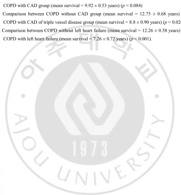
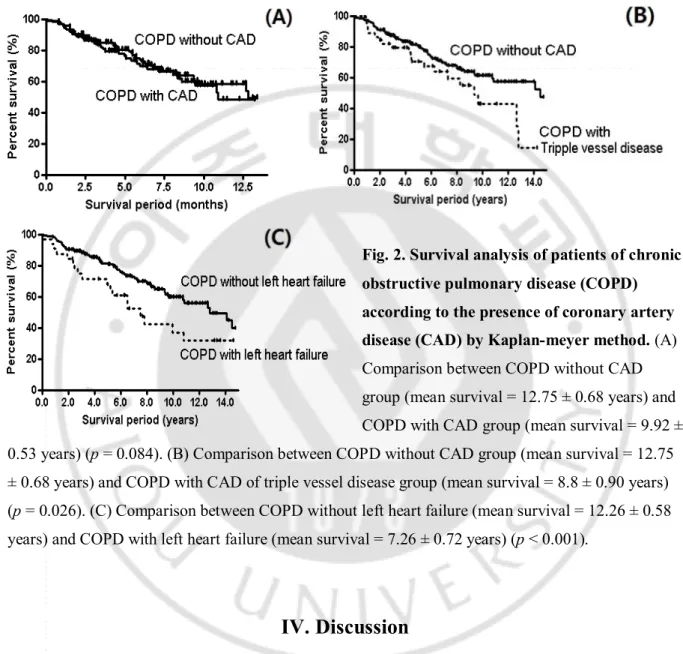
Fig. 2. Survival analysis of patients of chronic obstructive pulmonary disease (COPD) according to the presence of coronary artery disease (CAD) by Kaplan-meier method ··· 9 A. Comparison between COPD without CAD group (mean survival = 12.75 ± 0.68 years) and
COPD with CAD group (mean survival = 9.92 ± 0.53 years) (p = 0.084)
B. Comparison between COPD without CAD group (mean survival = 12.75 ± 0.68 years) and COPD with CAD of triple vessel disease group (mean survival = 8.8 ± 0.90 years) (p = 0.026) C. Comparison between COPD without left heart failure (mean survival = 12.26 ± 0.58 years) and
iv
Table 1. Baseline characteristics of COPD with CAD and without CAD ··· 4 Table 2. Comparison of laboratory findings and co-morbidities between COPD with CAD and
without CAD ··· 5
Table 3. Predictors of coronary artery disease in the patients with COPD by Multi-variate logistic regression analysis ··· 6
Table 4. Prognostic Factors for the Mortality in total COPD group by Univariate Cox Model ··· 7 Table 5. Prognostic Factors for the Mortality in Total COPD Group by Multivariate Cox Regression
1
I. Introduction
Chronic obstructive pulmonary disease (COPD) is an increasing cause of morbidity and mortality worldwide [1]. COPD is now recognized not as a single disease, but a syndrome with multiple phenotypes accompanied by many morbidities [2, 3]. Cardiovascular disease among the many co-morbidities of COPD is the main cause of death for COPD and atherosclerosis may occur in COPD regardless of smoking history [4-6].
Oxidative stress, proteolytic enzymes, inflammatory cytokines, and molecules related to the aging process are thought to contribute to the development of coronary artery disease (CAD) in COPD [7-12]. Clinically, the degree of emphysema and airflow limitation are reported to be associated with the development of CAD in COPD [13-15]. There is strong epidemiologic evidence that reduced lung function is a marker for cardiovascular mortality independent of age, gender, and smoking history [15]. However, clinical risk factors associated with CAD in patients with COPD are not easy to identify, since the data of angiography proven CAD in COPD are extremely difficult to obtain [16, 17]. So far, few reports clearly defined COPD without CAD as a control because angiography was not done in all the patients with COPD, although some studies did investigate the clinical course of COPD with CAD [18, 19].
Therefore, this study was performed to identify the prognostic factors associated with CAD in COPD and investigate the independent risk factors for mortality in COPD patients who underwent comprehensive cardiac evaluations
II. SUBJECTS AND METHODS
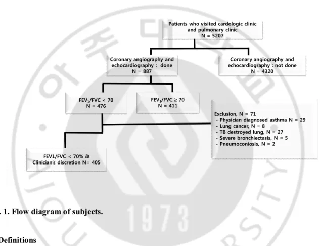
We retrospectively reviewed 5207 patients with airway disease who on an outpatient basis visited pulmonology clinic and cardiology clinic in Ajou University Hospital (a university affiliated 1,000-bed sized tertiary referral center in Suwon, Republic of Korea) from Jan 2000 to Dec 2012. Among them, we enlisted 405 patients with COPD (male 88.4%, mean age 68.9 ± 9.4 years) who had undergone coronary multi-detector computed tomography or coronary angiography. Based on the interpretation of chest radiographs by radiologists, 29 patients with physician diagnosed asthma, 8 patients with lung cancer, 27 patients with tuberculous destroyed lung, 5 patients with severe bronchiectasis, and 2 patients with pneumoconiosis were excluded from our study as in figure 1.
2
Absence of coronary artery disease was proven in 74 patients by coronary multi-detector computed tomography without coronary angiography and coronary angiography was performed by cardiologists in 331 patients. Clinical history, co-morbidities, demographics, medication history, laboratory findings, and radiological findings were acquired by reviewing medical records. Clinical and laboratory parameters were obtained at the time of cardiac evaluation. Survival period was calculated from the time of cardiac evaluation. Mean observation period was 5.5 ± 4.0 years. Survival status was obtained from telephone interview, medical records, or Statistics Korea (kostat.go.kr).
Exclusion, N = 71
- Physician diagnosed asthma N = 29 - Lung cancer, N = 8
- TB destroyed lung, N = 27 - Severe bronchiectasis, N = 5 - Pneumoconiosis, N = 2 Patients who visited cardologic clinic
and pulmonary clinic N = 5207
FEV1/FVC < 70 N = 476
Coronary angiography and echocardiography : done
N = 887
Coronary angiography and echocardiography : not done
N = 4320
FEV1/FVC < 70% & Clinician’s discretion N= 405
FEV1/FVC ≥ 70 N = 411
Fig. 1. Flow diagram of subjects.
A. Definitions
The diagnosis of COPD was determined by clinician’s judgement and previously documented airflow limitation (forced expiratory volume in 1 second [FEV1]/ forced vital capacity [FVC] <70%
that was not fully reversible) [1]. No or minimal abnormality on chest radiography. CAD was defined as more than 50% stenosis of at least one major epicardial coronary artery. The presence or absence of CAD was confirmed by cardiologists. Left heart failure was defined as ≤40% ejection fraction according to echocardiographic findings reported by cardiologists [20]. Data on duration and amount of cigarette smoking was obtained and an ex-smoker was defined as a subject who had not smoked for at least 1 year. Body mass index (BMI) was calculated as the body weight divided by the height squared (kg/m2). Acute exacerbation was defined according to the GOLD document [1]. If the
3
the clinician’s judgment, exacerbation was defined as moderate. If hospitalization was required at the discretion of the clinician, exacerbation was considered severe. We included only moderate and severe exacerbations in the exacerbation group of COPD as in other studies [21-22]. Spirometry (Elite-DX or CPFS; Medgraphics, MN, USA) was performed according to the American Thoracic Society guidelines [23].
B.Statistical analysis
SPSS version 12 (SPSS Inc., Chicago, IL, USA) was used for the analysis. All values were expressed as means ± standard deviation. The Chi-Square test and/or Fisher’s exact test were used for categorical data. The student T-test was used for continuous data. We constructed the 95% confidence interval (CI) of each variable. Simple and multiple logistic regression analyses of significant variables from previous analyses were performed. Clinically significant variables that showed statistically significant odds ratios from simple logistic regression models were selected for inclusion in multiple forward stepwise logistic regression models. Irrespective of the results of univariate analysis, age, gender, and smoking status were included in multiple logistic regression analysis. Survival curves were plotted using the Kaplan–Meier method, and the significance of differences between groups was analyzed using the log-ranks test. A Cox proportional hazards model was used for multivariate analysis to evaluate factors contributing to survival. A p-value less than 0.05 was deemed to indicate statistical significance.
C. Ethics Statement
The present study was approved by the Institutional Review Board of Ajou University Hospital.
III. RESULTS
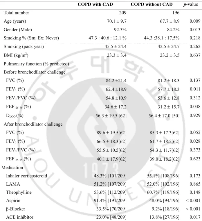
A. Baseline characteristics between COPD with CAD and COPD without CAD
COPD with CAD group was older and male predominant compared to COPD without CAD (p<0.05), although baseline pulmonary functions such as FEV1, FEF25-75%, and FVC after
bronchodilator challenge were better in COPD with CAD than COPD without CAD. However, there was no significant difference in smoking history and BMI. COPD patients with CAD used aspirin, angiotensin-converting enzyme inhibitors, angiotensin-receptor blockers and β-blockers more often than COPD patients without CAD (p<0.05) (Table 1).
4
Table 1. Baseline characteristics of COPD with CAD and without CAD
COPD with CAD COPD without CAD p-value
Total number 209 196
Age (years) 70.1 ± 9.7 67.7 ± 8.9 0.009
Gender (Male) 92.3% 84.2% 0.013
Smoking % (Sm: Ex: Never) 47.3 : 40.6 : 12.1 % 44.3 :38.1 : 17.5% 0.218
Smoking (pack year) 45.5 ± 24.4 42.5 ± 24.7 0.262
BMI (kg/m2) 23.3 ± 3.4 23.2 ± 3.5 0.637
Pulmonary function (% predicted) Before bronchodilator challenge
FVC (%) 84.2 ±21.4 81.2 ± 18.3 0.137
FEV1 (%) 62.4 ±18.9 57.7 ± 18.3 0.011
FEV1/FVC (%) 54.8 ±10.9 53.6 ± 12.8 0.312
FEF 25-75 (%) 34.6 ± 17.2 31.2 ± 15.7 0.038
DLCO (%) 56.3 ± 19.5 [62] 56.4 ± 17.0 [50] 0.929
After bronchodilator challenge
FVC (%) 89.6 ± 19.5[62] 85.3 ± 17.3[62] 0.052 FEV1 (%) 66.5 ± 18.3[62] 61.7 ± 18.5[62] 0.028 FEV1/FVC (%) 55.5 ± 10.5[62] 54.3 ± 11.7[62] 0.373 FEF 25-75 (%) 40.1 ± 17.9[62] 39.0 ± 18.2[62] 0.623 Medication Inhaler corticosteroid 48.3% [101/209] 55.1% [108/196] 0.173 LAMA 51.2% [107/209] 52.0% [102/196] 0.865 Theophylline 53.6% [112/209] 60.7% [119/196] 0.148 Aspirin 91.4% [191/209] 48.0% [94/196] < 0.001 β-Blocker 33.5% [70/209] 9.2% [18/196] < 0.001 ACE inhibitor 23.0% [48/209] 13.8% [27/196] 0.017
ACE inhibitor = converting enzyme inhibitor, BMI = body mass index, COPD = chronic obstructive pulmonary disease, CAD = coronary artery disease, DLCO = diffusion capacity of carbon monoxide, FEV1 = forced expiratory volume in 1 second, FEF 25-75% = forced expiratory flow 25–75%, FVC =
forced vital capacity, LAMA= long-acting muscarinic antagonist. Continuous variables are presented as means ± standard deviation. Categorical variables are expressed as numbers of subjects or percentages in parentheses. The number of participants is noted in square brackets, if any values for
5 each item were missing.
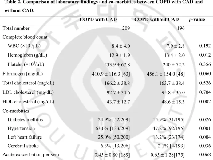
B. Comparison of laboratory findings and comorbidities between COPD with CAD and COPD without CAD (Table 2).
Hemoglobin level and serum HDL were lower in COPD with CAD than in COPD without CAD (p<0.05). Diabetes, hypertension, left heart failure, and/or cerebral stroke were combined more frequently in COPD with CAD, compared to COPD without CAD (p<0.05). However, no significant difference was found in the rate of acute exacerbation after enrollment between two groups.
Table 2. Comparison of laboratory findings and co-morbities between COPD with CAD and without CAD.
COPD with CAD COPD without CAD p-value
Total number 209 196
Complete blood count
WBC (×103/μL) 8.4 ± 4.0 7.9 ± 2.8 0.192 Hemoglobin (g/dL) 12.9 ± 1.9 13.4 ± 2.0 0.012 Platelet (×103/μL) 233.9 ± 67.8 240 ± 72.2 0.356 Fibrinogen (mg/dL) 410.9 ± 116.3 [63] 456.1 ± 154.0 [48] 0.060 Total cholesterol (mg/dL) 166.2 ± 38.8 163.7 ± 38.4 0.526 LDL cholesterol (mg/dL) 92.7 ± 34.6 95.8 ± 35.0 0.704 HDL cholesterol (mg/dL) 43.7 ± 12.7 48.6 ± 15.3 0.002 Co-morbities Diabetes mellitus 24.9% [52/209] 15.9% [31/195] 0.026 Hypertension 63.6% [133/209] 47.2% [92/195] 0.001
Left heart failure 25.0% [50/200] 13.2% [23/174] 0.004
Cerebral stroke 6.3% [13/206] 2.1% [4/193] 0.036
Acute exacerbation per year 0.45 ± 0.80 [189] 0.65 ± 1.28[175] 0.068 CAD = coronary artery disease, COPD = chronic obstructive pulmonary disease, HDL = high-density lipoprotein, hs-CRP = high-sensitivity C-reactive protein, LDL = low-density lipoprotein, WBC = white blood cell. Continuous variables are presented as the mean ± standard deviation. Categorical variables are expressed as percentages or numbers of subjects in square brackets .The number of participants is noted in square brackets, if there are missing data for each item.
6
C. Prognostic factors of CAD in COPD
Using simple logistic regression analysis, presence of CAD was associated with older age (OR=1.029), male gender (OR=2.266), diabetes (OR=1.752), hypertension (OR=1.959), cerebral stroke (OR=3.185), left heart failure (OR=2.188), lower HDL level (OR=0.975), and lower serum hemoglobin level (OR=0.876) (p<0.05) (Table 3). Multiple logistic regression analysis of risk factors significant by simple logistic analysis was performed to detect independent predictors. Male gender (OR=2.273), hypertension (OR=1.763), left heart failure (OR=2.096), lower HDL level (OR=0.981), and lower hemoglobin level (OR=0.842) were independently associated with the presence of CAD in total COPD group (p<0.05) (Table 4).
Table 3 Predictors of coronary artery disease in the patients with COPD by Multi-variate forward stepwise logistic regression analysis
Bi-variate model Multi-variate model Odds ratio (95% CI) p-value Odds ratio (95% CI) p-value Age (years) 1.029(1.007-1.051) 0.010 Male 2.266(1.197-4.290) 0.012 2.273(1.022-5.051) 0.044 Smoker 1.244(0.370-1.130) 0.126 BMI (kg/m2) 1.014(0.958-1.073) 0.637 Co-morbidities Diabetes Mellitus 1.752(1.067-2.876) 0.027 Hypertension 1.959(1.316-2.917) 0.001 1.764(1.064-2.925) 0.028 Left heart failure 2.188(1.271-3.767) 0.005 2.096(1.070-4.106) 0.031 Cerebral Stroke 3.185(1.019-9.901) 0.046 Laboratory findings HDL cholesterol (mg/dL) 0.975(0.959-0.991) 0.003 0.981(0.964-0.999) 0.041 WBC (x103/μL) 1.042(0.979-1.108) 0.198 Hemoglobin (g/dL) 0.876(0.789-0.972) 0.013 0.842(0.732-0.969) 0.016 Acute exacerbation(rate/year) 1.003(0.579-1.736) 0.993 FEV1 % 1.014(1.003-1.025) 0.012
BMI = body mass index, CI = confidence interval, COPD = chronic obstructive pulmonary disease, FEV1 = forced expiratory volume in 1 second, HDL = high-density lipoprotein, WBC = white blood
7
analyzed using simple and multiple logistic regression models.
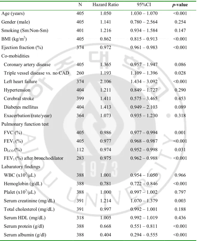
Table 4. Prognostic Factors for the Mortality in total COPD group by Univariate Cox Model.
N Hazard Ratio 95%CI p-value
Age (years) 405 1.050 1.030 – 1.070 <0.001 Gender (male) 405 1.141 0.780 – 2.564 0.254 Smoking (Sm:Non-Sm) 401 1.216 0.934 – 1.584 0.147 BMI (kg/m2) 405 0.862 0.815 – 0.913 <0.001 Ejection fraction (%) 374 0.972 0.961 – 0.983 <0.001 Co-mobidities
Coronary artery disease 405 1.365 0.957 – 1.947 0.086 Triple vessel disease vs. no CAD 260 1.193 1.109 – 1.396 0.028 Left heart failure 374 2.106 1.434 – 3.092 <0.001
Hypertension 404 1.211 0.849 – 1.727 0.290
Cerebral stroke 399 1.411 0.575 – 3.465 0.453
Diabetes mellitus 404 1.413 0.949 – 2.103 0.089
Exacerbation (rate/year) 364 1.073 0.935 – 1.230 0.318 Pulmonary function test
FVC (%) 405 0.986 0.977 – 0.994 0.001
FEV1 (%) 405 0.977 0.968 – 0.987 <0.001
DLCO (%) 112 0.974 0.952 – 0.998 0.031
FEV1 (%) after bronchodilator 283 0.975 0.962 – 0.988 <0.001
Labaratory findings WBC (x103/μL) 388 1.001 0.954 – 1.050 0.966 Hemoglobin (g/dL) 388 0.781 0.722 – 0.846 <0.001 Plalet (x103/μL) 388 1.000 0.997 – 1.002 0.797 Serum creatinine (mg/dL) 391 1.214 1.070 – 1.379 0.003 Total cholesterol (mg/dL) 391 0.997 0.992 – 1.001 0.188 Serum HDL (mg/dL) 318 1.005 0.992 – 1.019 0.436 Serum protein (g/dl) 388 0.668 0.551 – 0.811 <0.001 Serum albumin (g/dl) 388 0.404 0.294 – 0.555 <0.001 BMI = body mass index, CI = confidence interval, COPD = chronic obstructive pulmonary disease, Ejection fraction (%) = left ventricular ejection fraction, FEV1 = forced expiratory volume in 1 second,
8
D.Survival analysis
Among the 126 patients with COPD who died during the observation period, mortality was most frequently due to ischemic heart disease (27 patients, 21.4%). Other causes were respiratory failure including acute exacerbation of COPD (25 patients), cancers other than lung cancer (19 patients), lung cancer (11 patients), pneumonia (8 patients), cerebrovascular disease (6 patients), septic shock (4 patient), pulmonary embolism (1 patient) and unknown causes (1 patient), etc (24 patients). Only all-cause mortality was used for survival analysis, beall-cause the all-cause of death was not detected some cases of non-survivors. According to the univariate cox model, age, BMI, left heart failure, serum hemoglobin level, FVC, FEV1, DLCO, serum creatinine level, serum protein level, and serum albumin
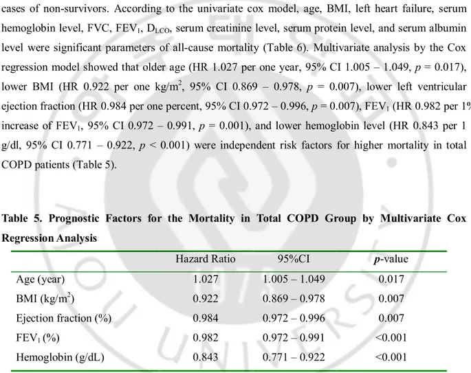
level were significant parameters of all-cause mortality (Table 6). Multivariate analysis by the Cox regression model showed that older age (HR 1.027 per one year, 95% CI 1.005 – 1.049, p = 0.017), lower BMI (HR 0.922 per one kg/m2, 95% CI 0.869 – 0.978, p = 0.007), lower left ventricular
ejection fraction (HR 0.984 per one percent, 95% CI 0.972 – 0.996, p = 0.007), FEV1 (HR 0.982 per 1%
increase of FEV1, 95% CI 0.972 – 0.991, p = 0.001), and lower hemoglobin level (HR 0.843 per 1
g/dl, 95% CI 0.771 – 0.922, p < 0.001) were independent risk factors for higher mortality in total COPD patients (Table 5).
Table 5. Prognostic Factors for the Mortality in Total COPD Group by Multivariate Cox Regression Analysis
Hazard Ratio 95%CI p-value
Age (year) 1.027 1.005 – 1.049 0.017
BMI (kg/m2) 0.922 0.869 – 0.978 0.007
Ejection fraction (%) 0.984 0.972 – 0.996 0.007
FEV1 (%) 0.982 0.972 – 0.991 <0.001
Hemoglobin (g/dL) 0.843 0.771 – 0.922 <0.001
BMI = body mass index, CI = confidence interval, COPD = chronic obstructive pulmonary disease, ejection fraction = left ventricular ejection fraction, FEV1 = forced expiratory volume in 1 second
In Kaplan –Meier survival analysis, COPD without CAD group (mean survival = 12.75 ± 0.68 years) has a tendency to survive longer than COPD with CAD group (mean survival = 9.92 ± 0.53 years, p = 0.084), though this parameter was not significant in multivariate analysis by the Cox regression model (Figure 2). COPD with CAD of triple vessel disease group (mean survival = 8.8 ±
9
0.90 years) had a shorter survival period, compared to the control group (mean survival = 12.75 ± 0.68 years) (p = 0.028) (Figure 2). In keeping with multivariate analysis, COPD without left heart failure (mean survival = 12.26 ± 0.58 years) survived longer than COPD with left heart failure (mean survival = 7.26 ± 0.72 years) (p < 0.001) (Figure 2).
Fig. 2. Survival analysis of patients of chronic obstructive pulmonary disease (COPD) according to the presence of coronary artery disease (CAD) by Kaplan-meyer method. (A)
Comparison between COPD without CAD group (mean survival = 12.75 ± 0.68 years) and COPD with CAD group (mean survival = 9.92 ± 0.53 years) (p = 0.084). (B) Comparison between COPD without CAD group (mean survival = 12.75 ± 0.68 years) and COPD with CAD of triple vessel disease group (mean survival = 8.8 ± 0.90 years) (p = 0.026). (C) Comparison between COPD without left heart failure (mean survival = 12.26 ± 0.58 years) and COPD with left heart failure (mean survival = 7.26 ± 0.72 years) (p < 0.001).
IV. Discussion
The aim of this study was to investigate the effect of cardiovascular comorbidities on the prognosis of COPD, and to identify the prognostic factors associated with the CAD in COPD. Our study demonstrated that older age, lower BMI, worse left ventricular ejection fraction, lower FEV1, and
lower hemoglobin level were independent risk factors for the mortality of COPD patients. Male gender, hypertension, left heart failure, and lower serum HDL level were independent predictors for
10
the presence of CAD in COPD. According to the Towards a Revolution in COPD Health (TORCH) trial, 27% of deaths that occurred during the observation period were due to cardiovascular causes [5]. One study reported that mortality for patients with COPD who had CAD reached 21 % at the 3 year follow up in a study of 4528 patients [24]. However, the true impact of CAD on the survival of COPD was not well explained because of the possibility of occult CAD in COPD which should be confirmed by coronary angiography was not identified in most studies [24-25]. Occult CAD is reported to be found in 59% of COPD patients and even severe CAD was detected in 15% of COPD group [16]. We are of the opinion that our study could verify the influence of CAD on the prognosis of COPD better than other studies because occult CAD was not included in the negative control which was confirmed by angiography carried out by cardiologists. The prevalence of CAD in COPD ranges from 4.7% to 60% [3, 19, 26]. However, our study could not precisely measure the true prevalence of CAD in COPD because only COPD patients who visited the cardiology clinic for evaluating CAD were enrolled.
We herein reported some noteworthy findings. Firstly, our study found that lower left ventricular ejection fraction and lower hemoglobin level were prognostic factors independently associated with the higher mortality of COPD, in addition to well known factors such as FEV1, age, and BMI,
whereas the presence of CAD was not. Interestingly, our data revealed that though COPD with CAD survived shorter than COPD without CAD, not the CAD presence but low left ventricular ejection fraction was proved to be an independent risk factor for the survival of patients with COPD in multivariate Cox analysis, regardless of airway obstruction. However, further studies with bigger cohort are required as to how the CAD of muti-vessel disease would influence the survival of COPD because our data showed the survival of CAD of triple vessel disease group had a shorter survival period, though not proven in multivariate analysis.
There are limited data yet suggesting that decreased left heart function marked by lower ejection fraction is independently associated with the mortality of COPD, regardless of airflow limitation. Recent report only suggested that in mechanically ventilated patients with severe exacerbation of COPD, unrecognized left ventricular failure is common and the early detection and appropriate treatment of left ventricular failure improves long-term quality of life, morbidity, and mortality [27]. Currently, it was reported that left ventricular ejection fraction was not associated with percent emphysema and airflow obstruction, although COPD and reduced FEV1 are markers for
cardiovascular mortality [15, 24, 28]. However, our survival data cannot be generalized until they are validated by future studies, because our cohort cannot represent general population of COPD, considering that out cohort enrolled COPD patients who visited cardiology clinic. Secondly, male gender, lower HDL level, lower hemoglobin level, hypertension, and left heart failure were independently associated with the presence of CAD in COPD. Defining prognostic factors associated
11
with the presence of CAD in COPD has been required for better management of patients with COPD. Our data revealed that hypertension and left heart failure as well as lower HDL level and lower hemoglobin level were independently factors associated with CAD in COPD. These findings suggest that multidisciplinary approaches are required in the care of COPD patients with CAD.
Our findings are supported by the recent evidence that low grade systemic inflammation is associated with an increased risk of major co-morbidities in COPD independent of smoking, because anemia is manifested as one of the co-morbidities due to chronic inflammation in COPD [29-30]. Furthermore, CAD in COPD is closely linked with low grade systemic inflammation, because low grade inflammation can lead to atherosclerosis which is the primary cause of CAD [25, 31]. Our data suggested that even in the COPD cohort, low HDL level should alert clinicians to medical intervention for prevention of possible CAD related to atherosclerosis. Significant association between HDL-C and the risk of CAD has been reported by other studies even when pharmacological intervention reduced LDL-C level in the general population [32-33]. However, we were unable to evaluate how the management of prognostic factors found in our study influence the clinical course of COPD because of the retrospective study design. Future investigation by a prospective design appears necessary to verify how modification of each risk factor for the presence of CAD, will affect the prognosis of COPD.
We acknowledge several limitations to the current study. Firstly, since the cohort of this present study was enrolled in single center where evaluation for CAD was performed, selection bias could be present. Secondly, diagnosis of COPD was made by pre-bronchodilator FEV1 and based on the
clinician’s judgment because post-bronchodilator test data were not obtained in 30.2% of our cohort. Thirdly, clinical assessments by COPD assessment test (CAT) score, Medical Research Council Dyspnea Scale (MRC score), and St. George Respiratory Questionnaire (SGRQ) were not obtained at the point of initial enrollment because of retrospective design of this study. Fourthly, COPD with CAD did not match COPD without CAD equally, in terms of FEV1 and gender.
V. CONCLUSION
Our study identified lower left ventricular ejection fraction and lower hemoglobin level along with older age, lower BMI, and lower FEV1 as independent risk factors for the mortality of COPD patients.
Our data suggest that multidisciplinary approaches are required in the care of CAD in COPD patients because male gender, hypertension, left heart failure, and lower serum HDL level were independent predictors for the presence of CAD in COPD.
12
REFERENCES
(1) Standard journal
1. Vestbo J, Hurd SS, Agusti AG, et al. Global strategy for the diagnosis, management, and prevention of chronic obstructive pulmonary disease: GOLD executive summary. Am J Respir
Crit Care Med. 2013;187(4):347-65.
2. Han MK, Agusti A, Calverley PM, et al. Chronic obstructive pulmonary disease phenotypes: the Future of COPD. Am J Respir Crit Care Med 2010; 182: 598-604.
3. Vanfleteren LE, Spruit MA, Groenen M, et al. Clusters of comorbidities based on validated objective measurements and systemic inflammation in patients with chronic obstructive pulmonary disease. Am J Respir Crit Care Med 2013; 187: 728-735.
4. McAllister DA, Maclay JD, Mills NL, et al. Arterial stiffness is independently associated with emphysema severity in patients with chronic obstructive pulmonary disease. Am J Respir Crit
Care Med 2007; 176: 1208-1214.
5. Calverley PM, Anderson JA, Celli B, et al. Salmeterol and fluticasone propionate and survival in chronic obstructive pulmonary disease. N Engl J Med 2007; 356: 775-789.
6. Preiss D, Thomas LE, Sun JL, et al. Predictors of cardiovascular events in a contemporary population with impaired glucose tolerance: an observational analysis of the Nateglinide and Valsartan in impaired glucose tolerance outcomes research (NAVIGATOR) trial. BMJ Open 2012;2.
7. Kameda K, Matsunaga T, Abe N, et al. Correlation of oxidative stress with activity of matrix metalloproteinase in patients with coronary artery disease. Possible role for left ventricular remodelling. Eur Heart J 2003; 24: 2180-2185.
8. Griendling KK, Ushio-Fukai M. Redox control of vascular smooth muscle proliferation. J Lab
Clin Med 1998; 132: 9-15.
9. Drager LF, Yao Q, Hernandez KL, et al. Chronic intermittent hypoxia induces atherosclerosis via activation of adipose angiopoietin-like 4. Am J Respir Crit Care Med 2013; 188: 240-248.
13
10. Savransky V, Nanayakkara A, Li J, et al. Chronic intermittent hypoxia induces atherosclerosis.
Am J Respir Crit Care Med 2007; 175: 1290-1297.
11. Baraldo S, Bazzan E, Zanin ME, et al. Matrix metalloproteinase-2 protein in lung periphery is related to COPD progression. Chest 2007; 132: 1733-1740.
12. Yasmin, McEniery CM, Wallace S, et al. Matrix metalloproteinase-9 (MMP-9), MMP-2, and serum elastase activity are associated with systolic hypertension and arterial stiffness.
Arterioscler Thromb Vasc Biol 2005; 25: 372.
13. Vestbo J, Edwards LD, Scanlon PD, et al. Changes in forced expiratory volume in 1 second over time in COPD. N Engl J Med 2011; 365: 1184-1192.
14. Barr RG, Ahmed FS, Carr JJ, et al. Subclinical atherosclerosis, airflow obstruction and emphysema: the MESA Lung Study. Eur Respir J 2012; 39: 846-854.
15. Sin DD, Wu L, Man SF. The relationship between reduced lung function and cardiovascular mortality: a population-based study and a systematic review of the literature. Chest 2005; 127: 1952-1959.
16. Reed RM, Eberlein M, Girgis RE, et al. Coronary artery disease is diagnosed and under-treated in advanced lung disease. Am J Med 2012; 125: 1228 e13-1228 e22.
17. Brekke PH, Omland T, Smith P, et al. Underdiagnosis of myocardial infarction in COPD - Cardiac Infarction Injury Score (CIIS) in patients hospitalised for COPD exacerbation. Respir
Med 2008; 102: 1243-1247.
18. Williams MC, Murchison JT, Edwards LD, et al. Coronary artery calcification is increased in patients with COPD and associated with increased morbidity and mortality. Thorax 2014. 19. Sidney S, Sorel M, Quesenberry CP, Jr., et al. COPD and incident cardiovascular disease
hospitalizations and mortality: Kaiser Permanente Medical Care Program. Chest 2005; 128: 2068-2075.
20. Lindenfeld J, Albert NM, Boehmer JP, Collins SP, Ezekowitz JA et al. Heart Fialure Society of America 2010 Comprehensive Heart Failure Practice Guideline. J Card Fail. 2010 Jun;16(6):e1-194
14
21. Wouters EF, Postma DS, Fokkens B, et al. Withdrawal of fluticasone propionate from combined salmeterol/fluticasone treatment in patients with COPD causes immediate and sustained disease deterioration: a randomised controlled trial. Thorax 2005; 60: 480-487.
22. Wedzicha JA, Seemungal TA. COPD exacerbations: defining their cause and prevention. Lancet 2007; 370: 786-796.
23. Standardization of Spirometry, 1994 Update. American Thoracic Society. Am J Respir Crit Care
Med 1995; 152: 1107-1136.
24. Berger JS, Sanborn TA, Sherman W, et al. Effect of chronic obstructive pulmonary disease on survival of patients with coronary heart disease having percutaneous coronary intervention. Am J
Cardiol 2004; 94: 649-651.
25. Sin DD, Man SF. Why are patients with chronic obstructive pulmonary disease at increased risk of cardiovascular diseases? The potential role of systemic inflammation in chronic obstructive pulmonary disease. Circulation 2003; 107: 1514-1519.
26. Mullerova H, Agusti A, Erqou S, et al. Cardiovascular comorbidity in COPD: systematic literature review. Chest 2013; 144: 1163-1178.
27. Matamis D, Tsagourias M, Papathanasiou A et al. Targeting occult heart failure in intensive care unit patients with acute chronic obstructive pulmonary disease exacerbation: effect on outcome and quality of life. J Crit Care. 2014 Apr;29(2):315.e7-14. 26.
28. Barr RG1, Bluemke DA, Ahmed FS, et al. Percent emphysema, airflow obstruction, and impaired left ventricular filling. N Engl J Med. 2010 Jan 21;362(3):217-27.
29.Agusti A, Edwards LD, Rennard SI, et al. Persistent systemic inflammation is associated with poor clinical outcomes in COPD: a novel phenotype. PLoS One 2012; 7: e37483.
30.Thomsen M, Dahl M, Lange P, et al. Inflammatory biomarkers and comorbidities in chronic obstructive pulmonary disease. Am J Respir Crit Care Med 2012; 186: 982-988.
31.Ross R. Atherosclerosis--an inflammatory disease. N Engl J Med 1999; 340: 115-126.
15 A Single-Center Cohort Study. Angiology 2013.
33.Acharjee S, Boden WE, Hartigan PM, et al. Low levels of high-density lipoprotein cholesterol and increased risk of cardiovascular events in stable ischemic heart disease patients: A post-hoc analysis from the COURAGE Trial (Clinical Outcomes Utilizing Revascularization and Aggressive Drug Evaluation). J Am Coll Cardiol 2013; 62: 1826-1833.
16 - 국문요약 -