Journal of Pharmaceutical Investigation Vol. 41, No. 6, 371-376 (2011)
Solid State of Tulobuterol : Characterization, Dissolution, Transformation
Eui Seon Do1 and Young Taek Sohn2†1Research Laboratory, Dong-A Pharmaceutical Co., Ltd. 47-5 Sanggal-Ri, Kiheung-Up, Yongin-Si, 449-905 Kyungki-Do, Korea 2College of Pharmacy, Duksung Women’s University, 419, Ssangmun-Dong, Dobong-Gu, 132-714 Seoul, Korea
(Received November 11, 2011·Revised December 12, 2011·Accepted December 19, 2011)
ABSTRACT−The objective of this work was to investigate the existence of new crystal forms of tulobuterol which is used to prevent morning asthma attacks by β2 agonist and the transformation of crystal forms. Three crystal forms of tulobuterol
have been isolated by recrystallization and Form 2 was transformed to Form 4 at 52% RH and 95% RH and these four crys-tal forms are characterized by differential scanning calorimetry (DSC), powder X-ray diffractometry (PXRD) and ther-mogravimetric analysis (TG). The DSC and PXRD patterns of four crystal forms of tulobuterol were different respectively. The dissolution patterns of these three crystal forms of tulobuterol were studied and they showed significant differences in the dissolution rate. After storage of 2 months at 0% RH (silica gel, 20oC), 52% RH (saturated solution of Na2Cr2O7·2H2O
/ 20oC) and 95% RH (saturated solution of Na2HPO4 / 20oC), Form 1 and Form 3 were not transformed. But Form 2 was
transformed to Form 4 at 52% RH and 95% RH.
Key words−Tulobuterol, Polymorphism, Crystal form, DSC, PXRD, Dissolution
Polymorphism defines as the ability of a substance to exist as two or more crystalline phases that have different arrange-ments and/or conformations of the molecules in the crystal lat-tice. Polymorphs share the same chemical composition but have different crystal structures. Because of their structural dif-ferences, polymorphs may have different physicochemical properties. For example, polymorphs can have different den-sity, habit, melting properties, vapor pressure, solubility, dis-solution rate, tableting and mechanical properties (Haleblian et al., 1969; Haleblian, 1975; Hüttenrauch, 1988; Song et al., 2010).
Crystal form includes polymorphs, solvates, and amorphous forms as defined in the International Conference on Harmo-nization (ICH) Guideline Q6A : Test Procedures and Accep-tance Criteria for New Drug SubsAccep-tances and New Drug Products: Chemical Substances. Federal Register, 2000.
Crystal form affects properties such as drug absorption, rate of dissolution, elimination rate and stability in galenic prep-arations (Opalchenova et al., 1997; Sohn, 2007; Lee et al., 2008).
Because different polymorphs and pseudopolymorphs exhibit significantly different pharmaceutically relevant prop-erties, discovery, preparation, and characterization of poly-morphs and pseudopolypoly-morphs are essential preformulation
steps in pharmaceutical research and development (Haleblian et al., 1969).
The compound tulobuterol (Figure 1), (RS)-2-tert-Buty-lamino-1-(2-chlorophenyl)ethanol, is used to prevent morning asthma attacks by β2 agonist. Caira et al. reported two poly-morphs of tulobuterol (Caira et al., 2004). The aim of this study was to investigate the existence of new crystal forms of tulobuterol, the influence of crystal form on dissolution, and the transformation of these crystal forms. In the present study, the identification, solid state stability, and dissolution behavior of tulobuterol crystalline forms were examined. Powder X-ray diffractometry (Bachet, 1997; Yamamura et al., 2001)and ther-mal analysis (Giron, 1995; Campeta et al., 2010) are clearly useful for the study of polymorphism and pseudopolymor-phism. A stability test at various relative humidities was also performed to provide an early and rapid method for predicting stability. In the present study, dissolution patterns of crystal forms of tulobuterol were studied.
†Corresponding Author :
Tel : +82-2-901-8385, E-mail : [email protected] DOI : 10.4333/KPS.2011.41.6.371
0 - 2oC condition. Form 2
Form 1 (1 g) was dissolved in acetic acid (1.5 mL) and the suspension was heated to 40oC for 30 minutes. The solution was filtered to remove most nuclei and then left undisturbed for 5 days at room temperature. The resulting solid was filtered and dried for 3 days in the desiccators to give Form 2.
Form 3
Form 1 ( 1g) was dissolved in formic acid (1 mL) and the suspension was heated to 40oC for 30 minutes. The solution was filtered to remove most nuclei and then left undisturbed for 5 days at room temperature. The resulting solid was filtered and dried for 3 days in the desiccators to give Form 3.
Form 4
Form 2 was transformed to Form 4 at 52% RH and 95% RH.
Powder X-ray diffraction (PXRD)
Powder X-ray diffraction patterns under ambient conditions were collected on Rigaku DMAX-IIIA (Japan) diffractometer using graphite monochromatized CuKα radiation (λ=1.54178 Å). The isothermal measurement conditions were; target, Cu; voltage, 30 kV, current, 20 mA. The PXRD patterns of the samples were compared with regard to peak position and rel-ative intensity, peak shifting, and the presence of lack of peaks in certain angular regions.
Thermal analysis
Thermal analysis methods used in this study included dif-ferential scanning calorimetry (DSC), thermogravimetric anal-ysis (TG). DSC patterns were recorded with a Mettler DSC 30 (Mettler, Switzerland). The temperature was usually scanned from 35 to 300oC at 10oC/min. 5 mg of sample was used for each study. TG analysis was performed on all samples indi-cated by DSC as being possible solvates or hydrates. TG
pat-intervals, an aliquot (1 mL) was withdrawn with a syringe and filtered with 0.45µm syringe filter. And then it was analyzed spectrophotometrically at 212 nm.
Transformation
A certain amount (20 mg) of crystal forms was taken and placed in weighing dish. They were stored in desiccator of 0% RH (relative humidity) (silica gel, 20oC), 52% RH (saturated solution of Na2Cr2O7·2H2O/20oC) and 95% RH (saturated solution of Na2HPO4/20oC). The transformation behavior of crystal forms was monitored by PXRD analysis, DSC and TG.
Results and Discussion
UV/Vis spectrophotometer was used for crystal form’s chemical identity. The UV/Vis spectrum shows same absorp-tion peak of four crystal forms in wavelength 212 nm.
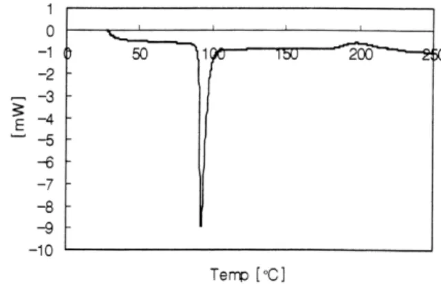
DSC curves of three crystal forms of tulobuterol are illus-trated in Figures 2-4. The DSC curve of Form 1 shows a single melting endothermic peak at 91-92oC The DSC curve of Form 2 shows two endothermic peaks, one endothermic melting peak at 150-151oC and the second broad endothermic peak at 190-210oC. The DSC curve of Form 3 shows three endot-hermic peaks, one melting endotendot-hermic peak at 116-117oC and two endothermic peaks at 175-210oC and 210-255oC. TG
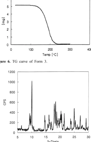
ysis represents a powerful adjunct to DSC, since a combination of a DSC study with a TG determination can be used in the assignment of observed thermal events. TG curves of Form 2 - 3 of tulobuterol are illustrated in Figures 5-6 and Form 2 and Form 3 were found to be nonsolvates.
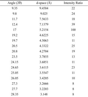
The Powder X-ray diffraction patterns of three crystal forms of tulobuterol are illustrated in Figures 7-9 and they showed distinct differences. Table I - III list 2θ angles, d-spaces and relative intensities of characteristic diffraction peaks up to 30o of three crystal forms.
Caira et al. reported the existence of two polymorphs of tulobuterol (Caira et al., 2004). The DSC patterns and PXRD patterns of Form 2, Form 3 and Form 4 are different from those of Form 2 of Caira et al.
Figure 3. DSC curve of Form 2.
Figure 4. DSC curve of Form 3.
Figure 5. TG curve of Form 2.
Figure 6. TG curve of Form 3.
Figure 7. Powder X-ray diffraction pattern of Form 1.
After storage of 2 months at 0% RH (silica gel, 20oC), 52% RH (saturated solution of Na2Cr2O7.2H2O / 20oC) and 95% RH
(saturated solution of Na2HPO4 / 20oC), Form 1 and Form 3
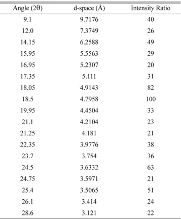
showed no change in DSC, TG and PXRD patterns. But Form 2 was transformed to Form 4 at 52% RH and 95% RH. The DSC, TG and PXRD patterns of Form 4 are illustrated in Fig-ures 10-12 and 2θ angles, d-spaces and relative intensities of characteristic diffraction peaks up to 30o of Form 4 are given in Table IV. Form 4 was found to be nonsolvate.
The dissolution patterns of three crystal forms of tulobuterol are illustrated in Figure 13. In the dissolution studies in dis-tilled water at 37±0.5oC, the solubility of Form 1 was the high-Figure 9. Powder X-ray diffraction pattern of Form 3.
Table I. Characteristic diffraction peaks of Form 1 up to 30oC Angle (2θ) d-space (Å) Intensity Ratio
9.3 9.5091 37 9.6 9.2126 33 10.05 8.8011 100 14.65 6.0463 33 16.1 5.5049 34 16.3 5.4378 24 18.0 4.9279 56 18.45 4.8087 60 19.4 4.5753 27 19.9 4.4615 27 20.2 4.3959 35 20.35 4.3638 41 20.6 4.3114 36 21.5 4.1329 37 23.7 3.754 37 25.0 3.5617 47 27.15 3.2843 32 29.15 3.0634 28 20.5 4.3322 25 20.8 4.2704 19 23.5 3.7855 13 24.15 3.6851 11 24.65 3.6115 23 25.05 3.5547 11 26.05 3.4205 10 27.3 3.2666 8 27.7 3.2203 8 28.35 3.148 8
Table III. Characteristic diffraction peaks of Form 3 up to 30oC Angle (2θ) d-space (Å) Intensity Ratio
9.7 9.1178 19 10.05 8.8011 100 13.3 6.6569 29 14.25 6.2151 97 15.85 5.5912 49 17.3 5.1257 36 18.6 4.7702 8 20.05 4.4284 20 21.3 4.1713 21 22.1 4.0221 64 22.6 3.9342 26 23.65 3.7618 35 24.55 3.6259 16 24.8 3.59 14 25.35 3.5133 27 26.5 3.3634 20 26.6 3.351 17 26.75 3.3325 17
est. And the solubility in water decreased in rank order: Form 1>Form 3>Form 2.
Conclusion
Three crystal forms of tulobuterol were prepared by recrys-tallization from different solvents. Form 2 was transformed to Form 4 at 52% RH and 95% RH. These four crystal forms were characterized by DSC, TG and PXRD. After storage of 2 months at 0% RH, 52% RH and 95% RH, Form 1 and Form 3 showed no transformation. But Form 2 was transformed to Form 4 at 52% RH and 95% RH. In the dissolution studies in
distilled water at 37±0.5oC, three crystal forms showed dif-ference. These four crystal forms were found to be nonsol-vates.
References
Bachet, B., 1997. X-ray characterization of the triclinic polymorph of carbamazepine. J. Pharm. Sci. 86, 1062-1065.
Caira, M.R., Bourne, S.A., and Oliver, C.L., 2004. Thermal and structural characterization of two polymorphs of the bron-Figure 10. DSC curve of Form 4.
Figure 11. TG curve of Form 4.
Figure 12. Powder X-ray diffraction pattern of Form 4.
Figure 13. Dissolution patterns of three crystal forms of tulobuterol.
Table IV. Characteristic diffraction peaks of Form 4 up to 30oC Angle (2θ) d-space (Å) Intensity Ratio
9.1 9.7176 40 12.0 7.3749 26 14.15 6.2588 49 15.95 5.5563 29 16.95 5.2307 20 17.35 5.111 31 18.05 4.9143 82 18.5 4.7958 100 19.95 4.4504 33 21.1 4.2104 23 21.25 4.181 21 22.35 3.9776 38 23.7 3.754 36 24.5 3.6332 63 24.75 3.5971 21 25.4 3.5065 51 26.1 3.414 24 28.6 3.121 22
Haleblian, J.K., 1975. Characterization of habits and crystalline modification of solids and their pharmaceutical applications. J. Pharm. Sci., 64, 1269-1288.
Hüttenrauch, R., 2008. Fundamentals of pharmaceutics. Acta Pharm. Technol., 34, 1-10.
ting procedure using X-ray powder diffraction data. Int. J. Pharm., 212, 203-212.