서 론
코끼리조개 (Geoduck clam, Genus Panopea)는 우럭목 (Order Myoida) 족사부착쇄조개과 (Family
Hiatellidae)에 속하는 이매패류로서 9종이 알려져 있으며, 이 중 5종이 북반구에 분포한다 (Leyva- Valencia et al., 2015). 국내에 분포하는 코끼리조개 는 Japanese geoduck (Panopea japonica)로서 강원 도 고성군에서부터 경상북도 울진군의 동해안 연 안에 걸쳐 서식하고 있으며, 각장이 13 cm 전후, 체중이 500 g 내외에 달하는 한해성 대형 이매패류
코끼리조개(
Panopea japonica
)에서 분리되는
비브리오속 세균의 동정
서현준
1・ 남우화
2・ 김정호
1,2† 1강릉원주대학교 동해안생명과학연구원 2강릉원주대학교 해양자원육성학과Identification of Genus Vibrio bacteria isolated
from geoduck clam (Panopea japonica)
Hyun-Joon Seo
1, U-Hwa Nam
2and Jeong-Ho Kim
1,2† 1The East Coast Research Institute of Life Science, Gangneung-Wonju National University, Gangneung 25457, Korea
2
Department of Marine Bioscience, Gangneung-Wonju National University, Gangneung 25457, Korea
We attempted to isolate and identify potentially pathogenic bacteria from geoduck clam (Panopea japonica) larvae, juvenile and adult, focusing on Vibrios. The isolates were identified by molecular approach and biochemical characterization. In particular, we applied MLSA (multilocus sequence anal-ysis) to the isolated Vibrios for clear identification and phylogenetic relationships, by combining 16s rDNA and several houskeeping genes (pyrH, recA, rpoA). We obtained 141 isolates; 10 from healthy adults, 52 from moribund adults with blisters and 79 from larvae. 46 from the moribund adults and 39 from the larvae were identified as Vibrio species, while the rest of these samples and all the isolates from healthy adult were identified as marine general bacteria. Among Vibrio species, Vibrio splendidus was the most frequently identified from the moribund adults and clustered with the known V. splendidus in GenBank by MLSA. However, it was still unclear that V. splendidus was the cause of blisters because the artificial infection experiment was not conducted and V. splendidus was isolated also from the larvae. Further studies are necessary to clarify the etiological agent of the blisters found in geoduck clam in this study.
Key words: Panopea japonica, geoduck clam, Vibrio splendidus, Multilocus sequence analysis.
†
Corresponding author: Jeong-Ho Kim Tel: +82-33-640-2851, Fax: +82-33-640-2340 E-mail: [email protected]
로서 수심 10~50m 미사질층에 깊게 잠입하여 굵고 긴 입출수관을 표층으로 내어 해수를 빨아들여 동 ㆍ식물성 플랑크톤을 여과섭식한다 (Lee et al., 1998; Nam et al., 2014). 1980년대 고압분사기가 있 는 잠수기 어선을 이용하여 발견된 이후로 지속적 으로 채취되어 1996년에 176톤으로 가장 많이 어 획되었지만 이후 무분별한 남획으로 인하여 국내 코끼리조개 어획량이 급격하게 감소하여 최근에 는 생산량이 거의 없는 실정이며 매년 4월부터 7월 까지 자원보존의 목적으로 포획이 금지되어 있다 (Nam et al., 2015). 코끼리조개는 성체의 크기 및 가식부위의 양을 고려했을 때 식용 가치가 높아 양식 대상 종으로 적합하다 (You et al. 1993). 국외에서는 Pacific geo-duck (Panopea generosa)이 미국, 캐나다에서 상업 적으로 양식되고 있으며 (Beattie and Blake, 1999), New zealand geoguck (Panopea zelandica)은 뉴질랜 드에서 양식이 시도되고 있다 (Gribben and Heas-man, 2015). 국내에서는 코끼리조개(P. japonica)의 양식기술이 개발되어 있으나 상업적인 대량 생산 은 아직까지 시도되지 않았다 (Nam et al., 2014, 2015). 이매패류의 인위적인 대량생산 과정에 있어서 감염성 질병은 유생 및 치패의 성공적인 생산을 저해하는 가장 중요한 요소이며, 특히 Vibrio 속 세 균은 이매패류의 유생에서 성패에 이르기까지 생 활사의 전 단계에서 생존에 영향을 줄 수 있는 중 요한 병원체이다 (Dubert at al., 2017). 이매패류의 유생에서는 Vibrio alginolyticus, V. neptunius, V. splendidus, V. tubiashii, V. pectenicida 등이, 종묘 및 성패에서는 V. aestuarianus, V. chagasii, V. splen-didus, V. tapetis (Beaz-Hidalgo et al., 2010) 등이 각 각 대량 폐사의 원인이 될 수 있는 것으로 알려져 있다. 이매패류는 여과 섭식을 하므로 다양한 세균들 과 공생하고 있어 세균 감염에 의한 질병이 발생하 였을 때 원인이 되는 세균을 동정하는데 어려움이 있다 (Yoon et al., 2017). 특히 이매패류의 대표적 인 병원성 세균인 Vibrio 속 세균은 Photobacterium 속, Aliivibrio 속 등 다른 Vibrio 과 (Family Vibriona-ceae) 세균들과 유전형과 표현형이 매우 유사하여
종 수준까지 동정이 쉽지 않다 (Dorsch et al., 1992). Vibrio 과 세균을 종수준까지 분류, 동정하기 위해 서 16S rDNA gene의 염기서열만을 비교, 분석하는 방법은 한계가 있으며, 다양한 housekeeping gene (e.g., gapA gene, gyrB gene, rpoA gene, recA gene, pyrH gene)의 염기서열을 읽어내어 비교, 분석하는 방법이 널리 사용되고 있다 (Thompson et al., 2005). 특히, MLSA (Multilocus Sequence Analysis)는 16S rDNA gene과 rpoA, recA, gyrB genes 등 다수의 housekeeping gene을 조합하여 균주의 계통정보를 분석하는 방법으로 Vibrio과 세균을 동정하는데 유 용하게 사용되고 있다 (Sawabe et al., 2007; Thomp-son et al., 2009; Nam et al., 2019). MLSA는 16S rDNA gene 만으로는 한계가 있는 Vibrio 속 세균의 근연종간의 유연관계를 명확하게 밝힐 수 있으며, 기존에 기재되었던 종의 재분류 및 신종의 기재에 도 유용하게 쓰일 수 있다 (Urbanczyk et al., 2007). 본 연구에서는 코끼리조개 양식과정에 있어서 생존 및 성장에 영향을 미칠 가능성이 있는 병원성 세균들을 분리, 동정을 시도하였다. 특히 Vibrio 속 세균들을 대상으로 MLSA (Multilocus Sequence Analysis) 방법을 적용하여 균주를 종 수준까지 명 확하게 동정하고, 해당균주들의 생화학적, 생리학 적 특징을 조사하였다.
재료 및 방법
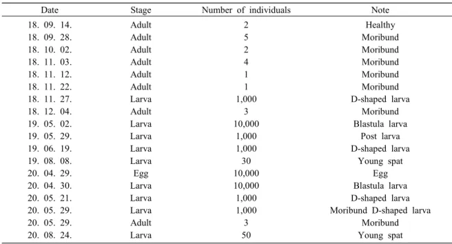
코끼리조개 샘플링 2018년 9월부터 2020년 8월에 걸쳐 강원도 강릉 시에 소재한 강릉원주대학교 해양생물연구센터에 서 사육 중인 코끼리조개를 대상으로 성패의 경우 건강한 개체와 수관부에 수포형태의 병변을 보이 는 빈사 개체를 총 8회 (21개체) 샘플링하였고, 유 생의 경우 수정란부터 치패까지 유생 단계별로 총 10회 샘플링을 실시하였다 (Fig. 1, Table 1). 코끼리 조개는 아이스박스에 해수와 같이 넣어 실험실로 운반한 후 즉시 세균 분리에 사용하였다. 세균 분리 코끼리조개를 샘플링한 직후 실험실로 운반하 여 세균분리를 시도하였다. 유생은 40, 80, 100, 200㎛의 Nylon screen mesh를 사용하여 단계별로 필터 링하여 이물질을 제거하고 멸균된 PBS (Phosphate buffered saline)로 수 회 세척한 후, 세척한 시료를 homogenizer를 사용하여 균질화한 후 멸균된 PBS 를 사용하여 10배씩 단계별로 희석하였다. 성패는 멸균된 해수로 세척하여 이물질을 제거한 후 패각 근을 절개한 뒤 멸균된 백금이로 중장선(midgut gland) 내부를 수 회 휘저었다. 일부 성패 시료는 수관부에 수포 형태의 병변이 발견되어 주사기를 사용하여 수포 내의 체액을 채취하여 세균분리에 사용하였다. 모든 샘플은 증균배지인 MA (Marine agar 2216, Difco, USA)배지와 Vibrio 속 세균의 선 택배지인 TCBS agar (Thioulphate citrate bile salts sucrose agar, Difco, USA)배지를 사용하여 코끼리
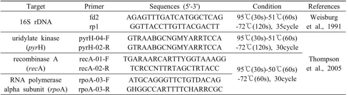
조개 사육 수온과 동일한 15℃에서 24~48시간 배 양하여 세균분리를 시도하였다. 세균 집락이 형성 된 배지는 육안으로 관찰하여 우점하고 있는 균주 를 선택하여 2회 이상 계대배양을 통해 순수분리 한 후에 MB (Marine broth 2216, Difco, USA)배지에 계대배양하여 15℃에서 24~48시간 배양하였다. 배 양된 세균은 80% glycerol과 동량으로 혼합하여 이 후 실험까지 -80℃에서 동결보관하였다. 분자생물학적 동정 MA 배지와 TCBS agar 배지에서 배양, 분리한 총 141개의 균주를 MB 배지에 계대배양하였다. 15 ℃에서 18~24시간 배양한 후 3000 rpm에서 5분간 원심분리하여 pellet 상태로 만든 후에 멸균된 PBS 로 수 회 세척한 다음 동일조건으로 다시 원심분리 하여 새로운 세균 pellet을 만들었다. 제작한 pellet 은 QIAamp DNA kit (QIAGEN, Germany) 매뉴얼에 따라 DNA를 추출하였다. 추출한 DNA는 16S rDNA primer를 사용하여 PCR을 진행하였고, TCBS agar 배지에서 분리한 균주는 Vibrio 과 세균의 house-keeping gene인 pyrH, recA, rpoA gene primer를 사 용하여 추가로 PCR을 수행하였다. 본 실험에 사용 Table 1. Sampling of geoduck clam, Panopea japonica
Date Stage Number of individuals Note
18. 09. 14. 18. 09. 28. 18. 10. 02. 18. 11. 03. 18. 11. 12. 18. 11. 22. 18. 11. 27. 18. 12. 04. 19. 05. 02. 19. 05. 29. 19. 06. 19. 19. 08. 08. 20. 04. 29. 20. 04. 30. 20. 05. 21. 20. 05. 29. 20. 05. 29. 20. 08. 24. Adult Adult Adult Adult Adult Adult Larva Adult Larva Larva Larva Larva Egg Larva Larva Larva Adult Larva 2 5 2 4 1 1 1,000 3 10,000 1,000 1,000 30 10,000 10,000 1,000 1,000 3 50 Healthy Moribund Moribund Moribund Moribund Moribund D-shaped larva Moribund Blastula larva Post larva D-shaped larva Young spat Egg Blastula larva D-shaped larva Moribund D-shaped larva
Moribund Young spat
한 primer sets의 정보는 Table 2에 나타내었다. 시퀀싱 및 분자계통 분석
증폭된 PCR 산물은 1.5% Agarose gel에서 전기 영동 한 후 UV transilluminator를 사용하여 target 밴드를 확인하였다. 생성된 밴드는 절단하여 Accu-prepⓇ Gel Purification kit (Bioneer, Korea)를 사용 하여 증폭된 산물을 정제, 회수하여 Solgent (Dae-jeon, Korea)에 sequencing을 의뢰하여 염기서열을 결정하였다. 결정된 염기서열은 GenBank에 등록 되어 있는 참고 균주들의 유전정보와 함께 MEGA 6 Program (Tamura et al., 2013)의 Kimura two-pa-rameter model (Kimura, 1980)을 사용하여 계통수를 제작하였다. 균주들 간의 염기서열 상동성 조사는 MEGA 6 Program의 Distance estimation을 사용하였다.
Multilocus Sequence Analysis (MLSA) 분리된 Vibrio 속 세균의 명확한 동정을 위해 16S rDNA gene을 비롯하여 Vibrio 과 세균의 hou-sekeeping gene으로 알려진 pyrH, recA, rpoA gene 의 염기서열을 조합하여 MLSA (Multilocus se-quence analysis)를 진행하였다. Vibrio 속에 속하는 참고 균주들의 각각의 유전자 염기서열은 Thomp-son et al. (2005), Dieguez et al. (2011), Balboa and Romalde (2013), Gabriel et al. (2014), Pérez-Cataluna et al. (2016)이 사용한 Vibrio 속 세균의 GenBank Accession number를 참고하였다 (Table 3). MLSA 진행시 발생하는 염기서열간의 gap이나 누락된 서 열들은 모두 제거하였다. Neighbor joining tree (Saitou and Nei, 1987)는 MEGA 6 Program (Tamura
et al., 2013)의 Kimura two-parameter model (Kimura, 1980)을 사용하여 계산하였으며, Bootstrap values 는 1,000번을 기준으로 하였다. 균주들 간의 염기 서열 상동성 조사는 MEGA 6 Program의 Distance estimation을 사용하였다.
생화학적, 생리학적 특징 조사
분자생물학적 방법으로 동정한 균주 (This study 1, 2, 5, 6, 7)를 대상으로 그람음성 간균의 동정에 사용되는 API 20E test (BioMeriux, France)와 Oxi-dase test (Becton Dickinson and company, USA)를 실시하여 생화학적 특징을 확인하였다. TCBS agar 배지에 형성된 집락을 MB 배지에 계대배양하여 15℃에서 24~28시간 배양 후 API 20E kit의 매뉴얼 에 따라 진행하였다. 또한 이들 균주를 대상으로 온도조건별, 염분조건별 배양을 진행하였다. TCBS agar 배지에 배양된 균주를 1.5% NaCl이 첨가된 TSB (Trypticase soybean broth, Difco, USA)배지에 계대배양하여 18~24시간 배양한 후 TSB 배지에 100 ㎕씩 접종하여 배양온도 4, 15, 25, 30, 40℃의 5가지 조건에서 각각 24~48시간 배양하여 각 균주 의 성장여부를 관찰하였다. 또한 염분조건은 TSB 배지에 NaCl을 첨가하여 염분농도 0.5, 3, 5, 8, 10% 의 TSB 배지를 제작 후 각 100 ㎕씩 접종하여 15℃ 에서 24~48시간 배양하여 각 균주의 성장여부를 관찰하였다.
결 과
분자생물학적 동정Table 2. Oligonucleotide primers used for PCR identification of isolated bacteria.
Target Primer Sequences (5'-3') Condition References
16S rDNA fd2 rp1 AGAGTTTGATCATGGCTCAG GGTTACCTTGTTACGACTT 95℃(30s)-51℃(60s) -72℃(120s), 35cycle Weisburg et al., 1991 uridylate kinase (pyrH) pyrH-04-F pyrH-02-R GTRAABGCNGMYARRTCCA GTRAABGCNGMYARRTCCA 95℃(30s)-51℃(60s) -72℃(120s), 30cycle Thompson et al., 2005 recombinase A (recA) recA-01-F recA-02-R TGARAARCARTTYGGTAAAGG TCRCCNTTRTAGCTRTACC 95℃(30s)-50℃(60s) -72℃(60s), 30cycle RNA polymerase
alpha subunit (rpoA)
rpoA-03-F rpoA-03-R
ATGCAGGGTTCTGTDACAG GHGGCCARTTTTCHARRCGC
코끼리조개 성패와 유생으로부터 세균분리를 한 결과 정상개체에서 10개, 빈사개체에서 52개, 유생에서 79개, 총 141개의 균주가 분리되었다. 이 들을 대상으로 16s rDNA sequencing을 실시한 결 과 정상개체에서 분리된 균주는 Glaciecola sp. (20%, 2/10), Kordia sp. (20%, 2/10) 등 모두 일반해 양세균으로 동정되었으며 빈사개체에서 분리된 균주 중 6개는 Photobacterium sp. (50%, 3/6), Pseudoalteromonas sp. (50%, 3/6)인 일반해양세균 으로, 46개는 Vibrio 속 세균으로 동정되었다. 유생 에서는 40개의 균주가 Pseudoalteromonas sp. (22.5 %, 9/40), Uncultured bacterium (17.5%, 7/40) 등 일 반해양세균으로, 39개의 균주가 Vibrio 속 세균으 로 동정되었다 (Table 4). 빈사개체와 유생에서 분
리된 Vibrio 속 균주를 대상으로 housekeeping gene 을 사용하여 PCR 및 sequencing을 실시한 결과 빈 사개체에서 분리된 46개의 Vibrio 속 세균은 V. splendidus (56.5%, 26/46), V. atlanticus (10.8%, 5/46), V. tapetis (8.7% 4/46) 등으로 동정되었고, 유 생에서 분리된 39개의 Vibrio 속 세균은 V. splen-didus (20.5%, 8/39), V. atlanticus (10.2%, 4/39), V. chagasii (7.7%, 3/39) 등으로 동정되었다 (Table 5A). 빈사개체 19 개체에서는 V. splendidus가 가장 빈번하게 분리되었으며 (73.7%, 14/19), V. atlanti-cus (21.1%, 4/19), V. tapetis (15.8%, 3/19), Vibrio sp. (15.8%, 3/19) 등의 순서로 분리되었다 (Table 5B). 또한, 19 개체 중 7 개체에서는 2종 이상의 Vibrio속 세균이 분리되었으며, 이들 7 개체에서 모 Table 3. GenBank accession numbers of bacterial strains used in this study
Species Strain pyrH recA rpoA
V. alginolyticus CECT 521T FM202578 AJ842373 AJ842558
V. anguillarum LMG 4437T Taxvibrio* AJ842375 AJ842561
V. atlanticus CECT 7223T FN582266 EU541589 EU541569
C 14.7 FN582268 FN582254 FN582259
Cmj 13.4 FN582270 FN582256 FN582257
C 2.4 FN582267 FN582253 FN582260
V. campbellii LMG 11216T EF596641 AJ842377 AJ842564
V. celticus CECT 7224T FN582244 EU541590 EU541570
V. cholerae CECT 514T FM202582 AM942078 AM942078
V. crassostreae LMG 22240T EU871948 EU541594 EU541574
V. gigantis LMG 22741T EU871951 EU541593 EU541573
V. harveyi LMG 4044T FM202541 DQ648369 AJ842627
V. lentus CECT 5110T EU871959 AJ842452 AJ842639
V. mediterranei LMG 11258T GU266288 AJ842459 AJ842644
V. orientalis LMG 7897T EU118243 EU130528 AJ842672
V. parahaemolyticus LMG 2850T EU118240 AJ842490 AJ842677
V. splendidus LMG 19031T EU118241 EU130529 AJ842725
R-14789 Taxvibrio* AJ842512 AJ842726
V. tapetis CECT 4600T HE795189 HE795219 HE795340
HH6087T HE795208 HE795238 HE795358
a200 HE795214 HE795244 HE795364
LP2 HE795218 HE795248 HE795368
B8.3 HE795200 HE795230 HE795351
GR0202RD HE795206 HE795236 HE795357
C0620701H HE795212 HE795242 HE795362
V. tasmaniensis LMG 20012T EU871961 AJ842515 AJ842731
두 V. splendidus가 분리되었다 (data not shown). Multilocus Sequence Analysis (MLSA) 동정된 Vibrio 속 세균들 중에서 무척추동물에 병원성이 있는 것으로 알려진 V. atlanticus (This
1~4), V. tapetis (This study 5), V. splendidus (This 6~14)를 선택하여 MLSA를 실시한 결과 This study 1과 3은 V. atlanticus C 14.7, This study 2와 4는 V. atlanticus C 2.4와 동일한 cluster를 형성하였으 며, This study 5는 V. tapetis a200, This study 6~14는 V. splendidus LMG 19031T와 동일한 cluster를 형성 하였다 (Fig. 2).
염기서열 상동성에서는 This study 1과 3은 V. atlanticus C 14.7, This study 2와 4는 V. atlanticus C 2.4와 상동성이 각각 99.30%, 99.57%이었으며, This study 5는 V. tapetis a200과 상동성이 99.79%, This study 6~14는 V. splendidus LMG 19031T와 상 동성이 99.57~99.95%이었다 (Table 6).
생화학적, 생리학적 특징 조사
분자생물학적 방법으로 동정된 10개의 균주 중 에서 중복된 균주를 제외한 5개 균주 V. atlanticus (This study 1~2), V. splendidus (This study 6~7), V. tapetis (This study 5)를 API 20E kit (BioMeriux, France)와 Oxidase test (Becton Dickinson and com-pany, USA)를 통해 생화학적 특징을 조사하였다. 그 결과, Oxidase test에서는 본 연구에 사용된 균주 들 모두 Vibrio 속 세균과 동일한 양성 반응을 나타 내었다. API 20E test에서는 본 연구에 사용된 균주 모두 ONPG (β-galractosidase) test, LDC (Lysine Decarboxylase) test, VP (Voges Proskauer) test, GLU (Glucose) test 등이 기존의 연구 결과와 유사하였 다. 그러나, GEL (Gelatinase) test와 AMY (Amygda-lin) test는 기존의 연구 보고와 다르게 음성 반응이 나타났다 (Borrego et al., 1996; Buller, 2004; Dieguez et al., 2011; Lee et al., 2018) (Table 7).
온도 조건별 배양 실험에서 본 연구에 사용한 5개의 균주는 4℃에서 25℃까지 모두 성장하였으 며 V. tapetis (This study 5)를 제외한 나머지 4개의 균주는 30℃에서도 성장하였다. 염분 조건별 배양 Table 4. Number of bacterial isolates in this study
Healthy adult Moribund adult Healthy larva Total Marine general bacteria
Vibrio spp. 10 -6 46 40 39 56 56 10 52 79 141
Table 5A. Species and Number of Vibrios Isolated in this study Moribund adult Healthy larva Total Vibrio spp. V. alginolyticus V. atlanticus V. chagasii V. cortegadensis V. crassostreae V. cyclitrophicus V. lentus V. mediterranei V. pelagius V. splendidus V. tapetis V. tasmaniensis V. toranzoniae 6 1 5 -2 1 -1 -26 4 -14 1 4 3 -2 1 3 1 8 -1 1 20 2 9 3 2 1 2 2 3 1 34 4 1 1 46 39 85
Table 5B. Percentage occurrence of each Vibrio species isolates from moribund geoduck clam individuals in this study
Species name % occurrence in moribund geoduck clam individuals (n=19)
V. alginolyticus V. atlanticus V. cortegadensis V. crassostreae V. lentus V. splendidus V. tapetis Vibrio spp. 5.3% 21.1% 10.5% 5.3% 5.3% 73.7% 15.8% 15.8% (1/19) (4/19) (2/19) (1/19) (1/19) (14/19) (3/19) (3/19)
실험에서는 5개 균주 모두 NaCl이 1.5%에서 5%까 지 첨가된 조건에서 성장하였다. 이상의 결과는 Buller (2004), Dieguez et al. (2011)와 유사하였다 (Table 7).
고 찰
코끼리조개 (Japanese geoduck)은 우리나라 동해 안, 일본, 중국 북부 연안에 분포하며 상품가치가 Fig. 2. Phylogenetic reconstructions based on 3 concatenated genes (pyrH, recA and rpoA). Alignment was conducted with all 3 genes. Gaps and missing parts in the sequence were eliminated to make 1,878 bp in total. The number after bacterial scientific name means strain number registered in the NCBI. The tree was constructed by neighbor-join-ing method. Numbers at nodes denote the level of bootstrap based on 1,000 replicates.
높아 최근 대량 생산을 목적으로 다양한 방면에서 연구가 진행되고 있으나, 아직 상업적인 대량 생산 은 불가능한 실정이다 (Nam et al., 2014, 2015; Huo et al., 2017). 이매패류의 인위적인 대량 생산에 있 어서 대규모의 폐사가 종종 발생하며, 수온, 염도, 먹이, 사육 밀도 등 다양한 환경 요인, 감염성 미생 물 들이 원인으로 지적되고 있다. 특히, 비브리오 속 세균은 해양 환경에 풍부하게 존재하며, 120 여 종 이상의 다양한 비브리오 속 세균이 해양 환경 내에서 자유생활을 하거나 다양한 해양 생물과 공 생하고 있다. 또한, 다양한 비브리오 속 세균이 해 양생물에 병원성을 가지고 있는 것으로 보고되어 있다 (Thompson et al., 2004).
Cáceres-Martínez et al. (2015)과 Dorfmeier et al. (2015)은 자연산 Pacific geoduck의 질병에 대해 조 사하였으며, 수 종의 기생성 갑각류, 미포자충류 및 리케치아를 보고하였다. 하지만 이들의 감염에 의해 유의할만한 조직병리학적 변화나 폐사는 확 인되지 않았다고 언급하였다. 코끼리조개 (Japanese geoduck)의 질병에 관해서는 아직까지 보고된 바 Table 6. Estimation of homology among Genus Vibrio species, based on the MLSA of 3 concatenated genetic sequences (pyrH, recA and rpoA genes)
A 1 2 3 4 5 6 7 8 1 2 3 4 5 6 7 8 This study 1 This study 2 This study 3 This study 4 V. atlanticus CECT 7223T V. atlanticus Cmj 13.4 V. atlanticus C 2.4 V. atlanticus C 14.7 -98.55% 99.73% 98.55% 98.65% 98.49% 98.60% 99.30% -98.82% 100.00% 99.68% 99.84% 99.95% 98.76% -98.82% 98.93% 98.76% 98.87% 99.57% -99.68% 99.84% 99.95% 98.76% -99.73% 99.73% 98.87% -99.89% 98.71% -98.82% -B 1 2 3 4 5 6 7 8 1 2 3 4 5 6 7 8 This study 5 V. tapetis CECT 4600T V. tapetis LP2 V. tapetis a200 V. tapetis B8.3 V. tapetis GR0202RD V. tapetis C0620701H V. tapetis subsp. britannicus HH6087T -99.73% 99.63% 99.79% 99.73% 99.73% 99.73% 94.91% -99.79% 99.73% 100.00% 99.79% 99.89% 94.91% -99.63% 99.79% 99.57% 99.68% 94.97% -99.73% 99.52% 99.63% 94.91% -99.79% 99.89% 94.91% -99.79% 94.68% -94.80% -C 1 2 3 4 5 6 7 8 9 10 11 1 2 3 4 5 6 7 8 9 10 11 This study 6 This study 7 This study 8 This study 9 This study 10 This study 11 This study 12 This study 13 This study 14 V. splendidus LMG 19031T V. splendidus R-14789 -99.68% 99.73% 99.79% 99.79% 99.95% 99.63% 99.68% 99.73% 99.84% 99.73% -99.95% 99.57% 99.57% 99.73% 99.52% 99.68% 99.41% 99.63% 99.63% -99.63% 99.63% 99.79% 99.47% 99.73% 99.47% 99.68% 99.68% -99.79% 99.84% 99.52% 99.68% 99.52% 99.73% 99.73% -99.84% 99.52% 99.57% 99.52% 99.95% 99.73% -99.68% 99.73% 99.68% 99.89% 99.79% -99.41% 99.36% 99.57% 99.52% -99.41% 99.63% 99.52% -99.57% 99.47% -99.68% -A: V. atlanticus strains, B: V. tapetis strains, C: V. splendidus strains. All positions containing gaps and missing data were eliminated. There were a total of 1,878 base pairs in the final dataset. Values means similarity of nucleotide sequences.
없으며, 본 연구에서 코끼리조개의 유생 및 성패를 채집하여 세균분리를 시도한 결과 총 141개의 세 균이 분리되었으며 16S rDNA gene sequencing에 의해 56개의 세균은 일반해양세균으로, 85개의 세 균은 비브리오 속 세균으로 동정되었다 (Table 4). 비브리오 속 세균은 housekeeping genes 및 MLSA 를 통해 V. splendidus, V. atlanticus, V. tapetis, V. chagasii 등 다양한 비브리오 속 세균으로 동정되 었다 (Table 5A).
V. splendidus는 참굴 (Crassostrea gigas)을 대량 폐사시키는 대표적인 Vibrio 속 세균으로 참굴 이 외에도 포목상 바지락 (Ruditapes decussatus)과 가 리비 (Patinopecten yessoensis) 등 다양한 이매패류 에서 폐사를 일으킨다고 보고되어 있다 (Lacoste et al., 2001; Gomez-Leon et al., 2005; Rojas et al.,
2015). 또한 V. tapetis는 바지락에서 Brown ring dis-ease (BRD)의 대표적인 원인세균으로 알려져 있으 며 (Paillard et al., 1994; Allam et al., 2000), V. atlan-ticus는 V. splendidus Clade에 속하는 균주로 이매 패류에 병원성을 보일 가능성이 있을 것으로 생각 되며 (Dieguez et al., 2011), 국내에서는 육상양식중 인 자주복 (Takifugu rubripes)에서 분리되어 보고 된 바 있다 (Lee et al., 2018).
또한 Bower and Blackbourn (2003)과 Cáceres- Martínez et al. (2015)은 Pacific geoduck의 입출수관 에서 수포성 병변을 보고하였다. 이 수포형 병변은 맑은 액체로 충만되어 있으며, 이 병변과 연관된 감염성 병원체는 발견되지 않았다고 하였다. 본 논 문에서 조사한 Japanese geoduck에서도 수포성 병 변이 일부 개체에서 관찰되었다 (Fig. 1). 이러한 Table 7. Differential phenotypic characteristics of Vibrio species isolated in this study and other Vibrio species
1 2 3 4 5 6 7 8 9 10
Growth at (℃) Growth in NaCl (%) β-Galactosidase (ONPG) Arginine dehydrolase (ADH) Lysine decarboxylase (LDC) Ornithine decarboxylase (ODC) Citrate utilization (CIT) H2S production (H2S)
Urease (URE)
Tryptophane deaminase (TDA) Indole production (IND) Voges Proskauer (VP) Gelatinase (GEL) D-Glucose (GLU) D-Mannitol (MAN) Inositol (INO) D-Sorbitol (SOR) L-Rhamnose (RHA) D-Sucrose (SAC) D-Melibiose (MEL) Amygdalin (AMY) L-Arabinose (ARY) Oxdiase (OX) 4-30 1.5-5 + -+ -+ + -+ + 4-30 1.5-5 -+ -+ -+ + -+ -+ 4-25 1.5-5 + -+ + -+ 4-30 1.5-5 -+ -+ -+ + -+ -+ 4-30 1.5-5 -+ -+ -+ + -+ -+ 4-30 1-7 + + -+ -+ + + -+ -+ -+ 4-30 1-7 -+ -+ + -+ + + -+ 4-22 1-5 + -+ -+ + -+ -+ 4-22 1-5 + -ND -ND -+ + ND -ND -ND -ND 4-30 3-6 + -ND -ND + -+ + + -+ -ND + 1: This study 1, 2: This study 2, 3: This study 5, 4: This study 6, 5: This study 7, 6: V. splendidus ATCC 33125, 7: V. splendidus NCMB 2251, 8: V. tapetis B1090T, 9: V. tapetis, 10: V. atlanticus Cmj 13.4. All the other data
of reference strains were adopted from Borrego et al. (1996), Buller (2004), Dieguez et al. (2011) and Lee et al. (2018). ND: no data, +: Positive, -: Negative.
병변이 발견된 개체는 검사 당시 빈사 상태이거나 결과적으로 폐사하는 것으로 보아 개체의 생존과 관련이 있는 것으로 생각되어 해당 병변에서 채취 한 맑은 액체를 사용하여 세균 분리를 시도하였으 며, Vibrio 속에 속하는 수 종의 세균이 분리되었다. 본 연구에서는 V. splendidus가 가장 많이 분리되 어 코끼리조개에서 발견된 수포 형태의 병변과 관 련이 있는 것으로 의심할 수 있었다. 하지만 이 세 균은 유생에서도 분리되었으며, 성패를 대상으로 인위감염실험을 실시하지 않아 본 연구에서 분리 된 Vibrio 속 세균이 수포 형태의 병변을 일으킨 1차적인 원인인지, 혹은 2차적으로 침입하여 증식 한 결과인지는 확인할 수 없었다. 차후 이들 Vibrio 속 세균을 사용한 인위감염을 실시하여 수포 형태 의 병변과의 연관성을 밝힐 필요가 있으며, 세균 이외의 병원체가 원인일 가능성도 검토해 볼 필요 가 있다. MLSA에 의해 코끼리조개에서 분리된 14개의 균주는 V. atlanticus (This study 1~4), V. tapetis (This study 5), V. spledidus (This study 6~14)로 각각 동정 되었다. 16S rDNA 유전자 단편만을 사용하여 염기 서열을 분석한 경우에는 분리된 대다수의 Vibrio 속 균주들을 종 수준까지 동정하지 못하였다. Hou-sekeeping gene (pyrH, recA, rpoA genes) 1개만을 사용하여 염기서열을 분석하면 대부분 Vibrio 속 세균을 종 수준까지 동정이 가능하였지만 분석하 는 housekeeping gene에 따라 다른 균주로 동정된 경우가 있었다 (data not shown). 따라서 이러한 불 일치를 줄이고 Vibrio 속 세균을 명확하게 동정하 기 위해서는 MLSA 방법을 적용할 필요가 있다고 생각된다.
감사의 글
이 논문은 2020년 해양수산부 재원으로 한국해 양과학기술진흥원의 지원을 받아 수행된 연구입 니다 (과제명: 코끼리조개 양식산업화 기술개발 (2018-0377)).요 약
코끼리조개 성패 및 유생으로부터 잠재적 병원 성 세균 분리 및 동정을 수행하였다. 분리된 균주 는 분자생물학적 기법 및 생화학적 검사를 통해 동정하였으며, 명확한 동정 및 계통수 분석을 위해 16S rDNA와 하우스키핑 유전자 (pyrH, recA, rpoA) 를 결합하여 분석하는 MLSA를 적용하였다. 총 141개의 균주가 분리되었으며, 건강한 성패에서는 10개, 수포 병변을 보이는 빈사 상태의 성패에서 52개, 유생에서 79개가 분리되었다. 이 중 빈사상태 의 성패에서 46개, 유생에서 39개의 균주가 Vibrio 속 세균으로 동정되었으며, 나머지 균주들은 모두 일반해양세균으로 동정되었다. Vibrio 속 세균 중 에서는 수포 병변을 보이는 성패에서 Vibrio didus가 가장 많이 분리되었으며 기존의 V. splen-didus와 함께 동일한 클러스터를 형성하였다. 하지 만, 인위감염실험을 실시하지 않았고 건강한 유생 에서도 V. splendidus가 분리되어 성패에서 나타난 수포와 V. splendidus와의 관계는 확실하지 않으며, 차후 추가 연구가 필요할 것으로 생각된다.References
Allam B., Parillard C., Howard A. and Pennec M. L.: Isolation of the pathogen Vibrio tapetis and defense parameters in brown ring diseased Manila clams
Ruditapes philippinarum cultivated in England. Dis.
Aquat. Org., 41: 105-113, 2000.
Balboa S. and Romalde J. L.: Multilocus sequence anal-ysis of Vibrio tapetis, the causative agent of Brown Ring Disease: Description of Vibrio tapetis subsp.
britannicus subsp. nov. Syst. Appl. Microbiol., 36(3):
183-187, 2013.
Beattie H. and Blake B.: Development of culture meth-ods for the geoduck clam in the USA (Washington State) and Canada (British Columbia). J. World Aquac. Soc., 30(3): 50–53, 1999.
Beaz-Hidalgo R., Balboa S., Romalde J. L. and Figuersa M. J.: Diversity and pathogenecity of Vibrio species in cultured bivalve molluscs. Environ. Microbiol. Rep., 2(1): 34-43, 2010.
Borrego J. J., Castro D., Luque A., Paillard C., Maes P., Garcia M. T. and Ventosa A.: Vibrio tapetis sp. nov., the causative agent of the Brown Ring Disease affecting cultured clams. Int. J. Syst. Bacteriol., 46(2): 480-484, 1996.
generosa): anatomy, histology, development,
pathol-ogy, parasites and symbionts: lesions of unknown cause on geoduck clams. Available at: https://www. dfo-mpo.gc.ca/science/aah-saa/species-especes/ shellfish-coquillages/geopath/warts-eng.html. 2003. Buller N. B.: Bacteria from fish and other aquatic
ani-mals: a practical identification manual. CABI Pub-lishing, Oxfordshire, 171-183, 2004.
Cáceres-Martínez J., Vásquez-Yeomans R. and Cruz- Flores R.: First description of symbionts, parasites, and disease of the Pacific Geoduck Panopea
gen-erosa from the pacific coast of baja california,
Mex-ico. J. Shellfish Res., 34(3): 751-756, 2015. Dieguez A. L., Beaz-Hidalgo R., Cleenwerck I., Balboa
S., Vos R. D. and Romalde J. L.: Vibrio atlanticus sp. nov. and Vibrio artabrorum sp. nov., isolated from the clams Ruditapes philippinarum and
Rudi-tapes decussatus. Int. J. Syst. Evol. Microbiol.,
61(10): 2406–2411, 2011.
Dorfmeier E. M., Vadopalas B., Frelier P. and Friedman C. S.: Temporal and spatial variability of native Geoduck (Panopea generosa) endosymbionts in the Pacific northwest. J. Shellfish Res., 34(1): 81–90, 2015.
Dorsch M. D., Lane D. and Stackebrandt E.: Towards a phylogeny of the Genus Vibrio based on 16s rRNA sequences. Int. J. Syst. Bacteriol., 42(1): 58-63, 1992. Dubert J., Barja J. L. and Romalde J. L.: New Insights
into pathogenic Vibrios affecting bivalves in hatch-eries: present and future prospects. Front. Microbiol., 8: 762, 2017.
Gabriel M. W., Matsui G. Y., Friedman R. and Lovell C. R.: Optimization of multilocus sequence analysis for identification of species in the Genus Vibrio. Appl. Enviro. Microbiol., 80(17): 5359-5365, 2014. Gomez-Leon J., Villamil L., Lemis M. L., Novoa B. and
Figueras A.: Isolation of Vibrio alginolyticus and
Vibrio splendidus from aquacultured carpet shell
clam (Ruditapes decussatus) larvae associated with mass mortalities. Appl. Environ. Microbiol., 71(1): 98-104, 2005.
Gribben, P. E., and Heasman, K. G.: Developing fish-eries and aquaculture industries for Panopea
zeland-ica in New Zealand. J. Shellfish Res., 34(1), 5-10,
2015.
Huo, Z., Guan, H., Rbbani, M. G., Xiao, Y., Zhang, X., Fan, C., Li Z., Li Y., Wu Q., Yang F. and Yan, X.: Effects of environmental factors on growth, survival, and metamorphosis of geoduck clam (Panopea
ja-ponica A. Adams, 1850) larvae. Aquac. Rep., 8: 31-
38, 2017.
Kimura M.: A simple method for estimating evolutionary rates of base substitutions through comparative stud-ies of nucleotide sequences. J. Mol. Evol., 16(2): 111-120, 1980.
Lacoste A., Jalabert F., Malham S., Cueff A., Gelebart F., Cordevant C., Lange M. and Poulet S. A.: A
Vibrio splendidus strain is associated with summer
mortality of juvenile oysters Crassostrea gigas in the Bay of Morlaix (North Brittany, France). Dis. Aquat. Org., 46: 139-145, 2001.
Lee C. S., Baik K. K. and Hong K. E.: Ecological studies on the habitat of geoduck clam, Panope japonica. J. Aquaculture, 11(1): 105-111, 1998.
Lee N. S., Cho M. Y., Jung S. H. and Won K. M.: The first case report on Vibrio atlanticus infection in cul-tured tiger puffer, Takifugu rubripes. J. Fish Pathol., 31(2): 087-092, 2018.
Leyva-Valencia, I., Cruz-Hernández, P., Álvarez-Casta-ñeda, S. T., Rojas-Posadas, D. I., Correa-Ramírez, M. M., Vadopalas, B., & Lluch-Cota, D. B.: Phylogeny and phylogeography of the Geoduck Panopea (Bival-via: Hiatellidae). J. Shellfish Res., 34(1), 11-20, 2015.
Nam M. M., Lee C., Kim M. K., Kim J. W. and Kim Y. D.: Development and growth in fertilized eggs and larvae of the Japanese geoduck, Panopea
japon-ica reared in the laboratory. Kor. J. Malacol., 30(4):
303-309, 2014.
Nam M. M., Lee J. Y., Lee C. Kang. H. W., Kim Y. D., Byun S. G. and Yoo H. K.: Effect of water tem-perature condition on growth and survival of juve-nile Geoduck (Panopea japonica A. Adams, 1850). Kor. J. Malacol., 31(4): 263-266, 2015.
Nam W. H., Seo H. J., Jang S. R., Kim M. R. and Kim J. H.: Multilocus sequence analysis of the genus
Aliivibrio: Identification and phylogeny of Aliivibrio
species isolated from cultured walleye pollock (Gadus
chalcogrammus) in Korea. J. Fish Pathol., 32(2):
69-80. 2019.
Paillard C. Maes P. and Oubella R.: Brown ring disease in clams. Annu. Rev. Fish Dis., 4: 219-240, 1994. Pérez-Cataluna A., Lucena T., Tarazona E., Arahal D.
R., Macián M. C. and Pujalte M. J.: An MLSA ap-proach for the taxonomic update of the Splendidus clade, a lineage containing several fish and shellfish pathogenic Vibrio spp. Syst. Appl. Microbiol., 39(6): 361–369, 2016.
Rojas R., Miranda C. D., Opazo R. and Romero J.: Characterization and pathogenicity of Vibrio
splen-didus strains associated with massive mortalities of
commercial hatchery-reared larvae of scallop
Argo-pecten purpuratus (Lamarck, 1819). J. Invertebr.
Pathol., 124, 61-69, 2015.
Saitou N. and Nei M.: The neighbor-Joining Method-a new method for reconstructing phylogenetic trees. Mol. Biol. Evol., 4: 406-425, 1987.
Sawabe, T., Kita-Tsukamoto, K. and Thompson, F. L.: Inferring the evolutionary history of vibrios by means of multilocus sequence analysis. J. Bacteriol., 189(21): 7932-7936, 2007.
Tamura K., Stecher G., Peterson D., Filipski A. and Kumar S.: MEGA6: molecular evolutionary genetics analysis version 6.0. Mol. Biol. Evol., 30(12): 2725- 2729, 2013.
Thompson C. C., Vicente A. C., Souza R. C., Vasconce-los A. T., Vesth T., Alves N., Ussery D. W., Iida T. and Thompson F. L.: Genomic taxonomy of Vibrios. BMC Evol. Biol., 9(1): 258, 2009. Thompson F. L., Gevers D., Thompsom C. C., Dawyndt
P., Naser S., Hoste B., Munn C. B. and Swings J.: Phylogeny and Molecular Identification of Vibrios on the Basis of Multilocus Sequence Analysis. Appl. Environ. Microbiol., 71(9): 5107-5115, 2005.
Thompson F. L., Iida T. and Swings J.: Biodiversity of vibrios. Microbiol. Mol. Biol. Rev., 68(3): 403-431, 2004.
Urbanczyk H., Ast J. C., Higgins M. J., Carson J. and Dunlap P. V.: Reclassification of Vibrio fischeri,
Vibrio logei, Vibrio salmonicida and Vibrio wodanis
as Aliivibrio fischeri gen. nov., comb. nov., Allivibrio
logei comb. nov., Allivibrio salmonicida comb. nov.
and Aliivibrio wodanis comb. nov. Int. J. Syst. Evol. Microbiol., 57(12): 2823-2829, 2007.
Weisburg W. G., Barns S. M., Pelletier D. A. and Lane D. J.: 16S Ribosomal DNA amplification of phylo-genetic study. J. Bacteriol., 173(2): 697-703, 1991. Yoon J. S., Park M. S., Kim K. Y., Won N. I. and Won
K. M.: Distribution of Vibrionaceae isolated from cultured bay scallop, Argopecten irradians. Kor. J. Malacol., 33(4): 285-297, 2017.
You B. J., Jeong I. H., Lee K. H. and Chol H. G.: Quality and storage stability of frozen Geoduck (Panope
ja-ponica A. Adams). Bull. Kor. Fish. Soc., 26(6): 549-
556, 1993.
Manuscript Received : Nov 4, 2020 Revised : Nov 29, 2020 Accepted : Nov 30, 2020