https://doi.org/10.20307/nps.2021.27.1.1
1
Effect of Curcuma xanthorrhiza Gel on Methicillin-Resistant
Staphylococcus aureus-Infected Second-Degree Burn Wound in Rats
Irfan Kesumayadi1,*, Ayyasi Izaz Almas1, Ilham Nur Hakim Rambe2, and Rebriarina Hapsari11Faculty of Medicine, Universitas Diponegoro, Semarang, Indonesia 2Faculty of Engineering, Universitas Diponegoro, Semarang, Indonesia
Abstract Methicillin-resistant Staphylococcus aureus (MRSA) infection often complicates burn wounds. Mupirocin is the antibiotic of choice for superficial MRSA infection, and its resistance is on the rise due to its frequent and widespread use. This study aimed to develop and evaluate Curcuma xanthorriza extract (CXE)-containing gel as a topical agent against MRSA-infected second-degree burn wound in rats. CXE was obtained using maceration with 96% ethanol. Xanthorrhizol level, antibacterial, and antioxidant activity were evaluated using a standardized method. In vivo, the wound’s healing and bacterial load were evaluated every three days, whereas the histopathology of the wound was examined on day 12 of treatment. One-Way ANOVA and Kruskal-Wallis test were used to analyze the data. In this study, 27.0% and 7.10% of the obtained CXE were xanthorrhizol and curcumin, respectively. Additionally, an IC50 of 64.27 ppm was shown in antioxidant activity
measurement, and MIC against MRSA was 5 mg/ml. Treatment with CXE-containing gels showed a significant reduction in bacterial load and proliferation of connective tissue in a dose-dependent manner. In conclusion, CXE-containing gel showed a greater reduction of bacterial load and more advanced wound healing phase than mupirocin.
Keywords Curcuma xanthorrhiza, Xanthorrhizol, MRSA, Burn Wound
Introduction
Burns has led to increased mortality and morbidity in low- and middle-income countries, with the highest incidence occurs at homes and workplaces.1 Burns ranked fourth as the cause of trauma-related deaths and second place as a cause of accidental death for children aged 1-4.2 World Health Organization estimates that nearly 11 million people get severe burns that require medical attention, and approximately 180,000 deaths attributed to burns, annually.1 Second-degree burns contribute for approximately 60% of burns incidence.3
Patients who suffer from burns are at a higher risk for local and systemic infections. The disruption of the skin barrier and the following immunocompromised state make burns easy to get colonized by bacteria.4 Staphylococcus aureus is the most common gram-positive early colonizer of burn wounds and has been one of major etiologic agents that cause burn wound infections that leads to increased morbidity and mortality among burn patients.5
Methicillin-resistant S. aureus (MRSA), which is resistant to almost all beta-lactam antibiotics, was found in 11% burn wound infections.6 The risk of MRSA infections also increased if the patients are admitted to intensive care unit.7
The therapy for MRSA infections is limited. For superficial MRSA infections, mupirocin has been the therapy of choice for decades. However, with the widespread of mupirocin use, resistance rate against mupirocin among MRSA isolates is also increasing. A study showed that 31.3% of MRSA showed resistance to mupirocin.8 Unsuccessful treatment of MRSA infections can lead to burn wound sepsis, invasive infection, septicemia, multi-organ failure, and death.9
There are various studies evaluating potential herbal plants as new antibiotic agents to overcome drug resistance in S. aureus. Curcuma xanthorriza Roxb. contains xanthorrhizol which has antibacterial activity with Minimum Inhibitory Concentration (MIC) of 25 µg/mL against MRSA.10 Xanthorrhizol can also be used as a pain reliever by reducing cyclooxygenase-2 (COX-2) and inducible nitric oxide synthetase (iNOS) which eventually reducing the production of prostaglandin and nitric oxide.11
*Author for correspondence
Irfan Kesumayadi, Faculty of Medicine, Universitas Diponegoro, Semarang, Indonesia
2 Natural Product Sciences Moreover, xanthorrhizol is also a potential anti-aging
agent, which significantly reduces the expression of Metalloproteinase-1 (MMP-1) and increases in type 1 procollagen synthesis.12 Excessive of MMPs induces chronic wounds. Therefore, regulation of MMPs may help to improve wound healing.13
This study, therefore, aimed to develop and evaluate Curcuma xanthorriza extract (CXE)-containing gel as a topical agent against MRSA-infected second-degree burn wound in rats.
Experimental
Materials The ingredients for the curcuma extract were Curcuma xanthorrhiza Roxb., ethanol 96%, and distilled water. The materials for MIC determination against MRSA were MRSA ATCC 4330, MRSA clinical strain 7097, and Mueller-Hinton agar. Carbopol, trie-thanolamine, methyl paraben, and propylene glycol were used to produce CXE-containing gel. Male Wistar rats (Rattus novergicus) and 2% mupirocin ointment were used for in vivo comparison study.
Maceration Process and Extract Characterization Curcuma xanthorrhiza Roxb. was cleaned, dried, and ground to make dry powder of it. These treatments were carried out in room temperature. One hundred grams Curcuma xanthorrhiza Roxb. dry powder was macerated using 300 mL ethanol 96% for 24 hours. The resulting extract, i.e. CXE, was concentrated by vacuum evaporator in 40oC. Identification of functional groups of the extract was made by FTIR (Fourier Transform InfraRed) (Spectrum two FT-IR, PerkinElmer). High-Performance Liquid Chromatography (HPLC) system (D-7000, Merck Hitachi, Tokyo, Japan) was used to determine xanthorrhizol content quantitatively. A 2,2-diphenyl-1-picryl-hydrazyl-hydrate (DPPH) method was used to determine anti-oxidant activity (IC50) in the extract using a spectro-photometer (Shimadzu, Kyoto, Japan) at 517 nm wavelength. Besides xanthorrhizol, curcumin content was also quantified using spectrophotometry method (530 nm wavelength). FTIR analysis was carried out in the Integrated Research Laboratory, Universitas Diponegoro. HPLC and spectro-photometry was carried out in the Indonesian Spice and Medicinal Crops Research Institute.
MIC Determination MIC determination was done by the agar dilution method on Mueller-Hinton Agar. Two milliliters CXE solution containing gradual concentration from 0.0006 mg/mL to 5 mg/mL was added and homo-genized in sterile liquid Mueller-Hinton Agar at 40-50oC and left to solidify. CXE-containing Mueller-Hinton Agar
was inoculated with 104 CFU MRSA ATCC 4330 and MRSA clinical specimens 7097 at 12 different points. MIC was determined if the reduction in bacteria growth was greater than 80% compared to control.14
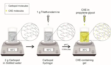
CXE Gel Production Distilled water was heated at 50oC in a beaker glass containing magnetic stirrer. Carbopol and triethanolamine were added to distilled water. Meanwhile, CXE and methylparaben was dissolved in another beaker glass containing propylene glycol. Both blends were then mixed together. Detailed composition of the CXE gel was shown in Table 1 and the process was depicted in Fig. 1.
In Vivo Study A total of 25 male Wistar rats weighing 200-300 grams at 2-3 months of age were acclimated for seven days. Local anesthesia was carried out using 3 mL 0.5% lidocaine injected intradermally and subcutaneously covering a diameter of 2.5 cm. According to Rodent Anesthesia Analgesia Formulary Guideline from University of British Columbia, 2% lidocaine HCL, diluted to 0.5% with a maximum dose of 7 mg/kg subcutaneously or intra-incisional, can be used as local anesthesia in rats. To ensure the local anesthesia, we pinched rat’s skin using toothed forceps. A 2-cm diameter iron plate was heated at 210oC, and then inducted for 5 seconds on rat’s shaved back skin to make a second-degree burn wound. After 6 hours, 107 CFU/mL MRSA ATCC 4330 was inoculated topically on the burn wound.15 Rats were kept in separate cages individually and fed with commercial food and water. Rats were randomly assigned into five groups: (i) negative control (C) group, in which rats were treated with placebo gel therapy, twice daily; (ii) Mupirocin (M) group, in which rats were treated with 2% mupirocin topical ointment, twice daily, as a positive control group; (iii) 1% CXE group, in which rats were treated with 1% CXE-containing gel therapy, twice daily; (iv) 3% CXE group, in which rats were treated with 3% CXE-containing gel therapy, twice daily; (v) 5% CXE group, in
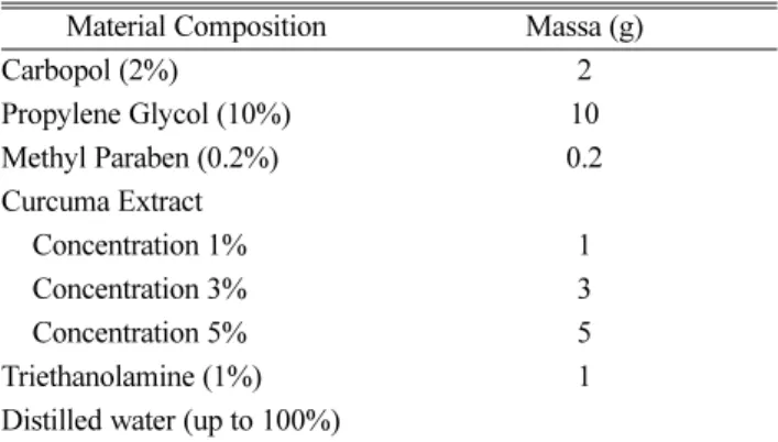
Table 1. CXE-containing gel composition on a basis of 100 g Material Composition Massa (g)
Carbopol (2%) 2 Propylene Glycol (10%) 10 Methyl Paraben (0.2%) 0.2 Curcuma Extract Concentration 1% 1 Concentration 3% 3 Concentration 5% 5 Triethanolamine (1%) 1 Distilled water (up to 100%)
which rats were treated with 5% CXE-containing gel therapy, twice daily. Duration of treatment of all rats was 12 days.
Semiquantitative Bacterial Load Bacterial load was counted semi quantitatively using the 4-quadrant streak plate method. A sterile cotton was swabbed on the burn wound and directly inoculated onto Mueller-Hinton Agar and incubated at 37oC for 24 hours. Swabs were performed every three days during treatment.
Wound Closure Measurement To evaluate wound closure, the diameter of the wound was measured horizontally, diagonally, and vertically every three days. Wound closure was indicated by scar and hair growth.
Skin Histopathology Histopathological assessment was carried out in 5 fields of view using 100x magni-fication for connective and granulation tissue in order to cover the total area of microscopic specimen. A magnification of 40x was used to measure the length of re-epithelization in the junctional area of normal skin and burned skin. The following indicators were used in the analysis: (i) Percentage of connective tissue in one field of view; (ii) Percentage of granulation in one field of view; (iii) The length of re-epithelization in millimeters. New epithelial cell on rat skin was observed by measuring the length of re-epithelial area without hair follicle.
Data Analysis Data were analyzed using One Way Analysis of Variance (ANOVA) and Kruskal-Wallis tests, with p values <0.05 was considered significant.
Ethics Statement The present study had obtained approval for the animal experiment from the Ethical Committee for Health Research Universitas Diponegoro (approval number 36/EC/FK-UNDIP/V/2019 date of
release May 3rd, 2019) before the commencement of the experiment.
Result and Discussion
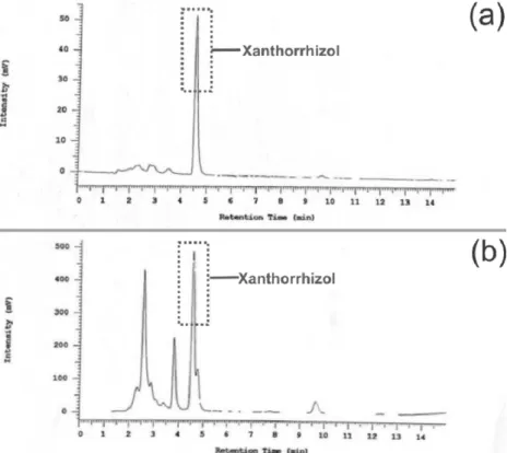
Curcuma xanthorrhiza Roxb. extract was obtained from 100 g dry powder and 300 mL 96% ethanol by maceration method. The extract of Curcuma xanthorrhiza Roxb was then evaporated in vacuum condition at 45oC. Approxi-mately 23 grams of concentrated extract was obtained and then analyzed using HPLC to identify and quantify xanthorrhizol substances. To confirm the presence of xanthorrhizol in the extract, we compared the chromato-gram of the produced extract and xanthorrhizol standard solution (Fig. 2a and 2b). HPLC chromatogram of the produced extract shows two peaks appeared at the retention time of 4.54 minutes and 4.75 minutes, whereas the xanthorrhizol standard solution shows a peak at 4.56 minutes retention time. It can be assumed that the produced extract contained xanthorrrhizol (Fig. 2a and 2b). CXE’s functional group was showed in Fig. 3. Based on the quantitative analysis result, the xanthorrhizol content in the sample was 27%. Beside xanthorrhizol, 7.10% curcumin was found in the extract (data not shown). Furthermore, the extract was classified as a strong antioxidant with IC50 value of 64.27 ppm.
MIC of CXE against MRSA ATCC 4330 and MRSA clinical specimens 7097 was 5 mg/mL (Fig. 4). Previous studies assessing the MIC of CXE against MRSA clinical specimens found MICs of 2048 µg/ml16 and 512 µg/ml.17 This difference of MIC values was presumably due to different composition and method of CXE production, the
4 Natural Product Sciences
concentration of xanthorrhizol produced, the origin of Curcuma xanthorriza, and the nature of different MRSA isolates.
Based on the MIC value obtained during in-vitro study, gels-containing 1, 3, and 5% of CXE were produced (Table 1). Rats receiving topical 3% and 5% CXE-containing gel showed significant reduction of bacterial
load on their wound after 3 days of treatment compared to negative control and mupirocin group, p = 0.014 and p = 0.001, respectively (Fig. 5a and 5b). Whereas none of the rats in the treatment group had abscesses, Three and one rats in negative control and mupirocin group, respectively, had abscesses that might indicate serious MRSA infection of the wound (Fig. 6).
Fig. 2. HPLC Chromatogram of Xanthorrhizol (a) Standard; and (b) CXE of this present study.
Fig. 3. FT-IR spectra in the frequency region 4000-450 cm1.Xanthorrizol functional group presumably identified in frequency region
Wound closure rates among groups did not differ until day-9 after treatment. They showed differences on the day-9 after treatment, in which 1% and 5% CXE-containing gel group showed more advanced wound closure compared to the negative control group, p = 0.029 and p = 0.019, respectively (Fig. 5c and 5d).
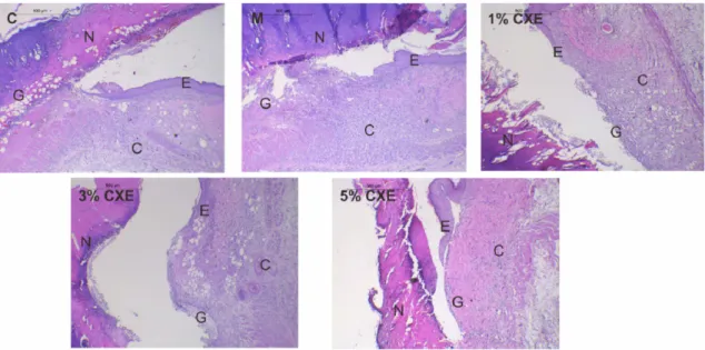
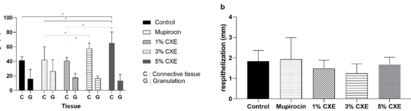
From the histopathological examination (Fig. 7), rats receiving 3% and 5% CXE-containing gel had higher percentage of connective tissue compared mupirocin group, p = 0.041 and p = 0.012, respectively. While the granulation tissue of the burn wound did not differ among
groups (p = 0.677), the ratio of connective tissue and granulation tissue was higher in rats receiving 3% and 5% CXE-containing gel compared to negative control and mupirocin group (Fig. 8a). These histopathological findings indicated that 3% and 5% CXE-containing gel gave a more advanced proliferative healing phase.
Healing processes are classically divided into four phases: (1) Hemostasis phase; (2) Inflammatory phase; (3) Proliferative phase; and (4) Maturation and remodeling phase, although these four phases are usually sequential, they can overlap each other.18 An advanced proliferative
Fig. 4. Dilution agar of CXE (a) 0.0006 mg/mL (b) 2.5 mg/mL (c) 5 mg/mL. No growth was observed in plates containing 5 mg/mL of Mueller-Hinton CXE-containing agar.
Fig. 5. Effect of CXE-containing gel on (a) zone of bacterial growth and (b) its significance per 3 day, (c) wound closure and (d) its significance per 3 day. Data are presented as mean ± SD, *p<0.05, **p<0.01, ***p<0.001.
6 Natural Product Sciences
healing phase of the treatment groups might have been
stimulated by xanthorrhizol substances on CXE-containing gel. The hypothetical mechanism is depicted in Fig. 9.Xanthorrhizol has the activity to reduce the expression of
Fig. 6. Wound closure appearance per 3 days.
Fig. 7. Histopathology of skin examination, C indicates connective tissue, G indicates granulation, E indicates re-epithelization, and N indicates necrosis.
MMP-1 and induce an increase in type 1 procollagen synthesis.12 Moreover, in the present study, CXE was shown to have a strong antioxidant activity. An excessive reactive oxygen species (ROS) was generated post-burn wound injury to kill pathogen entering through the skin.19 If an array of sophisticated antioxidant mechanisms did not control ROS, it leads to inflammatory tissue injury.20 Best ratio of antioxidant and ROS in burn wounds remains unclear. Abnormally, a low level of antioxidant level has been associated with impaired wound healing.21 A reduction of ROS (such as H2O2 that are present in normal skin wounds) induces upregulation of VEGF (vascular endothelial growth factor) for angiogenesis which is good for wound healing.22,23 The CXE, however, contains curcumin, which are reported to have dual effect
on VEGF, upregulate VEGF at low dose (5-10 µM) and downregulate at high dose (20-40 µM).24 In the present study, the net effect was an increase on angiogenesis. It was proven by an advanced healing phase of the treatment groups.
The negative control and mupirocin groups seemed to show higher re-epithelization compared to the treatment groups but the difference was not significant, p = 0.373 (Fig. 8b). There was no antioxidant content in mupirocin ointment and gel as a negative control group that might produce higher level of ROS in these groups. Higher level of ROS might induce epidermal proliferation. The hypothetical mechanism is depicted in Fig. 9. H2O2 triggers the activation of receptors for an epidermal growth factor (EGF) and the keratinocyte growth factor
Fig. 8. CXE-containing gel’s effect on (a) tissue proliferation (b) reepithelization after 12 days treatment, C indicates connective tissue, G indicates granulation. Data are presented as mean ± SD, *p<0.05.
8 Natural Product Sciences (KGF) and induces the production of TGFα (a member of
EGF) in fibroblasts.25,26 Thus, H
2O2 can support the migration and proliferation of epidermal cells. Meanwhile, the higher concentration of CXE-containing gel might have higher antioxidant activity which reduce levels of ROS. The treatment groups showed that 5% CXE-containing gel produced the highest re-epithelization among treatments group (1.67 ± 0.36 mm), followed by 1% CXE (1.49 ± 0.4 mm), and 3% CXE (1.24 ± 0.47 mm). The re-epithelization on rats receiving 5% CXE-containing gel presumably due to its advanced wound healing phase despite its ROS activity, while the rats receiving 1% and 3% CXE-containing gel might have affected by ROS activity.
Prolonged inflammation often interferes wound healing process. Anti-inflammatory effect is needed to balance inflammatory process to achieve good therapeutic target. Xanthorrhizol has an anti-inflammatory effect through the inhibition of cyclooxygenase-2 (COX-2) and inducible nitric oxide synthase (iNOS).11 A reduction of COX-2 can reduce pain, while inhibition of iNOS can induce the rolling and adhesion of the neutrophils on the endothelium, and prevent apoptosis in migrated neutrophils.27
The mechanism of the antibacterial activity of xanthorrhizol remains unclear. Xanthorrhizol is classified as a phenolic compound. Most of the phenolic compound plants provide antibacterial activities through membrane disruption in Gram-positive and Gram-negative bacteria.28 A study of xanthorrhizol on bacteria via electron micros-copy revealed disintegration of bacterial cells, indicating that xanthorrhizol disrupted membrane structure or induce cell rupture.29 Disrupted bacterial membrane structure and cell wall rupture will kill the bacteria resulting in a lesser bacterial load which sequentially reduce the risk for tissue necrosis due to bacterial toxins.
In conclusion, the present study showed that rats receiving 3% and 5% CXE-containing gel had lower MRSA load and more advanced wound healing in MRSA-infected second-degree burn wound compared to rats receiving 2% mupirocin ointment. Future animal studies on the effect of CXE-containing gel in remodelling phase, as well as clinical trials are warranted to demonstrate the safety and efficacy of CXE-containing gel as a topical antibacterial and wound healing agent in human.
Acknowledgments
This research was supported by the Ministry of Research, Technology, and Education of the Republic of Indonesia in the Student Creativity Program Grant 2019
(Grant number B/81/BBK/KM.02.01/2019). We thank Vega Karlowe, MD, Ph.D, who greatly helped in the histopathology examination.
References
(1) Anenden, H. Burns, 2018, https://www.who.int/news-room/fact-sheets/detail/burns. Acccessed 14 January, 2021.
(2) Schaefer, T. J.; Tannan, S. C. Thermal Burns; StatPearls Publishing: U.S.A, 2021, In StatPearls [Internet].
(3) Almarghoub, M. A.; Alotaibi, A. S.; Alyamani, A.; Alfaqeeh, F. A.; Almehaid, F. F.; Al-Qattan, M. M.; Kattan, A. E. J. Burn Care Res. 2020, 41, 1122-1127.
(4 ) Branski, L. K.; Al-Mousawi, A.; Rivero, H.; Jeschke, M. G.; Sanford, A. P.; Herndon, D. N. Surg. Infect. 2009, 10, 389-397.
(5) de Macedo, J. L.; Santos, J. B. Mem. Inst. Oswaldo Cruz. 2005, 100, 535-539.
(6) Amissah, N. A.; van Dam, L.; Ablordey, A.; Ampomah, O. W.; Prah, I.; Tetteh, C. S.; van der Werf, T. S.; Friedrich, A. W.; Rossen, J. W.; van Dijl, J. M.; Stienstra, Y. PloS one 2017, 12, e0181072.
(7) Khan, T. M.; Kok, Y. L.; Bukhsh, A.; Lee, L. H.; Chan, K. G.; Goh, B. H. Germs 2018, 8, 113-125.
(8) Antonov, N. K.; Garzon, M. C.; Morel, K. D.; Whittier, S.; Planet, P. J.; Lauren, C. T. Antimicrob. Agents Chemother. 2015, 59, 3350-3356.
(9) Church, D.; Elsayed, S.; Reid, O.; Winston, B.; Lindsay, R. Clin. Microbiol. Rev. 2006, 19, 403-434.
(10) Mustaffa, F.; Indurkar, J.; Ismail, S.; Shah, M.; Mansor, S. M. Molecules 2011, 16, 3037-3047.
(11) Chung, W. Y.; Park, J. H.; Kim, M. J.; Kim, H. O.; Hwang, J. K.; Lee, S. K.; Park, K. K. Carcinogenesis 2007, 28, 1224-1231.
(12) Oh, H. I.; Shim, J. S.; Gwon, S. H.; Kwon, H. J.; Hwang, J. K. Phytother. Res. 2009, 23, 1299-1302.
(13) Caley, M. P.; Martins, V. L.; O'Toole, E. A. Adv. Wound Care (New Rochelle). 2015, 4, 225-234.
(14) Weinstein, M. P.; Patel, J. B.; Burnham, C.A.; Campeau, S.; Coville, P.S.; Doern, C.; Eliopoulos, G. M.; Galas, M. F.; Humphries, R. M.; Jenkins, S. G. II.; Lewis, J. S.; Limbago, B.; Mathers, A. J.; Mazzulli, T.; Munro, S.D.; Danies, M.O.S.; Patel, R.; Richter, S. S.; Satlin, M.; Swenson, J. M.; Wong, A.; Wang, W. F.; Zimmer, B. L. CLSI document M07: Methods for Dilution Antimicrobial Susceptibility Tests for Bacteria that Grow Aerobically; Clinical and Laboratory Standards Institute; U.S.A, 2018, pp 24.
(15) Chopra, S.; Harjai, K.; Chhibber, S. Int. J. Med. Microbiol. 2016, 306, 707-716.
(16) Sukandar, E. Y.; Kurniati, N. F.; Anggadiredja, K.; Kamil, A. Int. J. Pharm. Pharm. Sci. 2016, 8, 108-111.
(17) Wikaningtyas, P.; Sukandar, E. Y. Asian Pac. J. Trop. Biomed. 2016, 6, 16-19.
(18) Gosain, A.; DiPietro, L. A. World J. Surg. 2004, 28, 321-326. (19) Miyazaki, H.; Kinoshita, M.; Ono, S.; Seki, S.; Saitoh, D. Shock 2015, 44, 252-257.
(20) Mittal, M.; Siddiqui, M. R.; Tran, K.; Reddy, S. P.; Malik, A. B. Antioxid. Redox Signal. 2014, 20, 1126-1167.
(21) Rasik, A. M.; Shukla, A. Int. J. Exp. Pathol. 2000, 81, 257-263. (22) Brauchle, M.; Funk, J. O.; Kind, P.; Werner, S. J. Biol. Chem. 1996, 271, 21793-21797.
(23) Roy, S.; Khanna, S.; Nallu, K.; Hunt, T. K.; Sen, C. K. Mol. Ther. 2006, 13, 211-220.
(24) Lee, C. K.; Chung, W. Y.; Park, K. K. Cancer Res. 2004, 64, 727. (25) Marchese, C.; Maresca, V.; Cardinali, G.; Belleudi, F.; Ceccarelli, S.; Bellocci, M.; Frati, L.; Torrisi, M. R.; Picardo, M. Oncogene. 2003, 22, 2422-2431.
(26) Vivekananda, J.; Lin, A.; Coalson, J. J.; King, R. J. J. Biol. Chem. 1994, 269, 25057-25061.
(27) Dal Secco, D.; Paron, J. A.; de Oliveira, S. H.; Ferreira, S. H.; Silva, J. S.; de Queiroz Cunha, F. Nitric Oxide 2003, 9, 153-164.
(28) Rempe, C. S.; Burris, K. P.; Lenaghan, S. C.; Stewart Jr, C. N. Front. Microbiol. 2017, 8, 422.
(29) Kim, J. E.; Kim, H. E.; Hwang, J. K.; Lee, H. J.; Kwon, H. K.; Kim, B. I. J. Microbiol. 2008, 46, 228-232.
Received August 22, 2020 Revised January 24, 2021 Accepted January 25, 2021