The change of subjective and objective
masticatory efficiency after implant prosthetic
rehabilitation of unilateral missing molars
Sang Soo Lee
The Graduate School
Yonsei University
The change of subjective and objective masticatory
efficiency after implant prosthetic rehabilitation of
unilateral missing molars
A Dissertation
Submitted to the Department of Dental Science
and the Graduate School of Yonsei University
in partial fulfillment of the
requirements for the degree of
Doctor of Philosophy of Dental Science
Sang Soo Lee
This certifies that the dissertation thesis of
Sang Soo Lee is approved.
Thesis Supervisor : Baek Il, Kim
Thesis committee : Ho Keun, Kwon
Thesis committee : Young Sik, Cho
Thesis committee : Jong Hoon, Choi
Thesis committee : Chung Ho, Choi
The Graduate School
Yonsei University
감사의 글
박사 과정에 들어온 지 5년이 지나도록 연구 논문을 쓰지 못했던 것은 ‘개업의로서 바빠서였다.’라고 말하곤 하였지만, 사실은 두려움 때문이었습니다. 아직 치과의사로서 도 부족한 사람이 치의학자로서 연구를 하고 논문을 쓴다는 것이 부족한 제겐 아직 요원한 일로만 여겨졌기 때문이었습니다. 그러나 두려움에 미루기만 한다면 어떠한 결과도 얻을 수 없다는 것을 경험을 통해 알고 있었기에 교수님의 지도와 주위 분들 의 도움을 의지하여 시작한 것이 오늘의 결실을 맺을 수 있게 만들었습니다. 오직 감사할 따름입니다. 무엇보다 지도교수로서 부족한 사람을 독려해 주시며 마 지막까지 나아갈 길을 제시해 주신 김백일 교수님께 감사의 마음을 전합니다. 항상 흐트러짐 없는 모습에 백 마디 말보다 더 강한 가르침을 주시는 교수님의 모습은 공 부하는 사람의 자세의 본보기가 되어 주셨습니다. 또한 교실의 어른이신 권호근 전 학장님께 감사를 드립니다. 부드러운 인품과 폭넓은 지혜로 늘 지식보다 더 큰 인생 의 지혜를 가르쳐 주시곤 하셨습니다. 두 분 교수님들께는 말로는 이루 다 표현할 수 없는 감사함을 다시금 전합니다. 연구의 시작부터 끝까지 함께 고생해 주신 교실의 연구원 선생님들 모두에게 감사 합니다. 초기 연구의 시작을 함께 논의해 주고 준비해 주신 정회인 선생님, 시간에 쫒 기며 받은 자료를 급히 통계 분석하고 정리하여 깔끔한 결과물로 제시해 주신 이은송 선생님, 완벽한 프리젠테이션 표를 만들어 주신 이형석 선생님, 그 외 함께 고생해 주 신 교실의 모든 선생님들께 감사드립니다. 특히 연구의 시작부터 끝까지 깊게 관여하 여 도와주시고 늦은 시간까지 자료를 검토하고 함께 고민을 해 주셨던 강시묵 선생님 께는 특별한 감사를 더해 드립니다. 강시묵 선생님의 자료에 대한 깊은 통찰과 분석 력이 있어서 본 연구의 수준이 한층 높아 질 수 있었다고 확신하는 바입니다. 본 연구를 임상에서 도와주신 우리 강북 예 치과병원 식구들에게 감사합니다. 바쁜 진료 일정 중에서도 환자분들에게 연구 내용을 일일이 설명하고 시편을 모아 주어 본 연구가 임상 연구로서의 가치를 높일 수 있는 결정적 역할을 해 주었습니다. 자신의진료 시간을 기꺼이 할애해 주신 각 과 원장님들, 실험이 원활히 진행되도록 설명하 고 관리해 주신 우리 위생사 선생님들, 내원하지 않는 환자분들의 내원을 지속적으로 요청해 주신 우리 상담실 선생님들 모두에게 감사드립니다. 아울러 귀찮고 불편할 수 도 있는 연구 과정에 흔쾌히 참여해 주신 환자분들에게 진심으로 감사의 말씀을 전합 니다. 사랑하는 우리 가족에게는 감사보다 미안함을 전합니다. 늘 일을 달고 사는 성격이 다보니 평상시에도 변변한 추억하나 만들어 주지 못하였는데, 본 연구를 시작 하게 되면서 근 1년여를 그 흔한 영화 한 편 보여 주지 못하였습니다. 미안하고 사랑합니 다. 성산동에 계신 홀어머니에게 감사를 전합니다. 일찍 혼자되시어 자식들 키우며 고 생하시었으니 이제 제가 학위를 받는 기쁨에 이를 수 있었던 것은 모두 그 고생에 뿌 리를 두고 양분을 빨아 먹었기 때문임을 다시금 기억합니다. 감사합니다. 방학동에 계 신 장인, 장모님에게도 감사를 전합니다. 늦은 나이에 학업을 재개한 사위의 학업 진 행을 누구보다 관심 갖아 주시고 물어 주시며 좋은 결과 있기를 격려해 주셨습니다. 두 분의 격려와 지속적인 관심에 감사 드립니다. 논문의 영문 번역을 도와준 동서 김 진련 교수에게도 감사합니다. 본인의 일도 늘 바쁘면서 불시에 청하는 도움에 싫은 내색 한번 없이 기꺼이 도움을 주었습니다. 이렇게 이 연구 논문이 나오기까지 주변을 둘러 보니 어느 것 하나 감사하지 않은 것이 없고, 어느 것 하나 저 스스로 해 낸 일이 없는 것 같습니다. 이 감사의 마음을 잊지 않고 살 수 있기를 바랍니다. 모든 분들께 진정 감사할 따름입니다. 감사합니다.
CONTENTS
LIST OF FIGURES ··· iii
LIST OF TABLES ··· iv
ABSTRACT ··· v
Ⅰ. INTRODUCTION ··· 1
1. Backgrounds ··· 1
2. Objective ··· 6
Ⅱ. MAERIALS AND METHODS ··· 8
1. Subjects ··· 8
2. Methods ··· 10
2.1. Assessment of Food Intake Ability ··· 12
2.1.1. Questionnaire ··· 12
2.1.2. Procedure ··· 12
2.2. Evaluation of Mixng Ability Index ··· 14
2.2.1. Artificial food wax ··· 14
2.2.2. Procedure ··· 15
2.2.3. Digital image analysis ··· 15
2.3. Statistical analysis ··· 18
Ⅲ. RESULTS ··· 19
3.1. Change of masticatory function implant treatment ··· 20
3.2. Gender-dependent recovery of masticatory function ··· 22
3.4. Edentulous area-dependent recovery of masticatory function ··· 25
3.5. Edentulous arch-dependent recovery in masticatory function ··· 27
3.6. Foodproperty-dependent masticatory ability ··· 28
3.7. Difference in MAI depending on edentulous states in masticatory function ··· 30
3.8. Correlatin between subjective evaluation and objective evaluation ··· 32
3.9. consistency of masticatory function ··· 33
Ⅳ. DISCUSSION ··· 34
Ⅴ. CONCLUSION ··· 40
REFERENCES ··· 41
LIST OF FIGURES
Fig. 1. Form of case report ··· 11
Fig. 2. Questionnaire for assessing ability of food intake ··· 13
Fig. 3. Wax cube used in this study ··· 14
Fig. 4. Chewed wax specimen by study subject ··· 15
Fig. 5. Image taking box for standardized condition ··· 15
Fig. 6. Schematic image from the chewed wax specimen ··· 16
Fig. 7. The FIA, KFIA, and MAI distribution of subjects before and after prosthodontic treatment ··· 21
LIST OF TABLES
Table 1. Distribution of subjects age group and gender ··· 9 Table 2. Distribution of subjects the edentulous area ··· 9 Table 3. Demographic characteristics of study subjects ··· 19 Table 4. Changes in masticatory efficiency before and after dental implant prosthesis of all
subjects ··· 20 Table 5. Changes in masticatory efficiency before and after dental implant prosthesis
according to gender (male = 22 ,female = 32) ··· 22 Table 6. Changes in masticatory efficiency before and after dental implant prosthesis
according to age groups (20∼39s = 11, 40∼59s = 26, over 60s = 17) ··· 24 Table 7. Changes in masticatory efficiency before and after dental implant prosthesis
according to edentulous area (upper right = 14, lower right = 11, upper left = 17, lower left = 12) ··· 26 Table 8. Changes in FIA before and after dental implant prosthesis according to food
property ··· 27 Table 9. Changes in masticatory efficiency before and after dental implant prosthesis
according to side of arch (upper = 31, lower = 23) ··· 29 Table 10. Changes in masticatory efficiency before and after dental implant prosthesis
according to MAI ··· 31 Table 11. Pearson correlation coefficients between the FIA, KFIA, and MAI according to
each groups ··· 32 Table 12. Changes in masticatory function in short and long interval according to MAI 33
ABSTRACT
The change of subjective and objective masticatory efficiency after
implant prosthetic rehabilitaiton in unilateral missing molars
Sang Soo Lee
Department of Dental Science The Graduate School, Yonsei University
(Directed by Professor Baek-Il Kim, D.D.S, M.D.S, Ph.D)
The tooth loss may compromise the chewing function, ultimately leading to decreased quality of life. For this reason, restoration of a lost tooth has been considered an important field in dentistry. Recently, dental implant has become widely available, serving as an important option for replacement of a missing tooth. In this study, I explored a combination of subjective and objective evaluations of chewing efficiency as an alternative, more reliable quantitative means for the determination of the success of the dental implant for the lost molar tooth in one side.
Specifically, I analyzed survey questionnaires on the Food Intake Ability (FIA) for 30 different food groups for subjective evaluation of chewing efficiencies, and then determined scores of the Key Food Intake Ability (KFIA) for chewing five hard foods. For objective
evaluation of chewing efficiencies, the Mixing Ability Index was used.
The participants were subject to dental examination before dental implant restoration. They were also asked to answer self-questionnaires and chew wax samples ten times before and after the completion of implant restoration treatment and I then collected the chewed wax samples for image analyses.
The conclusions drawn in this study are the following:
1. The dental implant treatment for the lost molar tooth in one side was found to improve both subjective and objective evaluation scores: the subjective evaluation scores, FIA and KFIA, increased by 7.4%** and 11.2%**, respectively, whereas the objective evaluation score did so by 8.6%. This improvement was statistically significant (**p<0.0001).
2. I compared changes in chewing efficiency according to gender of patients. FIA, KFIA and MAI of male patients increased by 9.2%*, 13.6%* and 8.4%*, respectively. In comparison, FIA, KFIA and MAI increased by 7.3%*, 11.2%* and 8.4%*, respectively, for female patients. The statistical analyses indicate the lack of a significant difference in changes of chewing efficiency between male and female patients(*p<0.05, **p<0.0001).
3. I compared FIA, KFIA and MAI scores according to age of patients. For patients at the age of 20-40, FIA and KFIA increased by 2.4%* and 6.2%**, respectively. The FIA and KFIA scores increased by 6.4%* and 10.0%**, respectively for patients at the age of 40-50. Similar statistical improvements were also found with patients over the age of 60 (i.e., 12.2%** and 16.2%* increases in FIA and KFIA, respectively). In contrast, a significant increase in the MAI score, which is an objective evaluation score of chewing
efficiency, was found with only a patient group at the age of 40-50 (*p<0.005,
**p<0.0001).
4. I compared changes in chewing efficiency according to locations where tooth losses occurred. For other locations than upper left as principal sites of tooth losses, I found no significant improvement in subjective evaluation scores, FIA and KFIA, after the treatment. In comparison, FIA and KFIA significantly increased by 6.0%** and 9.2%**,
respectively, after the treatment when the tooth loss occurred in upper left. An objective evaluation score, MAI, significantly increased by 7.4%* for upper right 13.4%* for lower right and 3.9%** for upper left. Changes in these scores after treatment for the loss of a tooth in lower left were found to be statistically insignificant (*p<0.005, **p<0.0001).
5. I compared changes in chewing efficiency according to arch where the lost tooth belongs to. I found no significant differences in these scores for upper arch after treatment.
6. I compared changes in chewing efficiency according to tested food groups and found that chewing efficiencies were improved by 10.6%* for food groups with high hardness, 5.6%* for those with medium hardness and 2.4%* for those with low hardness (*p<0.005).
7. I compared changes in FIA, KFIA and MAI scores for patient groups having different chewing efficiencies judged based on baseline MAI scores prior to implantation. For patients with relatively low chewing efficiency prior to treatment, FIA, KFIA and MAI increased by 9.4%**, 12.4%** and 14.0%**, respectively. In contrast, relatively small
improvements were observed with patients having relatively high chewing efficiency prior to treatment; FIA, KFIA and MAI scores increased by 5.6%**, 10.2%**, and 3.4%,
respectively. Differences in chewing efficiency changes between these two patient groups were statistically significant (**p<0.0001).
8. I explored correlations among chewing efficiencies evaluated subjectively and objectively through comparative analyses between FIA and MAI, and between KFIA and MAI. I found that FIA and KFIA (i.e., subjective evaluation scores) were inversely correlated with MAI (i.e., an objective evaluation score) (P<0.05).
In short, results from my study described here demonstrate that changes in chewing efficiency after dental implant restoration treatment were directly reflected by statistical improvement in subjective evaluation scores, which may also be indicative of increased quality of life. In addition, I found that FIA measurements based on self-questionnaire survey and MAI measurements using wax cubes enabled facile, clinical assessment of chewing efficiency of patients, thus allowing for reliable evaluation of outcomes of treatment. Altogether, my study presented here suggests that reliable and accurate assessments of a patient's condition based on the objective and subjective evaluation scores can provide an important guideline for future treatments and managements.
Key words : chewing efficiency, food intake ability, mixed ability index, wax cube
The change of subjective and objective masticatory
efficiency after implant prosthetic rehabilitation of
unilateral missing molars
Sang Soo Lee
Department of Dental Science
The Graduate School, Yonsei University
(Directed by Professor Baek-Il Kim)
Ⅰ. Introduction
1. Backgrounds
Oral cavity consists of lips, cheeks, pharynx, hard palate, soft palate, teeth, and tongue, and is responsible for masticatory, digestive, taste, respiratory and pronunciation functions. These functions affect many aspects of daily life, such as personal appearances and daily conversations (Watanabe, 1998). However, the most important feature of the oral cavity is masticatory function, which is very closely related with the quality of life (Miura, 2000).
As a first step in a series of processes to digest nutrients and absorb, the masticatory function plays a crucial role in maintenance and improvements of the systemic health through making the hard food to soft enough to swallow easily. If masticatory function is significantly compromised, the range of foods to ingest becomes narrow and balanced nutrient supply can be difficult to achieve. In this regard, masticatory function is very
closely related with systemic health (Miura, 1998). Osterberg et al. reported that the masticatory function was compromised in adults with loss of teeth (Osterberg, 2002). While the masticatory function is mediated with the teeth as well as soft tissue including muscle of mastication, cheeks, tongue, and lips, the teeth plays the most important role among other factors affecting mastication. First and second molars provided 36.7% and 28%, respectively, of a total effective masticatory area in complete dentures. These numbers for 1st and 2nd premolars were 8% each of the total area. Because a molar is a key part for the masticatory function, this tooth has gained significant interests in research on improvement of the masticatory function through the restoration of missing a molar.
Nowadays dental implants are quite popular, and the related clinical results have been reported to be very positive. In general, the success of recovery of missing teeth with dental implants has been assessed through survival rate, continuous prosthesis, stability, a radiographic bone loss, and the absence of infection in pre-implant soft tissues (Papaspyridakos et al. 2012). In comparison, the success of prosthodontic treatment depends on whether or not one can eat well and maintain the systemic health from a patient's point of view. Therefore, it will be more appropriate to evaluate the success of the treatment through examination of the change of masticatory efficiency rather than typical histological approaches.
Various methods have been used to assess the masticatory function of human. There are two main strategies to evaluate the mastication; subjective methods and objective methods. One can assess subjective and objective masticatory functions through evaluations of a masticatory ability and masticatory performance, respectively. The masticatory performance evaluation can be divided into static occlusal force measurement and dynamic masticatory efficiency measurement. Many researchers suggested various methods for objective evaluation of the mastication.
Masticatory ability which is used for subjective evaluation is defined as an individual’s own assessment of his or her masticatory function through questionnaires or interview.
Leaker (1990) investigated the masticatory ability with five foods, which have different hardness, by assessing their possibility of ingestion, and Locker et al. (1994) evaluated the masticatory functions of individuals by examining the degrees of their satisfaction of mastication. Miura et al. (1997) showed a relationship between quality of life of elderly persons and their capabilities of food ingestion, and Sugihara et al. (1989) investigated food intake abilities for various oral symptoms. Matsukubo et al. (2006) reported a relationship between the food acceptance ability for 31 different kinds of food and other factors including age, sex, caries status, tooth-contact area, occlusal force, and (full name)CPI.
As one of objective evaluation methods, measurement of an occlusal force has been employed. Helkimo et al. (1978) measured occlusal force using a bite fork-shaped pressure gauge and found that this force decreased as result of aging and a loss of teeth. Floystrand et al. (1982) reported a positive correlation between bite force and the number of teeth from data obtained using a mini bite force recorder, which has a measuring range from 10N to 10,000N. Charles et al. (1988) measured the extent of maximum cleaning using a strain-gauged transducer, which consisted of 2 stainless steel plates separated by a steel sphere and was balanced by occlusal forces imposed between right and left sides. They found that the extent of maximum cleaning was negatively correlated with the number of missing teeth. In 1990, Fuji company commercialized the Dental Prescale System, which allowed many researchers to study correlations between missing teeth and bite forces. The dental prescale system consisted of a pressure-sensitive, horse-shoe shaped bite foil and a computerized scanning system for analysis of the load. This system made simple, chairside examination of comfortable mouth shapes possible, while the previous pressure gauge system required a mouth to remain open during examination. Attempts to evaluate the occlusal force using muscle activity of mastication have also been reported. Devlin et al. (1985) explored a correlation between masseter muscle electromyograpic activity and bite force, but a simple linear relation was found to be inadequate to describe
the observations.
As one of objective evaluating methods, Manly and Braley (1950) developed the sieving method for measuring masticatory performance. This development represented an important breakthrough, which then served as a gold standard and is widely used nowadays. The method was based on determination of the volume percentage of chewed food (such as salted peanuts, shredded coconuts, carrots or raisins) which could pass through a sieve after a given number of masticatory strokes. They concluded that salted peanut was suitable for the sieve-based examination but carrot and other foods were not. Since then, many attempts to replace the peanuts, so gummy jelly, optosil silicone tablets, and coffee beans have been made for evaluation of the masticatory function. In addition, Vander der Bilt et al. (2004) revealed a multi sieve method was more reliable than a single sieve method if the sieve diameter is not close enough to the median particle size of the chewed food.
Liedberg and Owall et al. (1991) et al., reported a gum method for assessing the masticatory performance. Unfortunately, assessment using this method heavily relied on subjective evaluations by examiners, and outcomes from this assessment for partial denture patients were not consistent with masticatory performances measured using other methods. These findings directly indicated limited validity and reliability of the method developed by Liedberg and Owall. Meanwhile, Prinz (1999) reported that flattening of chew gums allowed more accurate assessment when two-color gums were used.
In 2000, some methods for measuring the masticatory efficiency were reported. Sato et al. (2003) proposed to use artificial food composed of two-color wax cubes (12 x 12 x 12 mm3) for evaluation of masticatory function, and formulated a mixing ability index (MAI), which was determined based on the degree of color mixing and a shape of wax after chewing. The MAI was calculated using several chewing-related quantities, such as a total projection area, a mixing color area, a maximum length, and a maximum width of chew wax. A statistical analysis using the MAI produced typical normal distribution with higher
validity when compared to sieving methods. Especially, the MAI-based method is easier to utilize in clinic than a sieving method, and more sensitive than a gum method. As Sato et al. pointed out usefulness of the MAI for evaluation of mastication in partial denture patients, the MAI will become widely used in dental fields. In Korea, Rhu et al. (2007) evaluated the MAI for a selected group of Korean adults, and showed that the MAI values were highly correlated with subjective data obtained from questionnaire. Although the MAI have high validity and reliability, there were still problems. To deduce the discriminant function for MAI calculation, examiner’s subjective intervention, i.e., classification of the chewed wax into three groups (good, medium, and poor), was required. To overcome such limitation, Lee et al. (2008) proposed to determine the MAI in a more objective manner through classification of three groups according to chewing stroke. They found that the MAI evaluated in this manner showed high validity and reliability similar to the original MAI. Using the new MAI, Ahn et al. (2011) reported
that masticatory efficiency was significantly low for patients suffering from
temporomandibular joint diseases.
A handful of research studies have been carried out to compare subjective and objective evaluating methods. Willits et al. (1988) noticed that some patients whose masticatory functions were diagnosed to be good could still feel uncomfortable in daily life. This result pointed out importance of combined uses of subjective and objective methods for more reliable evaluation of masticatory function. Hirai et al. (2001) showed that masticatory function efficiencies for full denture patients decreased with increasing age when determined using a sieving method and a questionnaire. Uchida et al. (2002) reported that results from evaluation using a questionnaire for 20 foods were significantly correlated with masticatory performances determined using a sieve method. Jo et al. (2006) reported that bite forces and food acceptance scores were significantly decreased as a result of increasing age and a loss of teeth. Kim et al. (2009) measured bite force and evaluated a food intake ability (FIA) using questionnaire for 30 general food items and 5
core food items. They found that results obtained using these subjective and objective methods displayed a high correlation with each other.
It is very important to evaluate adequately a patient’s condition before and after treatment in order to better determine the success of dental treatment. Moreover, the need is even more significant in the field of dental restoration treatment where a top priority is given to restoration of masticatory function. But it is impossible to control a position of missing teeth, and treatment methods should vary depending on a patient’s condition. For these reasons, the relevant research has been limited to simple monitoring of changes in masticatory function after treatment with fixed or removable restorative materials in different positions.
In this study, in regard to meet the unmet need, I attempted to quantitatively evaluate masticatory function changes using a combination of objective and subjective methods after dental implant treatment, and examine how these changes could affect patient’s quality of life. Throughout the evaluations, I aimed to demonstrate from a different perspective whether dental implant treatment is efficient for recovering masticatory function.
2. Objective
The objective of study described herein was to compare the change of masticatory function using objective and subjective methods for implant patients, who have one missing teeth at molars in each quadrant of the mouth, before and after implant treatment.
For subjective evaluation of mastication, FIA questionnaires for 30 foods suggested by Rhu et al. (2007) and key food intake ability (KFIA) questionnaires for 5 foods suggested by Kim et al. were used in this study. For objective evaluation of mastication, the Korea MAI modified by Lee et al. (2008) based on the MAI developed by Sato et al. was used. To better judge the success of implant treatment based on improved quality of life, I proposed novel examinations from a perspective of mastication recovery rather than that of previous histological bone fusion.
Null hypotheses of the present study were as follows;
1. There is no difference in subjective masticatory evaluation before and after implant treatment for patients who have one missing teeth at molars in each quadrant of mouths.
2. There is no difference in objective masticatory evaluation before and after implant treatment for patients who have one missing teeth at molar positions.
Ⅱ. Materials and Methods
1. Subjects
A total 56 adult patients who planned to receive dental implant treatment during the period between September 1, 2012 and June 31, 2013 at a local clinic, Seoul, Korea were invited to participate in the present study.
The protocol was approved by the institutional review board at the Yonsei Dental University Hospital (IRB 2-2012-0029). All subjects received verbal and written information and signed a consent form. The inclusion criteria were (i) ages of 20 through 70 years at initial records, (ii) only one missing teeth at a 1st or 2nd molar in mouth. Patients who had any fixed restorative materials were considered to have normal teeth. The following patients were excluded in thep resent study: those who (i) had abnormal mastication because of any oral pain, temporomandibular disorder, or severe malocclusion; (ii) had lost teeth on crown due to severe caries; (iii) were pregnant; (iv) had severe systemic disease such as AIDS, diabetes, cardiac disorder, and blood dyscrasia.
A study was designed based on the one-sample design, and a sample size was determined according to the previous study (Ahn et al. 2010). In the previous study, the mean difference and standard deviation between normal and temporomandibular disorder were 1.14 and 1.00, respectively. Given the consideration that the subjects who suffered from temporomandibular disorder had severe difficulty of mastication in the previous study, I assumed that the difference may further be decreased by about 40% in this study, and thus estimated the mean difference to be 0.46.
Thus sample size was initially estimated to be 50 from values of alpha and beta being 0.05 and 0.10, respectively, and the final sample size was then accordingly determined to
Age groups Male Female Total 20-39 4 7 11 40-59 12 14 26 More than 60 6 11 17 Total 22 32 54 Edentulous area Upper right Lower right Upper left Lower left Total Subject number 14 11 17 12 54
be 56 after taking into account that 20 % of the original participants might drop off in the middle of the study.
Of 56 subjects, 54 subjects finished the full examination, and total 216 waxes were collected.
Table 1. Distribution of subjects according to age group and gender
2. Methods
A dental implant screw (exclude abutment and manufactured crown) at missing tooth was embedded in every participant prior to this study, and the state of implant-bone integration was examined before participating in the present study.
The timing of implant and the period of the edentulous were different. One dentist examined each patient’s mouth and kept examination records. To assess the subjective masticatory ability, I asked all participating subjects to complete a questionnaire where they selected one through five levels for food intake of 30 food items. After completion of the questionnaire, participants were provided with wax cubes for chewing for 10 strokes in a habitual manner. Then, I collected feedbacks from all participating subjects and made prosthesis accordingly for 2 – 4 weeks. Then, I installed all other implant materials onto implant screws within mouths of patients. Then, a recall examination was administered two weeks later.
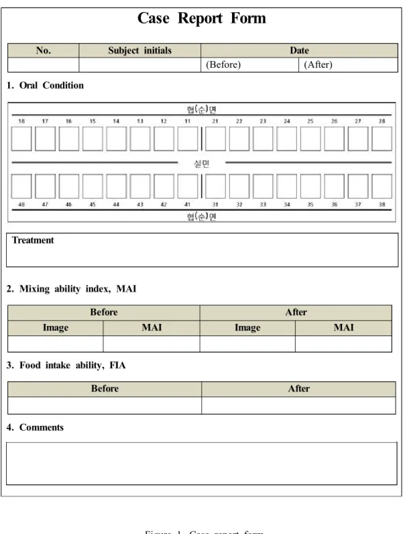
Treatment
Case Report Form
No. Subject initials Date
(Before) (After)
1. Oral Condition
2. Mixing ability index, MAI
Before After
Image MAI Image MAI
3. Food intake ability, FIA
Before After
4. Comments
2.1. Assessment of FIA
2.1.1. Questionnaire
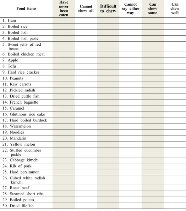
To assess the subjective masticatory ability, a 30 food items questionnaire was administered to all subjects. The food items were selected from Koreans favorite foods (13 items) and from Japan’s food questionnaire (17 items) suggested by the Tokyo dental school.
The response scales (5 point Likert) were as follows: ‘have never been eaten’ (0 point), ‘cannot chew at all’ (1 point), ‘difficult to chew' (2 points), ‘cannot say either was’ (3 ponits), ‘can chew some’ (4 points), and ‘can chew well’ (5 points). The total FIA score was calculated using the average score for 30 foods except for the case of ‘have never been eaten’. In addition, the average score for five key foods (dried cuttlefish, raw carrot, dried peanuts, cubed radish kimchi and caramel) selected from a previous study (Kim et al. 2009) was used for determination of the subjective KFIA.
2.1.2. Procedure
The subjects were requested to answer each question in the FIA questionnaire on a five-point Likert scale (Fig.2). An assistant was made available for subjects who had difficulty of completing the FIA questionnaire on his or her own.
Food items Have never been eaten Cannot chew all Difficult to chew Cannot say either way Can chew some Can chew well 1. Ham 2. Boiled rice 3. Boiled fish 4. Boiled fish paste 5. Sweet jelly of red beans
6. Boiled chicken meat 7. Apple
8. Tofu
9. Hard rice cracker 10. Peanuts
11. Raw carrots 12 .Pickled radish 13. Dried cuttle fish 14. French baguette 15. Caramel
16. Glutinous rice cake 17. Hard boiled burdock 18. Watermelon 19. Noodles 20. Mandarin 21. Yellow melon 22. Stuffed cucumber pickle 23. Cabbage kimchi 24. Rib of pork 25. Hard persimmon 26. Cubed white radish kimchi
27. Roast beef 28. Steamed short ribs 29. Boiled potato 30. Dried filefish
Questionnaire for food intake ability
2.2. Evaluation of MAI
To evaluate the objective masticatory efficiency, the mixing ability test (MAT) was performed using a wax cube as an artificial food, as previously suggested by Sato et al. (2003). Using this method, objective evaluation of masticatory function was possible through quantitative measurements of mixed color areas and flattened wax areas of two colored cubes.
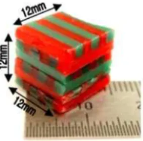
2.2.1. Artificial food wax
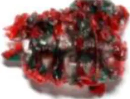
Red and green utility waxes were manufactured by Daedong Ind. (Daegu, Korea). The red and green colored rod shape utility waxes with 2 × 2 × 12 mm3 were put together side by side to make a sheet of 2×12×12mm3 with different color rods faced to each other. Then it was stacked into six identical sheets to make alattice cube of 12×12×12mm3 (Fig.3). To evaluate physical properties of the artificial food, the microhardness was measured for each wax rod using a microhardness tester (JTTOSHIINC.,Japan) and a melting point of the wax were determined using differential scanning calorimetry (STA 409 PC LuxxⓇ, NETZSCH Co.). The vickers hardness number (VHN) of red and green utility wax were 0.088±0.0004 and 0.3697±0.0673 respectively, and the melting temperature range were 63.4-66.3℃ and 59.3-69.9℃, respectively.
2.2.2. Procedure
The wax cubes were kept at 4℃ to maintain their properties until use. The subjects were required to chew a wax cube for 10 strokes in habitual manner. The chewed waxes were carefully removed from the mouth and washed with water, and then dried at room temperature (Fig 4).
Figure 4. A chewed wax specimen produced by one study subject
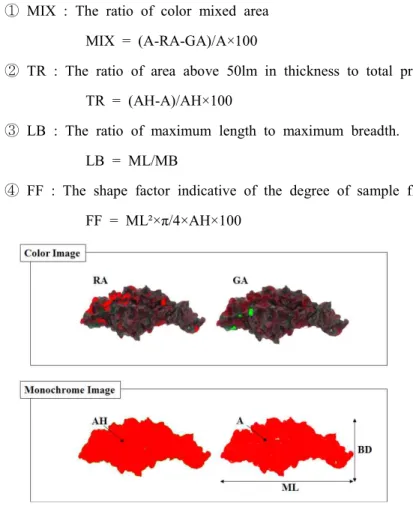
2.2.3. Digital image analysis
Digital images of dried waxes were captured using a DSLR camera (D80, Nikon Co., Tokyo, Japan) under standardized distance and light conditions (Fig. 5).
All images were saved as JPEG files and were analyzed using an image analyzer (Image-Proplus® v6.0, Media Media cybernetics Inc., Bethesda, MD, USA) to determine a color and a shape of the wax on each face. On each image, an examiner picked unmixed red and green colors using a eyedropper tool built in the analyzer. After that, each image was converted according to an 8-bit gray scale to measure the total projection area (AH), the projection area of under 50 um in thickness (A), the maximum length (ML), and the maximum breadth (MB).
These measured values were then converted into the following four parameters; ① MIX : The ratio of color mixed area
MIX = (A-RA-GA)/A×100
② TR : The ratio of area above 50lm in thickness to total projection area. TR = (AH-A)/AH×100
③ LB : The ratio of maximum length to maximum breadth. LB = ML/MB
④ FF : The shape factor indicative of the degree of sample flatness FF = ML²×π/4×AH×100
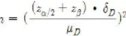
MAI was calculated from the discriminant function optimized for Koreans, as shown below (Lee et al. 2008)
MAI = 1.00 × 10
-1× MIX(50) - 1.50 × 10
-2× TR + 2.98 × 10
-1× LB
2.3. Statistical analysis
A frequency analysis was performed in order to confirm the distribution of each score. Two sample t-test was used to compare the mean value of each score obtained before and after implant treatment according to sex, side of arch (upper and lower), and MAI of baseline. A one-way analysis of variance (ANOVA) was applied to determine whether or not significant differences existed in each score according to age groups, implant positions in quadrant of the mouth, and food properties. For comparative examinations before and after implant treatment, the paired t-test was performed.
The Pearson’s correlation analysis was carried out to explore a relationship among FIA, KFIA and MAI. All statistical analyses were carried out using the PASW 18.0 (SPSS Inc., Chicago, IL, USA) statistical package program.
Ⅲ. Results
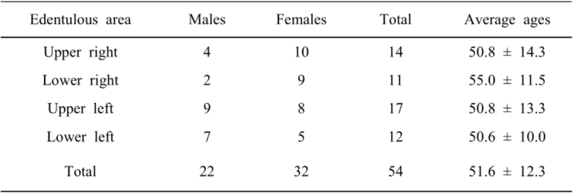
Of 56 subjects, 54 subjects were examined in the present study and the other two subjects did not complete the all process. Table 3 showed the average ages and numbers of male and female patients when classified according to the edentulous area in a quadrant of the mouth.
Table 3. Demographic characteristics of study subjects
Edentulous area Males Females Total Average ages
Upper right 4 10 14 50.8 ± 14.3
Lower right 2 9 11 55.0 ± 11.5
Upper left 9 8 17 50.8 ± 13.3
Lower left 7 5 12 50.6 ± 10.0
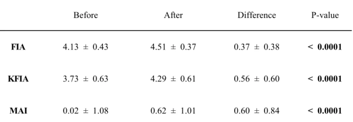
Before After Difference P-value
FIA 4.13 ± 0.43 4.51 ± 0.37 0.37 ± 0.38 < 0.0001
KFIA 3.73 ± 0.63 4.29 ± 0.61 0.56 ± 0.60 < 0.0001
MAI 0.02 ± 1.08 0.62 ± 1.01 0.60 ± 0.84 < 0.0001
1. Change of masticatory function after implant treatment
The FIA, KFIA, and MAI scores calculated before and after implant treatment were shown in Table 4.
Table 4. Changes in masticatory function before and after dental implant treatment of all subjects
FIA: Food intake ability index, KFIA: Key Food intake ability index, MAI: Mixing Ability Index All values are given as mean ± standard deviation.
P-values determined by the paired t-test.
Three scores (FIA, KFIA, and MAI) significantly increased after dental implant treatment as a result of recovery at the missing molar position (P<0.001). Compared with the edentulous state, the FIA and KFIA which is indicators of a subjective masticatory ability, were increased by 7.4% and 11.2%, respectively when examined 2 weeks after implant treatment. The MAI, an indicator of objective masticatory efficiency, was also increased by 8.6%.Histograms of FIA, KFIA, and MAI were obtained from the frequency analysis before and after implant treatment (Fig 7). In Fig 7, the histograms were shifted toward higher scores, indicating that masticatory function was improved after implant treatment.
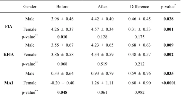
Gender Before After Difference p-value* FIA Male 3.96 ± 0.46 4.42 ± 0.40 0.46 ± 0.45 0.028 Female 4.26 ± 0.37 4.57 ± 0.34 0.31 ± 0.33 0.001 p-value** 0.010 0.128 0.175 KFIA Male 3.55 ± 0.67 4.23 ± 0.65 0.68 ± 0.63 0.009 Female 3.86 ± 0.58 4.34 ± 0.59 0.48 ± 0.57 0.002 p-value** 0.068 0.519 0.212 MAI Male 0.33 ± 0.64 0.93 ± 0.79 0.59 ± 0.76 0.035 Female -0.20 ± 0.40 1.26 ± 1.11 0.60 ± 0.90 <0.0001 p-value** 0.048 0.061 0.982
2. Gender-dependent recovery of masticatory function
While the subjective masticatory ability in the edentulous state was higher in female than male, the objective masticatory efficiency was lower in female than male (Table 5). No significant gender-dependent differences were observed in all masticatory function (P>0.05). However, the significant differences in masticatory function were detected before and after implant treatment in both male and female subjects (P<0.05).
Table 5. Changes in masticatory function before and after dental implant treatment examined according to genders of participating subjects (male = 22, female = 32)
FIA: Food intake ability index, KFIA: Key Food intake ability index, MAI: Mixing Ability Index All values are given as mean ± standard deviation.
*
p-values determined by the paired t-test.
3. Age-dependent recovery of masticatory function
The masticatory function is usually compromised with an increasing age in the edentulous state. This tendency was also observed after the treatment. Among the age groups, especially, more than 60 age group showed significantly low mean scores in FIA, KFIA, and MAI (P<0.05).
Compared with the edentulous state, the subjective masticatory ability increased by the following percent: FIA=2.4%, and KFIA=6.2% (20-39s age group), FIA=6.4%, and KFIA=10.0% (40-59s age group), and FIA=12.2%, and KFIA=16.2% (≥ 60s age group) when measured 2 weeks after implant treatment (P<0.05). Meanwhile, the MAI also increased by about 6.4% (20-39s age group), 11.0% (40-59s age group), and 6.0% (≥ 60s age group) when evaluated 2 weeks after implant treatment. Among them, only 40-50s age group showed a significant increase in masticatory function (P<0.05).
That is, the masticatory efficiency of 40-59s age group was dramatically increased but the other age groups showed a relative low increase in objective evaluation.
Age groups Before After Difference P-value FIA 20-39s 4.69 ± 0.15a 4.80 ± 0.07a 0.12 ± 0.09a 0.002 40-59s 4.20 ± 0.24a 4.52 ± 0.26a 0.32 ± 0.20a <0.0001 ≥ 60s 3.68 ± 0.27b 4.29 ± 0.49b 0.61 ± 0.56b <0.0001 KFIA 20-39s 4.33 ± 0.45a 4.64 ± 0.27a 0.31 ± 0.30a 0.007 40-59s 3.90 ± 0.39b 4.40 ± 0.40a 0.50 ± 0.37a <0.0001 ≥ 60s 3.09 ± 0.46c 3.91 ± 0.83b 0.81 ± 0.88a 0.002 MAI 20-39s 0.74 ± 0.40a 1.20 ± 0.45a 0.45 ± 0.68a 0.053 40-59s 0.15 ± 0.86a 0.92 ± 0.50a 0.77 ± 0.86a <0.0001 ≥ 60s -0.65 ± 1.30b -0.23 ± 1.32b 0.42 ± 0.88a 0.066
Table 6. Changes in masticatory efficiency before and after dental implant treatment examined according to different age groups (20-39s = 11, 40-59s = 26, over 60s = 17)
FIA: Food intake ability index, KFIA: Key Food intake ability index, MAI: Mixing Ability Index All values are given as mean ± standard deviation.
P-values determined by paired t-test.
Different letters within the same column indicate significant differences between groups by ANOVA and Bonferroni's post hoc test (p<0.05).
4. Edentulous area-dependent recovery of masticatory function
There were no significant differences in masticatory function before or after implant treatment (P>0.05). When subjective evaluation was performed depending on edentulous areas, no significant differences before and after the treatment were observed except for the upper left (P>0.05). In comparison, significant differences before and after the treatment were observed in objective evaluation for all examined edentulous areas except for the lower left. The observed MAI increase depended on the edentulous state (i.e., about 7.4%, 13.4 % and 3.9% for upper right, lower right and upper left, respectively), when measured 2 weeks after the treatment (P<0.05).
Edentulous
area Before After Difference P-value
FIA Upper right 4.18 ± 0.44 4.61 ± 0.30 0.43 ± 0.46 0.347 Lower right 4.11 ± 0.45 4.56 ± 0.39 0.45 ± 0.52 0.454 Upper left 4.10 ± 0.45 4.40 ± 0.46 0.30 ± 0.19 <0.0001 Lower left 4.16 ± 0.42 4.49 ± 0.28 0.34 ± 0.38 0.129 KFIA Upper right 3.76 ± 0.71 4.43 ± 0.51 0.67 ± 0.81 0.592 Lower right 3.84 ± 0.58 4.36 ± 0.61 0.53 ± 0.56 0.075 Upper left 3.67 ± 0.66 4.13 ± 0.78 0.46 ± 0.45 <0.0001 Lower left 3.70 ± 0.59 4.30 ± 0.44 0.60 ± 0.57 0.187 MAI Upper right 0.19 ± 0.84 0.70 ± 1.09 0.52 ± 0.89 0.022 Lower right -0.70 ± 1.43 0.24 ± 1.40 0.94 ± 1.08 0.015 Upper left 0.32 ± 1.17 0.59 ± 0.94 0.27 ± 0.61 <0.0001 Lower left 0.58 ± 0.44 0.89 ± 0.50 0.84 ± 0.70 0.731
Table 7. Changes in masticatory efficiency before and after dental implant prosthesis examined according to the edentulous area (upper right = 14, lower right = 11, upper left = 17, lower left = 12)
FIA: Food intake ability index, KFIA: Key Food intake ability index, MAI: Mixing Ability Index All values are given as mean ± standard deviation.
5. Edentulous arch-dependent recovery in masticatory function
There was no significant difference between upper and/or lower edentulous arches before or after implant treatment. However, the difference of MAI in a lower arch was significantly higher than in an upper arch (P<0.05).
Table 8. Changes in masticatory efficiency before and after dental implant prosthesis examined according to the side of arch (upper = 31, lower = 23)
Side of arch Before After Difference P-value*
FIA Upper 4.14 ± 0.44 4.49 ± 0.40 0.36 ± 0.34 <0.0001 Lower 4.14 ± 0.43 4.53 ± 0.33 0.39 ± 0.45 0.124 p-value** 0.998 0.761 0.767 KFIA Upper 3.71 ± 0.67 4.27 ± 0.68 0.55 ± 0.63 0.001 Lower 3.77 ± 0.57 4.33 ± 0.52 0.57 ± 0.55 0.018 p-value** 0.751 0.699 0.950 MAI Upper 0.26 ± 1.02 0.64 ± 0.99 0.38 ± 0.75 <0.0001 Lower -0.30 ± 1.09 0.58 ± 1.06 0.89 ± 0.88 0.001 p-value** 0.057 0.840 0.027
FIA: Food intake ability index, KFIA: Key Food intake ability index, MAI: Mixing Ability Index All values are given as mean ± standard deviation.
*P-values determined by the paired t-test. **P-values determined by the two-sample t-test.
6. Food property-dependent masticatory ability
To compare the subjective satisfaction of subjects before and after implant treatment, the following 30 foods item, which were divided into three groups (hard, medium, soft) based on the previous study (Rhu, 2006), were used;
Hard foods:
Dried filefish, Raw carrots, Dried cuttlefish, Peanuts, Pickled radish, Glutinous rice cake, French baguette, Hard persimmon, Roast beef, Caramel, Hard rice cracker, Steamed short ribs, Hard boiled burdock, Cubed white radish kimchi, Apple
Medium foods:
Yellow melon, Stuffed cucumber pickle, Watermelon, Noodles, Mandarin, Cabbage kimchi, Rib of pork, Boiled potato
Soft foods:
Ham, Tofu, Boiled fish, Boiled chicken meat, Boiled fish paste, Boiled rice, Sweet jelly of red beans
There were significant differences among three groups depending on food property before implant treatment. After implant treatment, FIA scores for the hard group became similar to those for the medium group. FIA scores for the soft food group were significantly different from those for the other groups (P<0.05). Mean values of all food groups were increased after implant treatment, and the biggest change was shown for the hard group.
Food group Before After Difference P-value
Hard 3.84 ± 0.24a 4.38 ± 0.18a 0.53 ± 0.10a <0.0001
Medium 4.15 ± 0.37b 4.43 ± 0.21a 0.28 ± 0.17b 0.003
Soft 4.76 ± 0.12c 4.88 ± 0.07b 0.12 ± 0.79b 0.006
Table 9. Changes in FIA before and after dental implant treatment examined according to food property
FIA: Food intake ability index, KFIA: Key Food intake ability index, MAI: Mixing Ability Index All values are given as mean ± standard deviation.
P-values determined by the paired t-test.
Different letter superscripts in the same column indicate significant differences between groups according to ANOVA and the Bonferroni's post hoc test (p<0.05).
7. Difference in MAI depending on edentulous states in masticatory function
To compare the objective masticatory efficiency before and after implant treatment, the subjects were classified into two groups according to the MAI baseline (MAIb≤0.0 and
MAIb>0). In the low MAI baseline group, FIA, KFIA, and MAI increased by about 9.4%,
12.4%, and 14.0%, respectively, after implant treatment when compared to the edentulous state. On the other hand, for the high MAI group, FIA, KFIA, and MAI increased by 5.6%, 10.2%, and 3.4%, respectively, when measured 2 weeks after treatment. Although FIA and KFIA were significantly different between low and high MAI groups in the edentulous state, these differences disappeared after implant treatment. In MAI, there were significant differences between low and high MAI groups in both before and after treatment. Moreover, greater MAI differences were found after dental implant with the low MAI group than the high MAI group.
MAI group N Before After Difference P-value* FIA Low MAIb 26 3.94 ± 0.31 4.42 ± 0.32 0.47 ± 0.41 <0.0001 High MAIb 28 4.31 ± 0.46 4.59 ± 0.40 0.28 ± 0.34 <0.0001 P-value** <0.0001 0.078 <0.0001 KFIA Low MAIb 26 3.52 ± 0.55 4.13 ± 0.52 0.62 ± 0.64 <0.0001 High MAIb 28 3.94 ± 0.64 4.44 ± 0.65 0.51 ± 0.56 <0.0001 P-value** 0.012 0.058 <0.0001 MAI Low MAIb 26 -0.88 ± 0.79 0.10 ± 1.18 0.98 ± 0.88 <0.0001 High MAIb 28 0.86 ± 0.43 1.10 ± 0.47 0.24 ± 0.62 0.05 P-value** <0.0001 <0.0001 0.001
Table 10. Changes in masticatory efficiency before and after dental implant prosthesis for different to MAI groups
FIA: Food intake ability index, KFIA: Key Food intake ability index, MAI: Mixing Ability Index All the values are given as mean ± standard deviation.
*
P-values determined by the paired t-test.
**
P-values determined by the two-sample t-test.
Low MAIb, MAI scores lower than 0.0 measured before treatment; High MAIb, MAI scores over
Group N FIA vs. MAI p-value KFIA vs. MAI P-value Ages 20-39 11 0.31 0.347 0.25 0.464 40-59 26 0.14 0.490 0.19 0.362 ≥ 60 17 -0.56 0.020 -0.63 0.007 Edentulous area Upper 31 0.28 0.132 0.23 0.224 Lower 23 0.38 0.073 0.32 0.140 MAI Low MAIb 28 -0.64 0.757 -0.07 0.754 High MAIb 26 -0.12 0.547 -0.76 0.699 Total 54 0.31 0.022 0.24 0.081
8. Correlation between subjective evaluation and objective evaluation
In the Pearson’s correlation analysis, the correlation coefficient between FIA and MAI was 0.31 (P<0.05). There were no significant correlations in sub-group except for the over 60 age group. In the over 60 age group, the correlation coefficient between MAI and FIA was -0.56, and that between MAI and KFIA was -0.63.
Table 11. Pearson’s correlation coefficients among the FIA, KFIA, and MAI scores measured before treatment for different groups
FIA: Food intake ability index, KFIA: Key Food intake ability index, MAI: Mixing Ability Index P-values determined by the Pearson's correlation analysis
Low MAIb, MAI scores lower than 0.0 measured before treatment; High MAIb, MAI scores over
MAI group N Short interval Long interval Difference P-value* △FIA Low MAIb 6 0.26 ± 0.14 0.86 ± 0.30 0.60 ± 0.32 0.005 High MAIb 13 0.26 ± 0.38 0.41 ± 0.43 0.16 ± 0.39 0.158 P-value** 0.929 0.033 0.028 △KFIA Low MAIb 6 0.20 ± 0.18 1.00 ± 0.68 0.80 ± 0.70 0.039 High MAIb 13 0.37 ± 0.55 0.29 ± 0.60 -0.08 ± 0.76 0.721 P-value** 0.480 0.035 0.029 △MAI Low MAIb 6 0.92 ± 0.87 2.39 ± 1.25 1.47 ± 1.91 0.118 High MAIb 13 0.44 ± 0.41 0.03 ± 0.92 -0.41 ± 0.75 0.072 P-value** 0.112 < 0.0001 0.006 9. Consistency of masticatory function
To evaluate the consistency of masticatory function after implant treatment, △FIA, △
KFIA, and △MAI were measured after shorter intervals (2 weeks after) and longer intervals (over than 3month after) and compared accordingly. Total 19 subjects were involved in this analysis because other subjects didn’t come again. They were classified into two groups (low MAIb, high MAIb) according to the MAI baseline measured initially
prior to the treatment. The improvement caused by implant treatment remained noticeable for more than 3 months. Especially, the more significant improvements were found with the low MAIb group when examined using subjective evaluation (P<0.05).
Table 12. Changes in masticatory efficiency at short and long interval according to MAI
FIA: Food intake ability index, KFIA: Key Food intake ability index, MAI: Mixing Ability Index All values are given as mean ± standard deviation.
*P-values determined by the paired t-test. **P-values determined by the two-sample t-test.
Low MAIb, MAI scores lower than 0.0 measured before treatment; High MAIb, MAI scores over 0.0 measured before treatment
Ⅳ. Discussions
Since Dr. Brnemark first introduced modern dental implantation for prosthetic care of patients in 1965, significant advances have been made in the implant treatment. Despite the fact that dental implant is so popular these days and considered as the primary option for a patient who has single missing teeth, effects of implant treatment on masticatory functions of patients and/or their quality of life have yet to be studied. In general, the success of the implant treatment has been judged mostly based on the extent of integration between bone and implant materials. The assessment of integration of implants requires extraction of tissues samples in vitro or examination using MRI in clinic.
However, it should be noted that the ultimate goal of dental treatment is to improve the quality of life of patient through comprehensive recovery of the masticatory function rather than simple restoration of missing teeth or esthetic appearance. Increasing treatment satisfaction through enhancing a patient’s understanding could improve the quality of treatment. To this end, we need to develop a scientific approach which allows for assessment of the quality of life through evaluations of subjective satisfaction and objective masticatory efficiency.
To address these issues, several methods to evaluate masticatory function were selected with the following considerations
First, the method should enable measurements of the satisfaction or inconvenience levels associated with food chewing
Second, the method should enable a quantitative and objective analysis. Third, the method should be easy and simple to use in clinic.
As one of the subjective evaluating methods, the FIA test proposed by Jo et al. (2006) was chosen. Using this method, it was possible to examine the change of masticatory function after implant treatment. For objective evaluation, the MAI test proposed by Sato
et al. was chosen because it was easy to use in clinic and helpful for communication with patient. Each participant had a single missing tooth in the first or second molar in any of four (i.e., left, right, upper and lower) positions. The FIA score, an indicator of a subjective masticatory ability, was 4.13 ± 0.43 before implant treatment, but it increased by 7.4% (4.51 ± 0.37) after the treatment. The KFIA score for 5 food items, which are a part of food groups considered in questionnaire for FIA increased about 11.2% after the treatment. The MAI, an indicator of objective masticatory efficiency increased about 8.6%. The FIA results shown in this study were lower than the previous study performed by Jeung et al. (2010), who explored a relationship between FIA and MAI. A possible explanation for this difference would be that the participating subjects in the previous study visited the dental hospital due to various reasons, including those having nothing to do with a tooth loss.
Although the masticatory ability determined using a subjective evaluating method was higher in female than male, the masticatory efficiency determined using an objective evaluation method was higher in male than female. This finding indicated that there could be a difference between objective and subjective masticatory function efficiencies. In other word, the males felt a greater decrease in their masticatory function even though the measured objective masticatory function was only slightly compromised. On the other side, the females felt less discomfort than a measured decrease in objective masticatory function. The previous study stated that the bite force was higher in male than female but masticatory function was higher in female. This observation indicated that the objective and subjective masticatory functions are not directly correlated with each other and the measured bite fore is not always proportional to masticatory efficiency. In other words, the results could be changed depending on methods used to evaluate the masticatory function.
Overall, the masticatory function was compromised with an increasing age. The results described in the present study are consistent with previous findings reported elsewhere. Okiyama et al. (2003) showed that the bite force decreased with increases in age and the
number of missing molar teeth. Matsukubo et al. (2006) reported that the food intake ability depended on numbers of remaining and missing teeth, maximum bite force, and an area of contacting teeth. In the present study, it was shown that increases in FIA and KFIA scores as a result of implant treatment were more significant when ages of patients increased. This result might have something to do with mental effects which could be originated from implant-mediated restoration of the edentulous state.
Among the quadrant of the mouth, the biggest change was shown at the upper left (P<0.0001). However, this result doesn't seem to be reasonable. It is because mastication is performed through contacts between the upper and lower teeth, and thus the missing tooth position doesn't seem to matter. The discrepancy might be attributable to nature of the samples used in the present study. Although the sample size of each quadrant was between 11 and 14, the upper left group’s sample size was 17 subjects (about 50%). Therefore, the observation might result from the asymmetrical and biased sample size. If sample sizes increase in other groups, the difference may be decreased. One of such evidence includes reduction of the edentulous area-dependent difference after implant treatment.
Meanwhile, the participants involved in the present study were restricted to those with a loss of the first or second molar. However, it was found that patients tended to chew on the opposite side even though they had a premolar on a lost side. This result suggests that most subjects had unilateral chewing habits. Based on this fact, it is expected that the FIA and MAI could be similar even if a premolar is lost. The FIA of persons who have one or two missing teeth was 4.89 ± 0.14, and the FIA of persons who have three to five missing teeth was 3.84 ± 1.42 (Jeung et al. 2010). The FIA of subjects who participated in the present study was 4.13 ± 0.43, and this value falls in between FIA scores for a group with one or two missing teeth and a group with three to five missing teeth. In this study, the MAI score was measured to be 0.12 ± 1.08 prior to implant. This masticatory efficiency was similar to the MAI value of persons who have a unilateral
chewing habit (0.20 ± 1.18) as described in a previous study (Rhu, 2008). Some of participating subjects tried to do bilateral chewing, and it was later found that these participants did so consciously. It was expected that the lower MAI score would result from the use of an unfamiliar side and limited times (i.e., 10) of chewing. Because the mastication movements are mediated with muscle of mastication, cheeks, tongue, and lips, the MAI had been affected by various factors.
Several studies have shown that mastication can better be examined using hard and tough foods rather than soft foods. One study using raw carrots showed a significant correlation between the rate of size breakdown and the number of chews required for swallowing in dentate subjects (Luke et al. 2007) [Is a food particle size a criterion for the initiation of swallowing?] . A significant correlation between bite fore and food intake ability was found in a previous study using a 30 food item questionnaire for three different food groups (hard, medium, soft) (Jo, 2006). Consistent with this finding, the biggest changes in FIA and MAI after implant treatment had been found with the hard food group.
In the Pearson’s correlation analysis, the correlation coefficient between the FIA and MAI was 0.31 (P<0.05). It means that there is a weak relationship between the subjective perception and objective efficiency for mastication. The result described in the present study was supported by findings of other previous studies. One clinical trial showed that the correlation coefficient of patients who have temporomandibular joint disease is 0.40 (Ahn et al., 2011). In another study, the correlation coefficient between FIA and MAI was 0.51 in Korean adults (Jeong et al., 2010). The difference between results from the present study and the previous study on Korean adults may be related with nature of participating subjects; while patients with only single missing tooth were subjected to examine in the present study, those in various edentulous states was included in the previous study on Korean adults.
all edentulous area groups and age groups studied except for the age over 60 group. Interestingly unexpected negative correlations between FIA and MAI (-0.56) and between KFIA and MAI (-0.63) were found with the age over 60 group. The reason for this observation is thought to be the fact that the MAI fell within negative values between -4.0 and 2.0. In the over 60 age group, the mean MAI was -0.65 (before) and -0.23 (after). It means MAI values in this age group were mostly negative regardless of implant treatment, and thus the negative correlation was found between MAI and FIA, and between MAI and FIA.
The improvement of masticatory function caused by implant treatment was maintained over 3 months. The greater improvement was found when MAI values were initially lower before implant treatment. The more significant improvement was found with subjective evaluation scores and this is due to physical adaptation of mouths and muscles over time since the implant treatment: a mouth and facial muscles were stiffened upon implant treatment and became relaxed over time. This relaxation might positively affect subject’s feeling for chewing. Note that this finding should not be generalized because the sample size used was rather limited and not all patients were examined during the same time period. However, monitoring of masticatory function will be useful for prediction of therapeutic effects and additional follow-up studies on a larger sample size over a longer time period will be needed for the relevant examination. There were other additional limitations in the present study. First of all, oral conditions, which can be reflected by the DMFT index community periodontal index (CPI), and teeth arrangement, were not considered in the present study. The lack of such consideration makes it difficult to generalize the findings described in the present study. The masticatory function is affected by the number and position of missing teeth (Kim et al. 2006) as well as restorative materials used (e.g., crowns and bridges)l. Although bite force showed little relation to the CPI, the bite force was found to be significantly dependent on age and the number of remaining teeth, gender, the DMFT index, and teeth arrangement (Rhu, 2006). It should