INTRODUCTION
Asthma is the most common chronic lung disease, affecting more than 300 million people of all ages, and the number of pa-tients is increasing by 50% per decade worldwide.1,2 This disease is characterized by airway hyperreactivity and mucous overse-cretion that result in intermittent airway obstruction.3,4 Asthma is considered to be an allergic, eosinophilic, and Th2-mediated disease.5 Th2-cell-derived cytokines such as IL-5 and IL-13 play a critical role in the type 2 immune response involved in aller-gen-induced airway inflammation. IL-5 affects the differentia-tion and maturadifferentia-tion of eosinophils, while IL-13 acts on epithe-lial and smooth muscle cells and plays a role in airway hyperre-sponsiveness in allergen-induced asthma.6,7 Serum IgE and IgG1 levels are important markers related to the pathogenesis of allergic asthma; asthma patients have increased serum levels of IgE and IgG1.8 Antigen-specific IgE can attach to receptors on mast cell, basophils, B lymphocytes, and eosinophils.9 This interaction results in the release of inflammatory mediators such as histamine, prostaglandins, leukotrienes, proteases, growth
Receptor Interacting Protein 2 (RIP2) Is Dispensable for
OVA-Induced Airway Inflammation in Mice
Tae-Hyoun Kim,
1Yeong-Min Park,
2Seung-Wook Ryu,
3Dong-Jae Kim,
4Jae-Hak Park,
1* Jong-Hwan Park
4*
1Department of Laboratory Animal Medicine, College of Veterinary Medicine, Seoul National University, Seoul, Korea
2Department of Microbiology and Immunology, School of Medicine, Pusan National University, Yangsan, Korea
3Department of Bio and Brain Engineering, KAIST, Daejeon, Korea
4Department of Biochemistry, College of Medicine, Konyang University, Daejeon, Korea
factors, cytokines, and chemokines; these are required for the development of allergic asthma.10 Interaction of antigen with IgE bound to receptors on the cell surface activates mast cells to release preformed mediators such as histamine, leukotrienes, prostaglandin D2, thromboxane B2, and platelet-activating fac-tors. These mediators induce airway smooth muscle contrac-tion, edema, and enhanced mucous secrecontrac-tion, leading to air-flow limitation.11
Nod-like receptors (NLRs) belong to a family of cytosolic re-Purpose: Asthma is a pulmonary chronic inflammatory disease characterized by airway obstruction and hyperresponsiveness. Pattern recognition receptors are known to play a key role in the development of allergic diseases as well as host defenses against microbial infection. Receptor inter-acting protein 2 (RIP2), a serine/threonine kinase, is an adaptor molecule of NOD1 and NOD2, and genetic variation in this receptor is known to be associated with the severity of allergic asthma in children. In this study, we examined the role of RIP2 in the development of allergic airway inflam-mation in a mouse model. Methods: Airway inflammation was induced in mice through intranasal administration of ovalbumin (OVA) after 2 intra-peritoneal immunizations with OVA. Lung inflammation and mucus hypersecretion were examined histologically and total cell infiltration in bron-choalveolar (BAL) fluids was determined. Levels of the Th2-related cytokines, IL-5 and IL-13, in lung extracts were measured by ELISA. Serum anti-gen-specific IgE and IgG1 levels were also assessed. Results: OVA-induced lung inflammation and mucus hypersecretion were not different be-tween WT and RIP2-deficient mice. The IL-5 and IL-13 levels in the bronchoalveolar (BAL) fluids were also not impaired in RIP2-deficient mice com-pared to WT mice. Moreover, RIP2 deficiency did not affect serum OVA-specific IgG1 and IgE levels. Conclusions: Our results suggest that RIP2 is not associated with the development of allergic airway inflammation.
Key Words: RIP2; ovalbumin; airway inflammation; Th2; IgE
This is an Open Access article distributed under the terms of the Creative Commons Attribution Non-Commercial License (http://creativecommons.org/licenses/by-nc/3.0/) which permits unrestricted non-commercial use, distribution, and reproduction in any medium, provided the original work is properly cited.
Correspondence to: Jong-Hwan Park, DVM, PhD, Department of Biochemistry, College of Medicine, Konyang University, 158 Gwanjeo-dong-ro, Seo-gu, Daejeon 302-718, Korea.
Tel: +82-42-600-6450; Fax: +82-42-600-6455; E-mail: jonpark@konyang.ac.kr Jae-Hak Park, DVM, PhD, Department of Laboratory Animal Medicine, College of Veterinary Medicine, Seoul National University, Gwanak-ro 1, Gwanak-gu, Seoul 151-742, Korea.
Tel: +82-2-880-1256; Fax: +82-2-880-1256; E-mail: pjhak@snu.ac.kr Received: January 30, 2013; Revised: April 16, 2013; Accepted: May 3, 2013
•There are no financial or other issues that might lead to conflict of interest.
Allergy Asthma Immunol Res. 2014 March;6(2):163-168.
http://dx.doi.org/10.4168/aair.2014.6.2.163
Sensitization Challenge Sacrifice Day 0 1 7 8 14 15 21 22 24 WT PBS OVA Rip2 -/-Histologic score 4 3 2 1 0 WT WT PBS OVA Rip2-/- Rip2 -/-WT PBS Rip2 PBS WT OVA Rip2 OVA % cell count 120 90 60 30 0
Macrophages Eosinophils Neutrophils Lymphocytes
Cells ( × 10 5) 7.5 5.0 2.5 0.0 WT WT PBS OVA Rip2-/- Rip2 -/-A D E B C
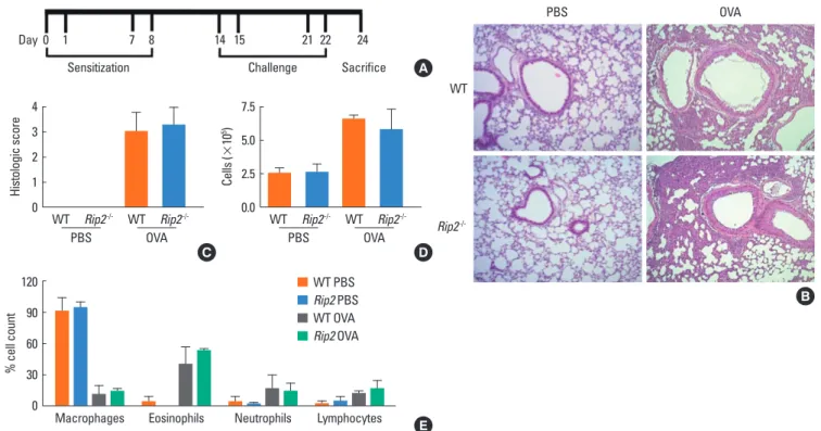
Fig. 1. OVA-induced airway inflammation in WT and RIP2-deficient mice. A schematic diagram of the experimental design (A). Mice were sensitized by i.p. adminis-tration of OVA mixed with adjuvant at days 0, 1, 7, and 8. On days 14, 15, 21, and 22, mice were challenged with OVA or PBS. Photographs of lung tissues were ob-tained from H&E-sob-tained sections (B) and histopathological scores were determined semi-quantitatively by microscopic examination (C). Total cell numbers in the BAL fluids were counted (D) and a differential cell count was performed using Diff-Quick staining (E). Data are expressed as means±SD.
ceptors that are responsible for the recognition of microbial molecules and danger signals. The first identified NLRs, NOD1 and NOD2, have an N-terminal caspase-recruitment domain (CARD), intermediate Nod, and C-terminal leucine-rich re-peats (LRRs). They sense the bacterial peptidoglycan compo-nents, meso-diaminopimelic acid (meso-DAP) and muramyl dipeptide, respectively.12 Following recognition, NOD1 and NOD2 recruit a serine/threonine kinase, receptor interacting protein 2 (RIP2 [also known as RICK and CARDIAK]), which has an N-terminal kinase domain and C-terminal CARDs linked by an intermediate region. Association between NOD1 or NOD2 and RIP2 mediated by CARD-CARD interactions induces the activation of NF-κB and mitogen-activated protein kinases (MAPKs), subsequently leading to the production of proinflam-matory mediators.13-15
Studies have shown that NOD1 and NOD2 signaling is involved in allergic disease. Genetic variations of NOD1 are associated with asthma and elevated IgE levels in humans.16 NOD1 poly-morphisms are also significantly associated with alteration in the strong protective effect which exposure to a farming envi-ronment has on allergies.17 In addition, children with the poly-morphic allele C2722 of the NOD2 gene are at greater risk of de-veloping allergic rhinitis and atopic dermatitis.18 There is also evidence of an association between the adaptor molecule RIP2 and asthma. Nakashima et al.19 suggested that genetic variants
of the RIP2 gene may be associated with the severity of asthma, even though these variants are not likely to be involved in asth-ma development. Moreover, blockade of RIP2 by the flavonoid aglycone, naringenin, contributed to the suppression of the production of thymic stromal lymphopoietin in mast cells, which play a pivotal role in allergic asthma.20 These findings suggest that RIP2 may be associated with the development of allergic asthma, and prompted us to determine the exact role of RIP2 in the development of allergic airway inflammation. We developed a mouse model of ovalbumin (OVA)-induced air-way inflammation using WT and RIP2-deficient mice and ex-amined the severity of lung inflammation, Th2-derived cyto-kine levels in lung extracts, and serum immunoglobulin levels. MATERIALS AND METHODS
Animals
Wild-type (WT) C57BL/6 mice, 6- to 8-weeks-old, were pur-chased from KOATECH (Pyeongtaek, Gyeonnggi-do, Korea). RIP2-deficient C57BL/6 mice were purchased from The Jack-son Laboratory (Bar Harbor, ME, USA). All animal experiments were approved by the Institutional Animal Care and Use Com-mittee of Konyang University.
Airway inflammation induction
Protocols are depicted schematically in Fig. 1A. Both WT and RIP2-deficient mice were sensitized with 40 μg OVA (Sigma-Al-drich, St. Louis, MO, USA) and 2 mg of adjuvant (Imject® Alum, Thermo scientific, Rockford, IL, USA) in 200 μL of PBS, or with PBS alone by intraperitoneal (i.p.) injection on days 0, 1, 7, and 8. On days 14, 15, 21, and 22, anesthetized mice were challenged intranasally (i.n.) with 200 μg of OVA in PBS or with PBS alone in a volume of 50 μL. Animals were sacrificed 2 days after the last challenge and bronchoalveolar lavage (BAL) fluids, serum, and lung tissues were collected for analysis.
Measurement of cytokine and serum OVA-specific antibody levels
Lung extracts were obtained using a tissue homogenizer. Ho-mogenates were centrifuged at 1,000×g for 10 minutes. Super-natants were collected, and then stored at -70°C for analysis. IL-5 and IL-13 levels were measured using a commercial en-zyme-linked immunosorbent assay (ELISA) kit (R&D Systems, Abingdon, U.K.). For the measurement of OVA-specific IgE lev-els, 96-well ELISA plates were coated with OVA (10 μg/mL) at 4°C overnight. Nonspecific binding was blocked with 1% BSA in PBS, and serum samples (at 1:20 dilution) were added to the plate. After incubation for 2 hours at room temperature, biotin rat anti-mouse IgE (BD Biosciences, San Jose, CA, USA) was ap-plied, followed by streptavidin HRP (BD Biosciences). After washing, the TMB substrate reagent set (BD Biosciences) was applied according to the manufacturer’s instructions and opti-cal density (OD) at 450 nm was measured. OVA-specific IgG1 levels were determined using the methods described above, ex-cept that peroxidase-conjugated rat anti-mouse IgG1 (Southern Biotech, Birmingham, AL, USA) was used.
Bronchoalveolar lavage (BAL)
After anesthesia by intraperitoneal injection of Zoletile (Virbac Laboratories, Carros, France), BAL fluid was obtained by lavage with 0.8 mL of PBS via a tracheal catheter. The BAL fluid was centrifuged at 200×g for 3 minutes at 4°C. After discarding the supernatant, we resuspended the cell pellet in cold RPMI 1640 medium. Total cell numbers in the lavage fluid were counted using a hemocytometer. A differential cell count was performed using Diff-Quick staining on the basis of morphological criteria.
Histopathology
To evaluate tissue inflammation, the left lung of each mouse was fixed in 10% neutral-buffered formalin for 48 hours and then embedded in paraffin. Tissue sections (2 μm thick) were prepared and stained with hematoxylin and eosin (HE) or peri-odic acid-Schiff (PAS), and examined under a light microscope. Tissue inflammation scoring was based on the presence or abundance of inflammatory lesions as follows: 0, non-specific; 1, mild; 2, mild to moderate; 3, moderate; 4, moderate to severe;
5, severe. To quantitate mucus staining, PAS-positive cells in the airways were counted, and the length of the basement mem-brane (BM) in each airway was measured using ImageJ version 1.44 (National Institutes of Health, Bethesda, MD, USA). The re-sults are reported as the mean numbers of PAS-positive cells per 100 micrometers of BM.21
Statistical analysis
The significance of differences in mean values of the groups was evaluated by t-tests, and values are expressed as means± SD. All statistical calculations were performed using GraphPad Prism version 4 (GraphPad Software, San Diego, CA, USA). Val-ue of P<0.05 was considered to indicate significance.
RESULTS
Role of RIP2 in the severity of OVA-induced inflammation in the mouse lung
Airway inflammation in mice was achieved by challenge with OVA 4 times i.n. after 4 immunizations by i.p. injection, as de-scribed in Fig. 1A. We first examined whether RIP2 affected the severity of airway inflammation. Intra-nasal challenge with OVA induced severe infiltration of inflammatory cells, consisting mostly of lymphocytes and granulocytes, around the bronchus, and increased the thickness of the alveolar walls in both WT and RIP2-deficient mice (Fig. 1B). However, when histopatho-logical scores were assessed, RIP2 deficiency did not appear to affect the severity of lung inflammation induced by OVA (Fig. 1C). In addition, total infiltrating cells were counted in the BAL fluids of mice with and without i.n. challenge by OVA. Com-pared to the PBS-treated mice, OVA challenge increased the number of infiltrating cells in the BAL fluids of both WT and RIP2-deficient mice, with no significant difference between the WT and RIP2-deficient mice (Fig. 1D). When a differential cell count was performed using Diff-Quick staining, intranasal challenge with OVA led to a decrease in the percentage of mac-rophages and an increase in eosinophils and neutrophils in the BAL fluids of mice (Fig. 1E). However, there was no significant difference between WT and RIP2-deficient mice (Fig. 1E).
Effects of RIP2 on goblet cell hyperplasia and mucus hypersecretion in the bronchi of OVA-challenged mice
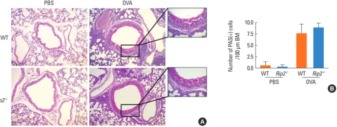
OVA-induced allergic airway inflammation is characterized by hyperplasia of goblet cells and mucus hypersecretion in the bronchus. To determine whether RIP2 deficiency influenced these phenomena, slide sections were stained with PAS and observed under a light microscope. As shown in Fig. 2 A and B, the number of PAS-positive cells as well as mucus secretion in the airway epithelial layer of mice was increased by intranasal challenge with OVA. However, there was no significant differ-ence in the number of PAS-positive cells or mucus secretion between WT and RIP2-deficient mice (Fig. 2 A and B).
Effect of RIP2 on Th2-derived cytokine levels in lung extracts of OVA-challenged mice
We examined OVA-induced IL-5 and IL-13 levels in lung ex-tracts of WT and RIP2-deficient mice. Results showed that i.n. challenge with OVA increased the IL-5 and IL-13 levels in the lungs of mice (Fig. 3 A and B). However, RIP2 deficiency did not affect OVA-induced IL-5 and IL-13 levels in lung extracts (Fig. 3 A and B).
Effect of RIP2 on serum antigen-specific IgE and IgG1 levels in OVA-challenged mice
Finally, we measured the serum antigen-specific IgE and IgG1 levels. As expected, antigen-specific IgE and IgG1 levels were increased by OVA challenge (Fig. 4 A and B). However, there were no significant differences in OVA-specific IgE and IgG1 levels between WT and RIP2-deficient mice (Fig. 4 A and B).
DISCUSSION
Allergic asthma is a chronic airway inflammatory disease re-lated to dysfunction of the airway caused by the release of in-flammatory mediators, and remodeling of the airway wall.22 In allergic asthma, exacerbation of the underlying airway inflam-mation is associated with symptoms and airway limitations.11 Pattern recognition receptors (PRRs) initiate immune respons-es by recognizing structurrespons-es of microorganisms and endoge-nous molecules released from damaged cells, and regulate the transcription of genes involved in inflammatory responses.23 In addition to their protective role in bacterial or viral infection, PRRs have been reported to be involved in allergic asthma. Toll-like receptors (TLRs), the best characterized PRRs, are known to mediate the induction of allergic airway inflammation. There is also evidence of a close association between polymorphisms in TLR genes and asthma.24-26 In addition, several studies have revealed that TLR stimulation exacerbates or alleviates aller-gen-induced asthma depending on the TLR type.27,28
Immuni-IL-5 (pg/mL) IL-13 (pg/mL) 300 200 100 0 800 600 400 200 0 WT WT WT WT (116.4) (110.1) (58.4) (52.8) (23.9) (9.0) (32.2) (23.2) OVA OVA PBS PBS Rip2-/- Rip2 -/-Rip2-/- Rip2 -/-A B
IgE (OD450) IgG1 (OD450)
1.5 1.0 0.5 0.0 1.5 1.0 0.5 0.0 WT WT WT WT OVA OVA PBS PBS Rip2-/- Rip2 -/-Rip2-/- Rip2 -/-A B WT PBS OVA Rip2 -/-A Number of P AS(+) cells /100 μm BM 10.0 7.5 5.0 2.5 0.0 WT WT PBS OVA Rip2-/- Rip2 -/-B
Fig. 2. Hyperplasia of goblet cells and mucus hypersecretion in the bronchus of lung tissue. Lung sections were stained with PAS and examined by light microscopy. Areas in the black boxes are shown at a higher magnification on the right of each picture (A). Numbers of PAS-positive cells in the airway epithelium were counted. In each airway studied, the length of the basement membrane (BM) was measured using image analysis software. The results are presented as mean numbers of PAS-positive cells per micrometer of BM (B). Data are expressed as means±SD.
Fig. 3. Cytokine production in the lung tissue of WT and RIP2-deficient mice. Lung extracts from the right lungs of sacrificed mice. IL-5 (A) and IL-13 (B) levels in lung extracts were measured by ELISA. Data are expressed as means±SD. Coefficients of variations (%) are shown in brackets.
Fig. 4. Levels of OVA-specific IgE and IgG1 in the serum. Serum was obtained from blood samples collected 48 hours after the last OVA challenge. Serum OVA-specific IgE (A) and IgG1 (B) levels were measured by ELISA. Data are ex-pressed as means±SD.
zation with OVA and a TLR2 agonist (Pam3Cys) induced Th2 immune responses such as the production of antigen-specific IgE in the serum, and IL-13 secretion by splenocytes.27 TLR2 ac-tivation promoted airway hyperresponsiveness.27 Double-stranded RNA increased lung inflammation, airway hyperre-sponsiveness, and antigen-specific Th2 responses in OVA-sen-sitized mice through the TLR3-TRIF (Toll/IL-1R domain-con-taining adaptor-inducing IFN-β) pathway.28 In contrast, intra-nasal administration of the TLR7 agonist, R848, suppressed ex-perimental asthma by inducing type Ι interferon production and inhibiting Th2 responses.29 Oral administration of CpG-ODN, a TLR9 agonist, prevented eosinophilic airway inflam-mation.30 These findings suggest that innate immune responses mediated by PRRs play a critical role in the development of al-lergic airway inflammation.
Similar to TLRs, NOD1 and NOD2 stimulation triggers the ac-tivation of NF-κB and MAPKs, which are critical factors for the production of proinflammatory cytokines; the adaptor mole-cule RIP2 is required for this.15 However, in contrast to TLRs, the association between NOD1 and NOD2 signaling and allergic asthma is poorly understood. Based on several indirect lines of evidence of its involvement in allergic asthma,19,20 we sought to determine the role of RIP2 in OVA-induced airway inflamma-tion. We found no significant differences between WT and RIP2-deficient mice in terms of the severity of lung inflamma-tion, total cell infiltration in BAL fluid, IL-5 and IL-13 levels in lung extracts, or serum antigen-specific IgE and IgG1 levels of mice challenged intranasally with OVA. A recent study showed that serum samples from normal mice, but not antibiotic-treat-ed mice, had NOD1- and NOD2-stimulating activity,31 suggest-ing that microbiota may steadily release NOD1- and NOD2-stimulatory factors (e.g., peptidoglycans) into the body fluid. Therefore, in this study, we compared various parameters be-tween WT and RIP2-deficient mice in the absence of NOD1 and NOD2 stimulation to mimic physiological conditions. Con-sistent with the findings of a previous study,32 we found that WT and RIP2-deficient mice immunized with OVA and alum with-out NOD1 and NOD2 ligands, did not show differences in se-rum antigen-specific IgG levels. Eosinophilic infiltration into the lung by OVA was also not impaired in RIP2-deficient mice compared with WT mice.33 In addition, RIP2 is not essential for T-cell proliferation and differentiation.34 Taken together, our re-sults indicate that under normal conditions, RIP2 deficiency is not associated with the development of allergic airway inflam-mation. Nevertheless, it is necessary to define the role of RIP2 in the development of allergic disease under NOD1 and NOD2 activation; Magalhaes et al.32 showed that RIP2 is required for NOD1- and NOD2-induced Th2 immunity. NOD1 and NOD2 ligands increased the number of OVA-specific cells producing IL-4 and IL-5 in the splenocytes of WT mice, but not in RIP2-deficient mice.32 In addition, RIP2 is essential for OVA-specific IgG1 production, mediated by NOD1 and NOD2 stimulation.32
These findings suggest that RIP2 may affect the development of allergic disease mediated by NOD1 and NOD2 activation.
Microbial infections are thought to affect the development or severity of allergic asthma through TLR-mediated signaling.33,35 NOD1 and NOD2 cooperate with TLRs to induce the innate im-mune response against microbial infections.36 Listeria-induced production of cytokines was impaired in NOD1/2 double- or RIP2-deficient macrophages after LPS exposure.37 RIP2 defi-ciency also led to decreased production of cytokines in TLR-to-lerized macrophages in response to Pseudomonas infection, and protected mice from lethality induced by the bacterial in-fection.38 Therefore, it is necessary to clarify whether NOD1/2 and RIP2 contribute to the control of the development of aller-gic diseases mediated by microbial infection.
ACKNOWLEDGMENTS
This study was supported by a grant from the Korean Health Technology R&D project, the Ministry of Health & Welfare, Re-public of Korea (Grant No. A111025).
REFERENCES
1. Masoli M, Fabian D, Holt S, Beasley R; Global Initiative for Asthma (GINA) Program. The global burden of asthma: executive summa-ry of the GINA Dissemination Committee report. Allergy 2004;59: 469-78.
2. To T, Stanojevic S, Moores G, Gershon AS, Bateman ED, Cruz AA, Boulet LP. Global asthma prevalence in adults: findings from the cross-sectional world health survey. BMC Public Health 2012;12: 204.
3. Busse WW, Lemanske RF Jr. Asthma. N Engl J Med 2001;344:350-62. 4. Shifren A, Witt C, Christie C, Castro M. Mechanisms of remodeling
in asthmatic airways. J Allergy (Cairo) 2012;2012:316049.
5. Wenzel SE. Asthma phenotypes: the evolution from clinical to mo-lecular approaches. Nat Med 2012;18:716-25.
6. Nakajima H, Takatsu K. Role of cytokines in allergic airway inflam-mation. Int Arch Allergy Immunol 2007;142:265-73.
7. Wills-Karp M, Luyimbazi J, Xu X, Schofield B, Neben TY, Karp CL, Donaldson DD. Interleukin-13: central mediator of allergic asth-ma. Science 1998;282:2258-61.
8. Shakib F, Sihoe J, Smith SJ, Wilding P, Clark MM, Knox A. Circulat-ing levels of IgG1 and IgG4 anti-IgE antibodies and asthma severi-ty. Allergy 1994;49:192-5.
9. Platts-Mills TA. The role of immunoglobulin E in allergy and asth-ma. Am J Respir Crit Care Med 2001;164:S1-5.
10. Kuhl K, Hanania NA. Targeting IgE in asthma. Curr Opin Pulm Med 2012;18:1-5.
11. Jarjour NN, Kelly EA. Pathogenesis of asthma. Med Clin North Am 2002;86:925-36.
12. Hasegawa M, Fujimoto Y, Lucas PC, Nakano H, Fukase K, Núñez G, Inohara N. A critical role of RICK/RIP2 polyubiquitination in Nod-induced NF-kappaB activation. EMBO J 2008;27:373-83.
13. Inohara N, Koseki T, Lin J, del Peso L, Lucas PC, Chen FF, Ogura Y, Núñez G. An induced proximity model for NF-kappa B activation in the Nod1/RICK and RIP signaling pathways. J Biol Chem 2000;
275:27823-31.
14. Kobayashi K, Inohara N, Hernandez LD, Galán JE, Núñez G, Jane-way CA, Medzhitov R, Flavell RA. RICK/Rip2/CARDIAK mediates signalling for receptors of the innate and adaptive immune systems. Nature 2002;416:194-9.
15. Chen G, Shaw MH, Kim YG, Nuñez G. NOD-like receptors: role in innate immunity and inflammatory disease. Annu Rev Pathol 2009; 4:365-98.
16. Hysi P, Kabesch M, Moffatt MF, Schedel M, Carr D, Zhang Y, Board-man B, von Mutius E, Weiland SK, Leupold W, Fritzsch C, Klopp N, Musk AW, James A, Nunez G, Inohara N, Cookson WO. NOD1 vari-ation, immunoglobulin E and asthma. Hum Mol Genet 2005;14: 935-41.
17. Eder W, Klimecki W, Yu L, von Mutius E, Riedler J, Braun-Fahrlän-der C, Nowak D, Holst O, Martinez FD; ALEX-Team. Association between exposure to farming, allergies and genetic variation in CARD4/NOD1. Allergy 2006;61:1117-24.
18. Kabesch M, Peters W, Carr D, Leupold W, Weiland SK, von Mutius E. Association between polymorphisms in caspase recruitment domain containing protein 15 and allergy in two German popula-tions. J Allergy Clin Immunol 2003;111:813-7.
19. Nakashima K, Hirota T, Suzuki Y, Akahoshi M, Shimizu M, Jodo A, Doi S, Fujita K, Ebisawa M, Yoshihara S, Enomoto T, Shirakawa T, Kishi F, Nakamura Y, Tamari M. Association of the RIP2 gene with childhood atopic asthma. Allergol Int 2006;55:77-83.
20. Moon PD, Choi IH, Kim HM. Naringenin suppresses the produc-tion of thymic stromal lymphopoietin through the blockade of RIP2 and caspase-1 signal cascade in mast cells. Eur J Pharmacol 2011;671:128-32.
21. Mäkelä MJ, Kanehiro A, Dakhama A, Borish L, Joetham A, Tripp R, Anderson L, Gelfand EW. The failure of interleukin-10-deficient mice to develop airway hyperresponsiveness is overcome by respi-ratory syncytial virus infection in allergen-sensitized/challenged mice. Am J Respir Crit Care Med 2002;165:824-31.
22. Holgate ST. Innate and adaptive immune responses in asthma. Nat Med 2012;18:673-83.
23. Takeuchi O, Akira S. Pattern recognition receptors and inflamma-tion. Cell 2010;140:805-20.
24. Basu S, Fenton MJ. Toll-like receptors: function and roles in lung disease. Am J Physiol Lung Cell Mol Physiol 2004;286:L887-92. 25. Cook DN, Pisetsky DS, Schwartz DA. Toll-like receptors in the
pathogenesis of human disease. Nat Immunol 2004;5:975-9. 26. Eder W, Klimecki W, Yu L, von Mutius E, Riedler J,
Braun-Fahrlän-der C, Nowak D, Martinez FD; ALEX Study Team. Toll-like receptor 2 as a major gene for asthma in children of European farmers. J Al-lergy Clin Immunol 2004;113:482-8.
27. Redecke V, Häcker H, Datta SK, Fermin A, Pitha PM, Broide DH,
Raz E. Cutting edge: activation of Toll-like receptor 2 induces a Th2 immune response and promotes experimental asthma. J Immunol 2004;172:2739-43.
28. Torres D, Dieudonné A, Ryffel B, Vilain E, Si-Tahar M, Pichavant M, Lassalle P, Trottein F, Gosset P. Double-stranded RNA exacerbates pulmonary allergic reaction through TLR3: implication of airway epithelium and dendritic cells. J Immunol 2010;185:451-9. 29. Xirakia C, Koltsida O, Stavropoulos A, Thanassopoulou A, Aidinis V,
Sideras P, Andreakos E. Toll-like receptor 7-triggered immune re-sponse in the lung mediates acute and long-lasting suppression of experimental asthma. Am J Respir Crit Care Med 2010;181:1207-16. 30. Kitagaki K, Businga TR, Kline JN. Oral administration of
CpG-ODNs suppresses antigen-induced asthma in mice. Clin Exp Im-munol 2006;143:249-59.
31. Clarke TB, Davis KM, Lysenko ES, Zhou AY, Yu Y, Weiser JN. Recog-nition of peptidoglycan from the microbiota by Nod1 enhances systemic innate immunity. Nat Med 2010;16:228-31.
32. Magalhaes JG, Lee J, Geddes K, Rubino S, Philpott DJ, Girardin SE. Essential role of Rip2 in the modulation of innate and adaptive im-munity triggered by Nod1 and Nod2 ligands. Eur J Immunol 2011; 41:1445-55.
33. Nembrini C, Sichelstiel A, Kisielow J, Kurrer M, Kopf M, Marsland BJ. Bacterial-induced protection against allergic inflammation through a multicomponent immunoregulatory mechanism. Tho-rax 2011;66:755-63.
34. Hall HT, Wilhelm MT, Saibil SD, Mak TW, Flavell RA, Ohashi PS. RIP2 contributes to Nod signaling but is not essential for T cell pro-liferation, T helper differentiation or TLR responses. Eur J Immu-nol 2008;38:64-72.
35. Conrad ML, Ferstl R, Teich R, Brand S, Blümer N, Yildirim AO, Pa-trascan CC, Hanuszkiewicz A, Akira S, Wagner H, Holst O, von Mu-tius E, Pfefferle PI, Kirschning CJ, Garn H, Renz H. Maternal TLR signaling is required for prenatal asthma protection by the non-pathogenic microbe Acinetobacter lwoffii F78. J Exp Med 2009;206: 2869-77.
36. Kim YG, Park JH, Reimer T, Baker DP, Kawai T, Kumar H, Akira S, Wobus C, Núñez G. Viral infection augments Nod1/2 signaling to potentiate lethality associated with secondary bacterial infections. Cell Host Microbe 2011;9:496-507.
37. Kim YG, Park JH, Shaw MH, Franchi L, Inohara N, Núñez G. The cytosolic sensors Nod1 and Nod2 are critical for bacterial recogni-tion and host defense after exposure to Toll-like receptor ligands. Immunity 2008;28:246-57.
38. Park JH, Kim YG, Núñez G. RICK promotes inflammation and le-thality after gram-negative bacterial infection in mice stimulated with lipopolysaccharide. Infect Immun 2009;77:1569-78.