Jung Min Lee, Department of Internal Medicine, Yonsei Uni-versity College of Medicine, Seoul 120-752, South Korea Sang Hoon Ahn, Department of Internal Medicine, Institute of Gastroenterology, Yonsei University College of Medicine, Seoul 120-752, South Korea
Sang Hoon Ahn, Liver Cirrhosis Clinical Research Center, Seoul 120-752, South Korea
Author contributions: Ahn SH designed this article and revised it critically; Lee JM acquired information and wrote this article. Supported by The Grant of the Bilateral International Collabora-tive R&D Program from the Ministry of Knowledge Economy and the Good Health R&D Project from the Ministry for Health, Welfare and Family Affairs, South Korea (A050021)
Correspondence to: Sang Hoon Ahn, MD, PhD, Professor, Department of Internal Medicine, Institute of Gastroenterology, Yonsei University College of Medicine, 250 Sungsanno, Seodae-moon-gu, Seoul 120-752, South Korea. ahnsh@yuhs.ac Telephone: +82-2-22281930 Fax: +82-2-3936884 Received: February 22, 2010 Revised: March 23, 2010 Accepted: March 30, 2010
Published online: January 21, 2011
Abstract
Hepatitis B surface antigen (HBsAg) is produced and secreted through a complex mechanism that is still not fully understood. In clinical fields, HBsAg has long served as a qualitative diagnostic marker for hepatitis B virus infection. Notably, advances have been made in the development of quantitative HBsAg assays, which have allowed viral replication monitoring, and there is an opportunity to make maximal use of quantitative HB-sAg to elucidate its role in clinical fields. Yet, it needs to be underscored that a further understanding of HBsAg, not only from clinical point of view but also from a viro-logic point of view, would enable us to deepen our in-sights, so that we could more widely expand and apply its utility. It is also important to be familiar with HBsAg variants and their clinical consequences in terms of im-mune escape mutants, issues resulting from overlap with corresponding mutation in the P gene, and detec-tion problems for the HBsAg variants. In this article, we review current concepts and issues on the quantification
of HBsAg titers with respect to their biologic nature, method principles, and clinically relevant topics.
© 2011 Baishideng. All rights reserved.
Key words: Hepatitis B virus; Hepatitis B surface anti-gen; Quantitative assay; Virology
Peer reviewers: Yukihiro Shimizu, MD, PhD, Kyoto Katsura Hospital, 17 Yamada-Hirao, Nishikyo, Kyoto 615-8256, Japan; Yogesh K Chawla, Professor, Department of Hepatology, Post-graduate Institute of Medical Education and Research, Chandi-garh 160012, India
Lee JM, Ahn SH. Quantification of HBsAg: Basic virology for clinical practice. World J Gastroenterol 2011; 17(3): 283-289 Available from: URL: http://www.wjgnet.com/1007-9327/full/ v17/i3/283.htm DOI: http://dx.doi.org/10.3748/wjg.v17.i3.283
INTRODUCTION
Hepatitis B virus (HBV) causes a wide range of clinical consequences, from acute and chronic infection to cirrho-sis and hepatocellular carcinoma, and represents a global public health problem[1,2]. Historically, HBV dates to 1967
when an unknown antigen in Australia was recognized to be associated with hepatitis type B, which was later referred to as the hepatitis B surface antigen (HBsAg)[3].
Since then, HBsAg has served as a qualitative diagnostic marker for HBV infection. Notably, advances have been made in the development of quantitative HBsAg assays, which have allowed viral replication monitoring. A num-ber of clinical studies have evaluated the clinical utility of HBsAg and suggested its potential roles. Yet, it needs to be underscored that a further understanding of HBsAg, not only from a clinical point of view but also from a virologic point of view, would enable us to deepen our insights, so that we could more widely expand and ap-ply its utility. Therefore, in this article, we review current concepts and issues on the quantification of HBsAg titers
© 2011 Baishideng. All rights reserved. doi:10.3748/wjg.v17.i3.283
TOPIC HIGHLIGHT
Quantification of HBsAg: Basic virology for clinical practice
Jung Min Lee, Sang Hoon Ahn
(qHBsAg) with respect to their biologic nature, method principles, and clinically relevant topics.
STRUCTURE AND MOLECULAR
VIROLOGY OF HBsAg
Components of the viral structure
HBV belongs to Hepadnaviridae and is composed of the
envelope, core, DNA genome, and viral polymerase. It has a circular form of partially double-stranded DNA and is approximately 3200 nucleotides in length[4,5]. A 42-45 nm
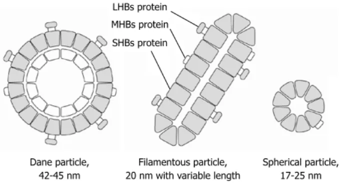
long HBV spherical form (Dane particle), which is the full virion with infectivity, can be visualized (Figure 1) under electron microscopy. It has two-layered shells. The outer shell is the envelope protein referred to as hepatitis B sur-face (HBs) protein, which is further divided into small, middle, and large HBs proteins (SHBs, MHBs and LHBs proteins, respectively), and the inner shell is a core protein referred to as the hepatitis B core protein in which viral polymerase and the HBV genome is enclosed. In addition to the abovementioned full virion, smaller non-infectious subviral particles are present in the serum; 17-25 nm spheri-cal particles, mainly composed of SHBs protein, constitute the most abundant form, which is as much as 10 000-fold in excess of the full infectious virion[4,6]. Filamentous (or
tubu-lar) particles are another form, with a 20 nm diameter and variable length, and are composed of SHBs, MHBs, and the LHBs protein. The form of the HBV particles appears to be determined by the proportion of LHBs protein[7]. All
three forms can be detected in serum with commercial as-says and are collectively referred to as HBsAg.
Synthesis and secretion
HBV has four distinct open reading frames (ORFs) that encode the envelope, core, polymerase, and X proteins. ORF S has three internal AUG codons encoding the SHBs, MHBs, and LHBs proteins, which correspond to the S, preS2 + S, and preS1 + preS2 + S domains, respec-tively (Figure 2). These proteins have a common carboxyl end but different amino ends[8].
Like all other proteins, mRNA transcription is the first event to occur. Two 2.1 kb mRNAs for the M/SHBs proteins and a 2.4 kb mRNA for the LHBs protein are formed, and take a separate pathway from viral replica-tion. Diverse transcription factors are involved and act on promoters, enhancers, and other regulatory elements, such as the glucocorticoid responsive element[9,10]. LHBs
and M/SHBs expression are thought to be independently regulated with different promoters; a typical TATA box is present in the LHBs promotor (S promotor Ⅰ, SPI), whereas the TATA-less promotor, which usually has mul-tiple initiation sites, is associated with the M/SHBs pro-moter, thus accounting for synthesis of distinct proteins from one mRNA. In patients with active viral replication, the protein expression pattern shows a predominance of the M/SHBs protein in contrast to a predominance of the LHBs protein in inactive carriers[11]. After
transcrip-tion, protein synthesis and glycosylation follows at the endoplasmic reticulum (ER) membrane resulting in a 226
amino acid SHBs protein, the MHBs protein with an ditional 55 amino acids, and the LHBs protein with an ad-ditional 108-119 amino acids. Although the LHBs mRNA includes the M/SHBs sequence, it does not translate into the M/SHBs protein, and the ratio between the MHBs and SHBs protein is controlled by a complex mechanism, which is not fully understood[12]. To form a full virion, a
mixture of HBs proteins in a well-balanced ratio is utilized to envelop core particles in which SHBs and LHBs pro-tein are indispensible[13]. The virion is transported to the
cell membrane through vesicles, and several conditions must be satisfied for successful secretion, because excess SHBs protein is required, whereas excess LHBs protein prevents secretion and causes dilatation of the ER with a ground-glass appearance[14-16].
Function
The primary function of the HBs protein as a virologic structure is to enclose the viral components. It also plays a major role in cell membrane attachment to initiate the infection process. Several studies have confirmed the idea that the peptide in the preS1 domain is essential in this process, showing that it specifically binds to the human liver plasma membrane and can be inhibited by a mono-clonal antibody[17,18]. However, participation of the SHBs
protein in attachment has also been suggested following identification of hepatocyte-bound endonexin Ⅱ, which
LHBs protein MHBs protein SHBs protein Dane particle, 42-45 nm Filamentous particle, 20 nm with variable length
Spherical particle, 17-25 nm
Figure 1 Schematic model of hepatitis B surface antigen structure. Three forms of hepatitis B surface (HBs) antigen (Dane particle, filamentous particle, and spherical particle) are visualized in serum by electron microscopy. These are composed of small, middle, and large hepatitis B surface proteins. LHBs: Large HBs proteins; MHBs: Middle HBs proteins; SHBs: Small HBs proteins.
2.4 kb mRNA LHBs protein 2.1 kb mRNA MHBs protein SHBs protein preS1 preS2 S
Figure 2 Schematic presentation of the S/preS1/preS2 gene, RNA tran-scripts, and translational products. Opening reading frame S has three in-ternal AUG codons. Transcription to produce the 2.1 kb and 2.4 kb mRNAs first occurs after translation into small hepatitis B surface proteins (SHBs), middle hepatitis B surface proteins (MHBs), and large hepatitis B surface proteins (LHBs) ensues with different promoters.
specifically binds the SHBs protein[19]. Additionally, from
the host perspective, the HBs protein has the major anti-genic components, including the a determinant, which is
important for host-activated immunity. However, from a virologic perspective, it is postulated that excess HBs pro-tein may divert such neutralizing antibody immune func-tion away from the infectious virion[20].
QUANTITATIVE HBsAg ASSAYS
Methods to detect HBsAg were first described in the 1970s using radioimmunoassays and enzyme immunoas-says[21,22]. Since then, various diagnostic techniques have
been developed, which are mostly confined to qualitatively diagnose HBV in clinical practice. Recently, quantitative assay of HBsAg has been developed, and two commer-cially available assays will be briefly introduced here.
The Architect HBsAg QT (Abbott Diagnostic, Wi-esbaden, Germany) is a chemiluminescent microparticle immunoassay, which is currently the method most widely used in clinical studies[23]. The Architect HBsAg QT assay
is a two-step immunoassay with flexible assay protocols, referred to as Chemiflex, for quantitatively determining human serum and plasma HBsAg concentrations. In the first step, the sample and hepatitis B surface antigen anti-body (anti-HBs) coated with paramagnetic microparticles are combined. HBsAg present in the sample binds to the anti-HBs coated microparticles. After washing, acridini-um-labeled anti-HBs conjugate is added. Following anoth-er wash cycle, pre-trigganoth-er and trigganoth-er solutions are added to the reaction mixture. The resulting chemiluminescent reaction is measured as relative light units (RLUs). A di-rect relationship exists between the amount of HBsAg in the sample and the RLUs detected by the Architect Im-munoassay System optics. The Architect HBsAg is a fully automated system and can detect as low as 0.2 ng/mL of HBsAg with a dynamic range of 0.05-250.0 IU/mL[24].
Elecsys HBsAg Ⅱ (Roche Diagnostics, Indianapolis, IN, USA) is another method for quantitatively determin-ing HBsAg[25]. In the first incubation step, the antigen
in the sample reacts with two biotinylated monoclonal HBsAg-specific antibodies and a monoclonal/polyclonal (sheep) HBsAg-specific antibody, labeled with a rutheni-um complex, to form a sandwich complex. In the second step, streptavidin-coated microparticles are added, and the complex binds to the solid phase via interaction with
bio-tin and streptavidin. The results are reported as a cutoff index (signal sample/cutoff), and the sample is considered reactive if the index is greater than 1.0.
CLINICAL APPLICATION OF
QUANTITATIVE HBsAg
Correlation with serum HBV DNAAlthough measuring serum HBV DNA is the gold stan-dard for monitoring viral load, it is relatively expensive and not yet readily available in some areas. By contrast, the technique for detecting qHBsAg is fairly easy and in-expensive, and the primary aim of initial clinical studies
was to determine the relationship between qHBsAg and serum HBV DNA (Table 1). In 2004, Deguchi et al[23] first
reported the clinical significance of a high qHBsAg in pa-tients who were hepatitis B e antigen (HBeAg) positive as opposed to those with an antibody positive to the hepatitis B e antigen (anti-HBe), and that qHBsAg correlated well with the serum HBV DNA level (r = 0.862). Although
there are some contradicting results on whether qHBsAg is correlated with serum HBV DNA[26,27], it seems that they
are correlated based on a number of studies[28-33]. Further
studies are required to investigate the possibility of using qHBsAg as an aid, if not an alternative, for HBV DNA. Correlation with covalently closed circular DNA
An important qHBsAg issue is its association with cova-lently closed circular DNA (cccDNA). cccDNA is a mini-chromosome and acts as a viral template and replenishing pool for maintaining a chronic HBV infection[34].
There-fore, it is essential to understand the biology of cccDNA when considering HBV therapy. However, to examine cccDNA, an invasive procedure is required, and qHBsAg has been suggested as a surrogate marker for cccDNA. Werle-Lapostolle et al[29] reported a significant decrease in
cccDNA, qHBsAg, and serum HBV DNA with adefovir (ADV) therapy, and that there was a strong correlation be-tween cccDNA and other variables. This observation was supported by subsequent studies; Wursthorn et al[35] and
Chan et al[36] also showed that cccDNA was significantly
correlated with qHBsAg, suggesting that serial monitoring of qHBsAg might act as an additional marker to evaluate treatment response during antiviral therapy.
Prediction of response to antiviral therapy
After the accumulation of data confirming that qHBsAg can be utilized as a viral monitor, qHBsAg has been evalu-ated as a predictor of virologic response. In a study by Chan et al[36] the sensitivity, specificity, and positive and
negative predictive values for sustained virologic response (SVR) in patients treated with pegylated interferon (Peg-IFN) + lamivudine (LAM) were 86%, 56%, 43%, and 92%, respectively, with baseline qHBsAg concentrations less than 10 000 IU/mL. According to the data of Manesis
et al[31] achieving the complete elimination of HBsAg would
probably require 10.6 years of effective LAM therapy or 5.4 years of a sustained response to interferon. Recently, the clinical usefulness of on-treatment qHBsAg in patients treated with Peg-IFN ± LAM has been suggested in both HBeAg positive and negative patients; a decline in qHBsAg of > 1 log IU/mL or specifically 0.5 and 1.0 log IU/mL at weeks 12 and 24, respectively, had high predictive value for SVR, and on-treatment HBsAg levels could be used as an early predictor of durable off-treatment response to Peg-IFN-based therapy[32,33,37]. Of note is a long-term study by
Marcellin et al[38] in which 35% of patients who had
qsAg < 1500 IU/mL at week 12 eventually cleared the HB-sAg by 4 years post-treatment, which supports the clinical utility of qHBsAg. Furthermore, qHBsAg was superior to cccDNA and serum HBV DNA for predicting SVR in patients undergoing Peg-IFN-based therapy with receiver
operating characteristic (ROC) curves of 0.769, 0.734, and 0.714, respectively[39].
MOLECULAR HBsAg VARIANTS
Much of our understanding of the biologic nature of the HBs protein has been gathered from various mutation and truncated protein experimental models[40,41], and it is
worthwhile to address the relevance and consequences of HBsAg variants from a clinical point of view. Besides the lack of HBV proof-reading capacity[42], the
develop-ment of an HBsAg mutation can be attributed to immune pressure from extensive vaccination programs, injections of hepatitis B immunoglobulin (HBIG) following liver transplantation, and the overlap with a mutation in the corresponding P gene.
Immune escape mutants
Since the introduction of an extensive vaccination pro-gram, concerns about HBsAg variants have increased after an HBV infection occurred in infants who had received an HBV vaccination and who had mounted an adequate anti-HBs response. This was presumed to be caused by immune selection pressure, because the HBsAg a
deter-minant is the major epitope for HBV vaccination[43,44].
Changes in the amino acids within the a determinant,
par-ticularly between 137-147, disable surface antigen domain
recognition by neutralizing antibodies. Of importance is the G145R mutant, because it is the most common and is replication competent with stability[45]. In a Taiwanese
epidemiological study, it was reported that the prevalence of the a determinant mutation had increased from 7.8%
to 28.1%, after 15 years of a universal vaccination pro-gram[46]. Fortunately, in the following years, neither the
percentage increase nor any significantly adverse events with an outbreak of HBV infection actually occurred; thus, a mass vaccination program is continuing with ad-equate justification[47].
In addition to the extensive vaccination program, the wide use of HBIG following liver transplantation adds selection pressure to HBV. Ten of 20 patients who de-veloped recurrent HBV infection despite hepatitis B im-munoglobulin prophylaxis had amino acid substitutions involving the a determinant, which were mostly absent in
pretransplantation clones[48]. Overlap and mutation in the P gene
A mutation in the P gene from prolonged oral nucleos(t)ide
therapy can cause an altered sequence in the correspond-ing S gene due to overlap of the two genes[49], which is
summarized in Table 2[50]. The nucleotide at rt204 in the
P gene is associated with resistance to LAM, telbivudine
(LdT), and entecavir (ETV), and the rtM204V/I mutation typically results in a sI195M, sW196S, sW196L or a
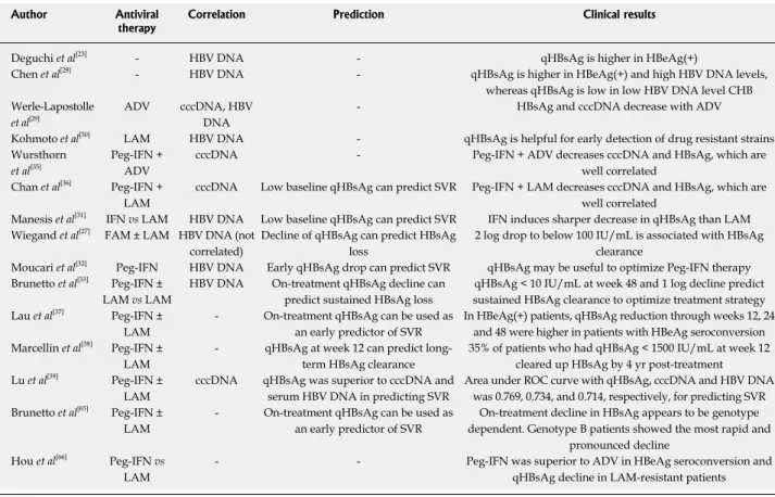
termi-Table 1 Recent clinical studies with quantification of hepatitis B surface antigen titers in hepatitis B virus infection
Author Antiviral
therapy Correlation Prediction Clinical results
Deguchi et al[23] - HBV DNA - qHBsAg is higher in HBeAg(+)
Chen et al[28] - HBV DNA - qHBsAg is higher in HBeAg(+) and high HBV DNA levels,
whereas qHBsAg is low in low HBV DNA level CHB Werle-Lapostolle
et al[29]
ADV cccDNA, HBV
DNA
- HBsAg and cccDNA decrease with ADV
Kohmoto et al[30] LAM HBV DNA - qHBsAg is helpful for early detection of drug resistant strains
Wursthorn
et al[35] Peg-IFN + ADV cccDNA - Peg-IFN + ADV decreases cccDNA and HBsAg, which are well correlated
Chan et al[36] Peg-IFN +
LAM
cccDNA Low baseline qHBsAg can predict SVR Peg-IFN + LAM decreases cccDNA and HBsAg, which are
well correlated
Manesis et al[31] IFN vs LAM HBV DNA Low baseline qHBsAg can predict SVR IFN induces sharper decrease in qHBsAg than LAM
Wiegand et al[27] FAM ± LAM HBV DNA (not
correlated)
Decline of qHBsAg can predict HBsAg loss
2 log drop to below 100 IU/mL is associated with HBsAg clearance
Moucari et al[32] Peg-IFN HBV DNA Early qHBsAg drop can predict SVR qHBsAg may be useful to optimize Peg-IFN therapy
Brunetto et al[33] Peg-IFN ±
LAM vs LAM
HBV DNA On-treatment qHBsAg decline can
predict sustained HBsAg loss
qHBsAg < 10 IU/mL at week 48 and 1 log decline predict sustained HBsAg clearance to optimize treatment strategy
Lau et al[37] Peg-IFN ±
LAM
- On-treatment qHBsAg can be used as
an early predictor of SVR
In HBeAg(+) patients, qHBsAg reduction through weeks 12, 24 and 48 were higher in patients with HBeAg seroconversion Marcellin et al[38] Peg-IFN ±
LAM
- qHBsAg at week 12 can predict
long-term HBsAg clearance
35% of patients who had qHBsAg < 1500 IU/mL at week 12 cleared up HBsAg by 4 yr post-treatment
Lu et al[39] Peg-IFN ±
LAM
cccDNA qHBsAg was superior to cccDNA and
serum HBV DNA in predicting SVR
Area under ROC curve with qHBsAg, cccDNA and HBV DNA was 0.769, 0.734, and 0.714, respectively, for predicting SVR Brunetto et al[65] Peg-IFN ±
LAM
- On-treatment qHBsAg can be used as
an early predictor of SVR
On-treatment decline in HBsAg appears to be genotype dependent. Genotype B patients showed the most rapid and
pronounced decline Hou et al[66] Peg-IFN vs
LAM
- - Peg-IFN was superior to ADV in HBeAg seroconversion and
qHBsAg decline in LAM-resistant patients
HBV: Hepatitis B virus; HBsAg: Hepatitis B surface antigen; qHBsAg: Quantification of HBsAg titers; HBeAg: Hepatitis B e antigen; CHB: chronic hepatitis B; ADV: Adefovir; LAM: Lamivudine; Peg-IFN: Pegylated interferon; cccDNA: Covalently closed circular DNA; FAM: Famciclovir; SVR: Sustained virologic response; ROC: Receiver operating characteristic.
nal codon in the overlapping S gene[50]. In previous
stud-ies, LAM selected HBsAg mutants with reduced anti-HBs binding capacity, and secretion of HBsAg was prevented with a mutant strain due to the stop codon[51,52]. rt181 is
another important site that confers resistance to ADV and/or LAM/LdT. Recently, Warner and Locarnini dem-onstrated that rtA181T caused a secretory defect and had a negative effect on secretion of the wild-type HBV virion because of a concomitant change in the envelope protein at sW172[53]. Similarly, ETV-associated rtI169T/sF161L
leads to a decrease in HBsAg immunoreactivity[54].
The clinical significance of overlap and a common mutational substitution in the S and P gene was further
extended by Kamili et al[55] who demonstrated a
success-ful experimental infection with the rtV173L, rtL180M, and rtM204V HBV mutants that resulted in sE164D and sI195M despite high anti-HBs levels in chimpanzees[55].
Furthermore, the possibility of a vice versa phenomenon
with respect to an extensive vaccination program might be postulated in that HBsAg mutants from selection pressure might harbor the corresponding P gene mutation, resulting
in primary resistance to antiviral agents and therapy failure with these agents.
Detection and variants of HBsAg
As described above, an HBsAg mutation leads to diverse effects, such as decreased secretion and reduced binding capacity to anti-HBs. Of note is that not only a mutation in the a determinant but also in the S promoter or a
dele-tion in the preS region can cause such effects[56,57]. These
effects may hamper the diagnostic performance of com-mercial assays, and several reports have pointed to the
problem of not being able to detect HBV with an a
deter-minant mutation[58,59].
An occult HBV infection is defined as the persistence of the HBV genome in HBsAg negative individuals, and one of the explanations for occult HBV infection is a mu-tation in HBsAg and undetectability by available assays[60].
Both the Architect HBsAg QT and Elecsys HBsAg Ⅱ
seem to reliably detect HBsAg mutants with high sensi-tivity and specificity[24,25,61]. However, further studies are
needed to validate such detection ability, because new or complex combinations of mutations can arise in this era of antiviral agents and extensive vaccination.
FUTURE PERSPECTIVES
Despite progress on qHBsAg, a number of unanswered questions still remain. Precise control mechanisms for HBsAg production in HBV are poorly understood. A discrepancy between qHBsAg and serum HBV DNA ex-ists, although a correlation has been documented. Further research on the virologic nature of HBV could answer these two questions. Meanwhile, the role of qHBsAg in the clinical field is being actively investigated, especially as a predictor to virologic response. Of particular interest is the potential role of qHBsAg for defining the end point of oral antiviral therapy. Current American Association for the Study of Liver Diseases and European Association for the Study of the Liver guidelines with respect to an end point for therapy are unsatisfactory, because reversion to HBeAg positivity does occur after terminating therapy, and the loss of HBsAg is infrequently encountered[62,63]. In this regard,
qHBsAg might be particularly helpful in patients with un-detectable HBV DNA, even with a highly sensitive poly-merase chain reaction assay[64]. In contrast to undetectable
HBV DNA, which provides no further information for the virologic responders, HBsAg is continuously shed and de-tected and, based on the observations of previous studies, qHBsAg with serial monitoring in patients with undetect-able HBV DNA may be utilized to determine the end point of therapy and validate the durability of antiviral agents.
CONCLUSION
HBsAg is produced and secreted through a complex me-chanism that is still not fully understood. Nevertheless, quantification of serum HBsAg is currently available and there is an opportunity to make maximal use of qHBsAg to elucidate its role in clinical fields. However, a deep un-derstanding of the virology is necessary, and it is also im-portant to be familiar with HBsAg variants and their clinical consequences in terms of immune escape mutants, issues resulting from overlap with corresponding mutation in the
P gene, and detection problems for the HBsAg variants.
Unanswered questions need to be resolved through further qHBsAg research.
REFERENCES
1 Lok AS. Chronic hepatitis B. N Engl J Med 2002; 346: 1682-1683 2 Ahn SH, Han KH, Park JY, Lee CK, Kang SW, Chon CY,
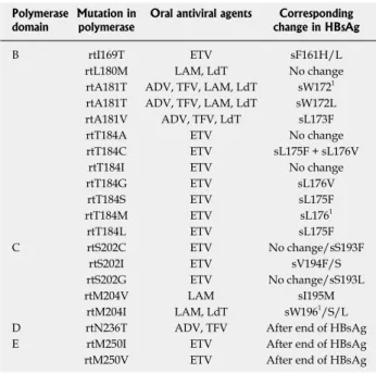
Table 2 Mutations in viral polymerase gene induced by oral antiviral agents and corresponding changes in hepatitis B sur-face antigen
Polymerase
domain Mutation in polymerase Oral antiviral agents change in HBsAgCorresponding
B rtI169T ETV sF161H/L
rtL180M LAM, LdT No change
rtA181T ADV, TFV, LAM, LdT sW1721
rtA181T ADV, TFV, LAM, LdT sW172L
rtA181V ADV, TFV, LdT sL173F
rtT184A ETV No change
rtT184C ETV sL175F + sL176V
rtT184I ETV No change
rtT184G ETV sL176V rtT184S ETV sL175F rtT184M ETV sL1761 rtT184L ETV sL175F C rtS202C ETV No change/sS193F rtS202I ETV sV194F/S rtS202G ETV No change/sS193L rtM204V LAM sI195M rtM204I LAM, LdT sW1961/S/L
D rtN236T ADV, TFV After end of HBsAg
E rtM250I ETV After end of HBsAg
rtM250V ETV After end of HBsAg
1Stop codon. Modified from reference[50]. HBsAg: Hepatitis B surface
anti-gen; ETV: Entecavir; LAM: Lamivudine; LdT: Telbivudine; ADV: Adefo-vir; TFV: Tenofovir.
Kim YS, Park K, Kim DK, Moon YM. Association between hepatitis B virus infection and HLA-DR type in Korea. Hepa-tology 2000; 31: 1371-1373
3 Blumberg BS, Gerstley BJ, Hungerford DA, London WT, Sutnick AI. A serum antigen (Australia antigen) in Down's syndrome, leukemia, and hepatitis. Ann Intern Med 1967; 66: 924-931
4 Thomas HC, Lemon S, Zuckerman AJ. Viral Hepatitis. In: Kann M, Gerlich WH, editors. Structure and molecular virol-ogy. 3rd ed. Oxford: Blackwell Publishing, 2005: 149-180 5 Scaglioni PP, Melegari M, Wands JR. Recent advances in the
molecular biology of hepatitis B virus. Baillieres Clin Gastro-enterol 1996; 10: 207-225
6 Ganem D, Prince AM. Hepatitis B virus infection--natural history and clinical consequences. N Engl J Med 2004; 350: 1118-1129
7 Marquardt O, Heermann KH, Seifer M, Gerlich WH. Cell type specific expression of pre S 1 antigen and secretion of hepatitis B virus surface antigen. Brief Report. Arch Virol 1987; 96: 249-256
8 Heermann KH, Goldmann U, Schwartz W, Seyffarth T, Baumgarten H, Gerlich WH. Large surface proteins of hepatitis B virus containing the pre-s sequence. J Virol 1984; 52: 396-402 9 Schaller H, Fischer M. Transcriptional control of
hepadna-virus gene expression. Curr Top Microbiol Immunol 1991; 168: 21-39
10 Tur-Kaspa R, Burk RD, Shaul Y, Shafritz DA. Hepatitis B vi-rus DNA contains a glucocorticoid-responsive element. Proc Natl Acad Sci USA 1986; 83: 1627-1631
11 Dienes HP, Gerlich WH, Wörsdörfer M, Gerken G, Bianchi L, Hess G, Meyer zum Büschenfelde KH. Hepatic expression patterns of the large and middle hepatitis B virus surface proteins in viremic and nonviremic chronic hepatitis B. Gas-troenterology 1990; 98: 1017-1023
12 Gallina A, De Koning A, Rossi F, Calogero R, Manservigi R, Milanesi G. Translational modulation in hepatitis B virus preS-S open reading frame expression. J Gen Virol 1992; 73 (Pt 1): 139-148
13 Bruss V, Ganem D. The role of envelope proteins in hepatitis B virus assembly. Proc Natl Acad Sci USA 1991; 88: 1059-1063 14 Ueda K, Tsurimoto T, Matsubara K. Three envelope proteins
of hepatitis B virus: large S, middle S, and major S proteins needed for the formation of Dane particles. J Virol 1991; 65: 3521-3529
15 Chisari FV, Filippi P, Buras J, McLachlan A, Popper H, Pinkert CA, Palmiter RD, Brinster RL. Structural and patho-logical effects of synthesis of hepatitis B virus large envelope polypeptide in transgenic mice. Proc Natl Acad Sci USA 1987; 84: 6909-6913
16 Persing DH, Varmus HE, Ganem D. Inhibition of secretion of hepatitis B surface antigen by a related presurface poly-peptide. Science 1986; 234: 1388-1391
17 Pontisso P, Ruvoletto MG, Gerlich WH, Heermann KH, Bar-dini R, Alberti A. Identification of an attachment site for hu-man liver plasma membranes on hepatitis B virus particles. Virology 1989; 173: 522-530
18 Ishikawa T, Ganem D. The pre-S domain of the large viral envelope protein determines host range in avian hepatitis B viruses. Proc Natl Acad Sci USA 1995; 92: 6259-6263
19 Hertogs K, Leenders WP, Depla E, De Bruin WC, Meheus L, Raymackers J, Moshage H, Yap SH. Endonexin II, present on human liver plasma membranes, is a specific binding protein of small hepatitis B virus (HBV) envelope protein. Virology 1993; 197: 549-557
20 Ganem D. Assembly of hepadnaviral virions and subviral particles. Curr Top Microbiol Immunol 1991; 168: 61-83 21 Engvall E, Perlmann P. Enzyme-linked immunosorbent
as-say (ELISA). Quantitative asas-say of immunoglobulin G. Im-munochemistry 1971; 8: 871-874
22 Wolters G, Kuijpers L, Kacaki J, Schuurs A. Solid-phase enzyme-immunoassay for detection of hepatitis B surface
antigen. J Clin Pathol 1976; 29: 873-879
23 Deguchi M, Yamashita N, Kagita M, Asari S, Iwatani Y, Tsu-chida T, Iinuma K, Mushahwar IK. Quantitation of hepatitis B surface antigen by an automated chemiluminescent mic-roparticle immunoassay. J Virol Methods 2004; 115: 217-222 24 Nguyen T, Desmond P, Locarnini S. The role of quantitative
hepatitis B serology in the natural history and management of chronic hepatitis B. Hepatol Int 2009; Epub ahead of print 25 Mühlbacher A, Weber B, Bürgisser P, Eiras A, Cabrera J,
Louisirirotchanakul S, Tiller FW, Kim HS, v Helden J, Bossi V, Echevarria JM. Multicenter study of a new fully automated HBsAg screening assay with enhanced sensitivity for the detection of HBV mutants. Med Microbiol Immunol 2008; 197: 55-64
26 Kuhns MC, Kleinman SH, McNamara AL, Rawal B, Glynn S, Busch MP. Lack of correlation between HBsAg and HBV DNA levels in blood donors who test positive for HBsAg and anti-HBc: implications for future HBV screening policy. Transfusion 2004; 44: 1332-1339
27 Wiegand J, Wedemeyer H, Finger A, Heidrich B, Rosenau J, Michel G, Bock CT, Manns MP, Tillmann HL. A decline in hepatitis B virus surface antigen (hbsag) predicts clearance, but does not correlate with quantitative hbeag or HBV DNA levels. Antivir Ther 2008; 13: 547-554
28 Chen CH, Lee CM, Wang JH, Tung HD, Hung CH, Lu SN. Correlation of quantitative assay of hepatitis B surface anti-gen and HBV DNA levels in asymptomatic hepatitis B virus carriers. Eur J Gastroenterol Hepatol 2004; 16: 1213-1218 29 Werle-Lapostolle B, Bowden S, Locarnini S, Wursthorn
K, Petersen J, Lau G, Trepo C, Marcellin P, Goodman Z, Delaney WE 4th, Xiong S, Brosgart CL, Chen SS, Gibbs CS, Zoulim F. Persistence of cccDNA during the natural history of chronic hepatitis B and decline during adefovir dipivoxil therapy. Gastroenterology 2004; 126: 1750-1758
30 Kohmoto M, Enomoto M, Tamori A, Habu D, Takeda T, Kawada N, Sakaguchi H, Seki S, Shiomi S, Nishiguchi S. Quantitative detection of hepatitis B surface antigen by chemiluminescent microparticle immunoassay during lami-vudine treatment of chronic hepatitis B virus carriers. J Med Virol 2005; 75: 235-239
31 Manesis EK, Hadziyannis ES, Angelopoulou OP, Hadzi-yannis SJ. Prediction of treatment-related HBsAg loss in HBeAG-negative chronic hepatitis B: a clue from serum HB-sAg levels. Antivir Ther 2007; 12: 73-82
32 Moucari R, Mackiewicz V, Lada O, Ripault MP, Castelnau C, Martinot-Peignoux M, Dauvergne A, Asselah T, Boyer N, Bedossa P, Valla D, Vidaud M, Nicolas-Chanoine MH, Mar-cellin P. Early serum HBsAg drop: a strong predictor of sus-tained virological response to pegylated interferon alfa-2a in HBeAg-negative patients. Hepatology 2009; 49: 1151-1157 33 Brunetto MR, Moriconi F, Bonino F, Lau GK, Farci P,
Yur-daydin C, Piratvisuth T, Luo K, Wang Y, Hadziyannis S, Wolf E, McCloud P, Batrla R, Marcellin P. Hepatitis B virus surface antigen levels: a guide to sustained response to pe-ginterferon alfa-2a in HBeAg-negative chronic hepatitis B. Hepatology 2009; 49: 1141-1150
34 Tuttleman JS, Pourcel C, Summers J. Formation of the pool of covalently closed circular viral DNA in hepadnavirus-infected cells. Cell 1986; 47: 451-460
35 Wursthorn K, Lutgehetmann M, Dandri M, Volz T, Buggisch P, Zollner B, Longerich T, Schirmacher P, Metzler F, Zankel M, Fischer C, Currie G, Brosgart C, Petersen J. Peginterferon alpha-2b plus adefovir induce strong cccDNA decline and HBsAg reduction in patients with chronic hepatitis B. Hepa-tology 2006; 44: 675-684
36 Chan HL, Wong VW, Tse AM, Tse CH, Chim AM, Chan HY, Wong GL, Sung JJ. Serum hepatitis B surface antigen quantitation can reflect hepatitis B virus in the liver and predict treatment response. Clin Gastroenterol Hepatol 2007; 5: 1462-1468
H, Button P, Batrla R. On-treatment HBsAg decline during peginterferon alfa-2a (40KD) ± lamivudine in patients with HBeAg-positive CHB as a potential predictor of durable off-treatment response. Hepatology 2008; 48: 714A
38 Marcellin P, Brunetto MR, Bonino F, Hadziyannis E, Kap-prell H, McCloud P, Batrla R. In patients with HBeAg-neg-ative chronic hepatitis B HBsAg serum levels early during treatment with peginterferon alfa-2a predict HBsAg clear-ance 4 years post-treatment. Hepatology 2008; 48: 718A 39 Lu L, Ye D, Wang Y, Kwok A, Wong A, Yueng Y, Zhang H,
Chen Y, Bowden S, Batrla-Utermann R, Locarnini S, Lau G. Correlation between HBV cccDNA and HBsAg levels and their reduction by peginterferon alfa-2a based therapy in pa-tients with chronic hepatitis B. Hepatology 2008; 48: 746A 40 Bruss V, Ganem D. Mutational analysis of hepatitis B
sur-face antigen particle assembly and secretion. J Virol 1991; 65: 3813-3820
41 Meyer M, Caselmann WH, Schlüter V, Schreck R, Hofsch-neider PH, Baeuerle PA. Hepatitis B virus transactivator MHBst: activation of NF-kappa B, selective inhibition by an-tioxidants and integral membrane localization. EMBO J 1992; 11: 2991-3001
42 Nowak MA, Bonhoeffer S, Hill AM, Boehme R, Thomas HC, McDade H. Viral dynamics in hepatitis B virus infection. Proc Natl Acad Sci USA 1996; 93: 4398-4402
43 Mimms L. Hepatitis B virus escape mutants: "pushing the envelope" of chronic hepatitis B virus infection. Hepatology 1995; 21: 884-887
44 Carman WF, Zanetti AR, Karayiannis P, Waters J, Manzillo G, Tanzi E, Zuckerman AJ, Thomas HC. Vaccine-induced escape mutant of hepatitis B virus. Lancet 1990; 336: 325-329 45 Zuckerman JN, Zuckerman AJ. Mutations of the surface
protein of hepatitis B virus. Antiviral Res 2003; 60: 75-78 46 Hsu HY, Chang MH, Ni YH, Chen HL. Survey of hepatitis B
surface variant infection in children 15 years after a nationwide vaccination programme in Taiwan. Gut 2004; 53: 1499-1503 47 Chen DS. Hepatitis B vaccination: The key towards
elimina-tion and eradicaelimina-tion of hepatitis B. J Hepatol 2009; 50: 805-816 48 Ghany MG, Ayola B, Villamil FG, Gish RG, Rojter S, Vier-ling JM, Lok AS. Hepatitis B virus S mutants in liver trans-plant recipients who were reinfected despite hepatitis B im-mune globulin prophylaxis. Hepatology 1998; 27: 213-222 49 Torresi J. The virological and clinical significance of
muta-tions in the overlapping envelope and polymerase genes of hepatitis B virus. J Clin Virol 2002; 25: 97-106
50 Zoulim F, Locarnini S. Hepatitis B virus resistance to nucleos-(t)ide analogues. Gastroenterology 2009; 137: 1593-1608.e1-e2 51 Torresi J, Earnest-Silveira L, Deliyannis G, Edgtton K,
Zhuang H, Locarnini SA, Fyfe J, Sozzi T, Jackson DC. Re-duced antigenicity of the hepatitis B virus HBsAg protein arising as a consequence of sequence changes in the over-lapping polymerase gene that are selected by lamivudine therapy. Virology 2002; 293: 305-313
52 Yeh CT, Chien RN, Chu CM, Liaw YF. Clearance of the
orig-inal hepatitis B virus YMDD-motif mutants with emergence of distinct lamivudine-resistant mutants during prolonged lamivudine therapy. Hepatology 2000; 31: 1318-1326
53 Warner N, Locarnini S. The antiviral drug selected hepatitis B virus rtA181T/sW172* mutant has a dominant negative se-cretion defect and alters the typical profile of viral rebound. Hepatology 2008; 48: 88-98
54 Sloan RD, Ijaz S, Moore PL, Harrison TJ, Teo CG, Tedder RS. Antiviral resistance mutations potentiate hepatitis B virus immune evasion through disruption of its surface antigen a determinant. Antivir Ther 2008; 13: 439-447
55 Kamili S, Sozzi V, Thompson G, Campbell K, Walker CM, Locarnini S, Krawczynski K. Efficacy of hepatitis B vaccine against antiviral drug-resistant hepatitis B virus mutants in the chimpanzee model. Hepatology 2009; 49: 1483-1491 56 Melegari M, Scaglioni PP, Wands JR. The small envelope
protein is required for secretion of a naturally occurring hepatitis B virus mutant with pre-S1 deleted. J Virol 1997; 71: 5449-5454
57 Sengupta S, Rehman S, Durgapal H, Acharya SK, Panda SK. Role of surface promoter mutations in hepatitis B surface antigen production and secretion in occult hepatitis B virus infection. J Med Virol 2007; 79: 220-228
58 Louisirirotchanakul S, Kanoksinsombat C, Theamboonlert A, Puthavatana P, Wasi C, Poovorawan Y. Mutation of the "a" determinant of HBsAg with discordant HBsAg diagnos-tic kits. Viral Immunol 2004; 17: 440-444
59 Gerlich WH. Diagnostic problems caused by HBsAg mu-tants-a consensus report of an expert meeting. Intervirology 2004; 47: 310-313
60 Raimondo G, Pollicino T, Cacciola I, Squadrito G. Occult hepatitis B virus infection. J Hepatol 2007; 46: 160-170 61 Coleman PF, Chen YC, Mushahwar IK. Immunoassay
detec-tion of hepatitis B surface antigen mutants. J Med Virol 1999; 59: 19-24
62 Lok AS, McMahon BJ. Chronic hepatitis B: update 2009. Hepatology 2009; 50: 661-662
63 EASL Clinical Practice Guidelines: management of chronic hepatitis B. J Hepatol 2009; 50: 227-242
64 Lee JM, Park JY, Ahn SH, Kim DY, Chon CY, Kim HS, Han KH. Clinical applicability of HBsAg titer in chronic hepatitis B patients with undetectable HBV DNA by real-time PCR assay. Hepatology 2009; 50: 517A
65 Brunetto MR, Bonino F, Marcellin P, Button P, Batrla R. Kinetics of HBsAg decline in patients with HBeAg-negative chronic hepatitis B treated with peginterferon alfa-2a accord-ing to genotype and its association with sustained HBsAg clearance 4 years post-treatment. Hepatology 2008; 48: 740A 66 Hou J, Sun J, Xie Q, Li X, Zhang J, Wang Y, Lai J, Chen S, Jia
J, Sheng J, Chan H, Wang J, Jiang M, Popescu M, Sung J. Effi-cacy and safety of peginterferon alfa-2a versus adefovir dip-ivoxil (ADV) in treating lamivudine resistant HBeAg posi-tive CHB: An interim analysis of a prospecposi-tive randomised study. Hepatology 2008; 43: 745A