DOI: http://doi.org/10.15205/kschs.2020.12.31.1436
흡연행태에 따른
Porphyromonas gingivalis
의 발현율과 유전형
차이
5) 김진경 대구보건대학교 치위생과Differences in the expression rate and genotype of
Porphyromonas gingivalis
according to smoking status
Jin-Kyoung Kim*
Department of Dental Hygiene, Daegu Health College
(Received October 22, 2020; Revised November 04, 2020; Accepted November 18, 2020)
Abstract
Purpose: The purpose of this study was to differences in the expression rate of Porphyromonas gingivalis
according to smoking status, smoking amount and period of smoking. Methods: At the time of investigation, 30 smokers and non-smokers were recruited among patients with periodontitis with a probing pocket depth(PPD) of 4 mm or more. General information was collected using a self-questionnaire, and the average value was used by a dentist to measure the probing pocket depth of three times each for the first or second molar. Plaque collection and analysis were performed by collecting only subgingival plaque using a conventional method, and the expression rate of P. gingivalis was confirmed using polymerase chain reaction (PCR). For statistical analysis, the SPSS Ver 25.0 program was used. Results: Smoking did not have a significant effect on the expression of P. gingivalis, but it did affect the expression of more type II genotypes (p<0.05). In addition, smokers had more slight periodontal pocket, and the amount and duration of smoking did not affect the expression of P. gingivalis. Conclusions: In the future, it is necessary to reinforce the group of smokers and non-smokers with healthy oral conditions, and to investigate the quantitative difference in the expression rate and genotype of P. gingivalis over time of harmful substances in smoking.
Key words: Expression rate, Genotype, Porphyromonas gingivalis, Smoking status
1. 서 론
WHO 보고에 따르면 우리나라 성인의 평균 흡연 율은 세계 1위이며1), 국민건강영양조사에 의하면, 2018년 기준 남자 36.7%, 여자 7.5% 가 흡연자이 다2). 흡연은 폐암 등 다양한 호흡기질환을 유발하 며, 뇌·신장-혈관질환을 유발함으로써 성인의 가 장 중요 한 사망요인으로 거론되고 있다3). 이와 같이 전신건강을 위협하는 것 이외에도 흡 연은 연기에 의해 구강조직, 특히 치주조직에 병 적인 변화를 초래하는 것으로 알려져 있다. 담배 연기 성분의 침착으로 인해 흡연자는 비흡연자에 비해 구강위생상태가 불량해질 수 밖에 없으며, 다양한 산화생성물들에 의해 치주조직에 직접 또 는 간접적인 영향을 미친다4). 이에 흡연은 치주질 환 유발이나 진행, 치과수술 후 치유과정 지연정 도와 연관성이 있는 것으로 보고되고 있으며5), 흡 연은 치주질환에 가장 중요한 환경인자로 간주되 고 있다6). 치주질환은 구강세균이 깊이 관여하고 있으며 P.gingivalis는 red complex에 속하는 대표적인 그람
음성혐기성 세균으로 성인 치주염을 포함하여 치 주질환과 연관되어 있다7). 또한 흡연은 면역기능 에 영향을 미치며, 구강은 흡연 시 담배연기에 가 장 먼저 접촉하는 곳이기 때문에 세균에 직접적인 영향을 미칠 것으로 추정된다. 그러나 세균이 치 주질환에 미치는 영향에 관해서는 견해가 일치하 고 있지 않다. Lie 등은 세균의 집합체인 치면세균 막은 흡연에 의해 영향을 받지 않으며8), Darby 등 은 치태의 이환율이나 분포에도 변화를 주지 않는 다고 하였다9). 반면, Shiloah 등은 흡연자가 비흡 연자에 비해 치주질환 원인균의 이환율과 분포율 이 높다고 하였다10). 이에 본 연구에서는 흡연행태에 따른 P. gingivalis 발현율 및 유전형 분포 차이를 알아보 고자 하였다.
2. 연구대상 및 방법
대상자는 2019년 9월부터 2020년 1월까지 OO치 과병원 치주과에 방문한 20세 이상 성인 중 연구 의 목적과 취지를 이해하고 자발적 참여에 동의한 사람에 대해 선정 기준11)을 만족하는 사람을 모집 하였으며(Table 1), 정규성 확보를 위해 흡연자와 비흡연자를 각 30명씩 최종 60명을 선정하였다. 본 연구는 생명윤리위원회의 승인을 받아 수행되 었다(KHUSD IRB 0804-07). 2.2. 연구방법 설문조사는 일반적인 특성, 흡연여부, 흡연양 및 흡연기간에 대한 정보를 수집하였다. 일반적인 특 성은 성별, 나이로 구성하였다. 흡연여부는 조사당 시에 흡연자와 비흡연자로 구분하였다. 흡연자는 최근 5년간 하루 10개피 이상 피우는 사람이었으 며, 비흡연자는 최근 5년간 비흡연했거나, 과거에 흡연한 경험이 없는 사람으로 정하였다. 흡연양은 하루에 피우는 양을 개피로 기록하였고, 흡연기간 은 총 흡연기간을 합하여 기록하였다. 치태수집은 검사치아에 대해 멸균된 치주기구를 이용하여 치은연하에서 보말수집하고, 즉시 PBS buffer에 담아 –20℃에 보관하였다. 치주세균 발현 유무를 확인하기 위해 통상적인 방법으로 DNA를 검출하여 PCR로 증폭시킨 후 170-8060 Gel Documentation System을 이용하여 확인하였다11). 2.3. 자료분석 수집된 자료는 SPSS Ver. 25.0 프로그램(SPSS Inc, Chicago, IL, USA)을 사용하여 분석하였으며, 구체적인 분석 방법은 다음과 같다. 첫째, 연령, 흡연양, 흡연기간, 치주낭 깊이의 평균을 구하기 위해 기술통계를 실시하였다. 둘째, 연구대상자의 흡연여부에 따른 성별, 연 령, 치주낭 깊이별 분포 차이와 흡연행태에 따른 P. gingivalis 발현율을 확인하기 위해 Chi-square test를 실시하였다.Inclusion criteria
4mm to 7mm pocket probing depth(PPD) Exclusion criteria
- pregnancy
- previous periodontal treatment
- a known systemic condition that could influence the periodontal condition
- history of systemic or local use of antimicrobials during the 6 months before sampling
- at least the past three months among patients in the tooth with periodontal care and professional teeth
Table 1. Inclusion and exclusion criteria
3. 연구결과
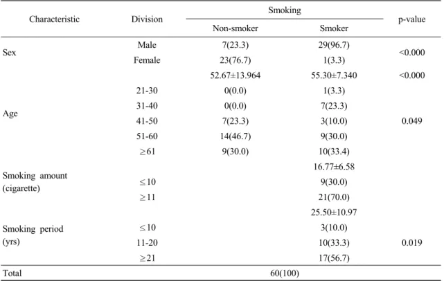
3.1. 일반적인 특성 대상자는 비흡연자 중 남자 7명(23.3%), 여자 23명(76.7%) 였으며, 흡연자 중 남자 29명 (96.7%), 여자 1명(3.3%)였다. 평균연령은 비흡연 자 52.67세, 흡연자 55.30세 였으며, 연령분포는 두 그룹 모두 50대 이상이 23명(76.7%), 19명 (63.4%)로 가장 많았다. 흡연양은 하루 평균 16.77 개피였으며, 10개피 이하 9명(30.0%), 11개피 이 상 21명(70.0%)였다. 흡연기간은 평균 25.5년이었 으며, 21년 이상이 17명(56.7%)로 가장 많았다 (Table 2).Characteristic Division Smoking p-value Non-smoker Smoker Sex Male 7(23.3) 29(96.7) <0.000 Female 23(76.7) 1(3.3) Age 52.67±13.964 55.30±7.340 <0.000 21-30 0(0.0) 1(3.3) 0.049 31-40 0(0.0) 7(23.3) 41-50 7(23.3) 3(10.0) 51-60 14(46.7) 9(30.0) ≥61 9(30.0) 10(33.4) Smoking amount (cigarette) 16.77±6.58 ≤10 9(30.0) ≥11 21(70.0) Smoking period (yrs) 25.50±10.97 ≤10 3(10.0) 0.019 11-20 10(33.3) ≥21 17(56.7) Total 60(100)
Data are presented as N(%) and mean±SD
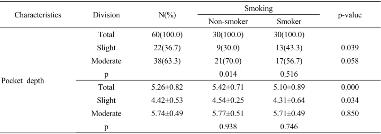
Characteristics Division N(%) Smoking p-value Non-smoker Smoker Pocket depth Total 60(100.0) 30(100.0) 30(100.0) Slight 22(36.7) 9(30.0) 13(43.3) 0.039 Moderate 38(63.3) 21(70.0) 17(56.7) 0.058 p 0.014 0.516 Total 5.26±0.82 5.42±0.71 5.10±0.89 0.000 Slight 4.42±0.53 4.54±0.25 4.31±0.64 0.034 Moderate 5.74±0.49 5.77±0.51 5.71±0.49 0.850 p 0.938 0.746
Data are presented as N(%) and mean±SD Slight: 3.0-4.99mm, Moderate: 5.0-6.99mm
Table 3. Differences in pocket depth between non-smoking and smoking
3.2. 흡연여부에 따른 치주낭 깊이의 차이 흡연여부에 따른 치주낭 깊이의 빈도 차이는 경 도에서는 흡연자의 비율이 13명(43.3%)로 높았고 (p<0.05), 중등도에서는 비흡연자의 비율이 약간 높았다. 치주낭 깊이의 전체 평균은 5.26±0.85로 비흡연자는 5.42±0.71, 흡연자는 5.10±0.89로 유의 한 차이가 있었다(p<0.01). 경도에서 비흡연자는 4.54±0.25, 흡연자는 4.31±0.64로 유의한 차이가 있었으며(p<0.05), 중등도에서는 유의한 차이가 없었다. 비흡연자와 흡연자의 치주낭 깊이 변화는 유의한 차이가 없었다(Table 3). 3.3. 흡연여부에 따른 P. gingivalis 발현율 및 유전형 분포 대상자 전체에서 P. gingivalis 발현이 확인되었 으나, 비흡연자와 흡여자간에 발현율 및 유전형 분포의 차이는 없었다(Table 4). 3.4. 흡연행태에 따른 P. gingivalis 유전형 분포 흡연양에 따른 P. gingivalis 유전형 분포의 경 우 type Ⅲ는 10개피 이하 그룹에서 높은 발현율 는 11개피 이상 흡연그룹에서 주로 발현되었으나, 두 그룹 간에 유의한 차이는 없었다. 흡연기간에 따른 P. gingivalis의 유전형 분포 차이는 없었다 (Table 5).
4. 고찰
흡연은 치주질환의 주요 위험요인이다. 또한, P.gingivalis는 red complex에 속하는 그람음성 혐기
성 세균으로 성인 치주염을 포함한 치주질환과 밀 접하게 관련되어 있다11). 따라서, 본 연구는 흡연 행태에 따른 P. gingivalis 발현유무 및 유전형 분 포 차이를 확인하고자 하였다. 본 연구 대상자의 흡연율은 우리나라 성인 흡연 율 43.1% 보다 높았다1). 이는 본 연구의 대상자를 흡연자이면서 치주염을 가지고 있는 사람으로 선 정하였기 때문이며, 남녀 간 흡연자 비율이 큰 차 이를 보이는 것은 여성 흡연을 바라보는 사회적인 편견 때문에 설문 응답 시 흡연을 숨기거나, 흡연 경력을 조작하는 등의 의도가 반영된 것이라고 생 각된다. 즉, 여자 흡연자의 실제 통계 수치는 보다 높을 것이라고 예상함에도 불구하고 사회적 욕구 편향이 반영된 것이라고 추정된다. 또한 흡연량은 11개피 이상을 피운다는 흡연자가 21명(70.0%)로
연기간이 20년 이상인 대상자가 가장 많았다1). 이 는 우리 연구의 대상자를 특정 조건으로 제한했기 때문이라고 생각된다. 흡연여부에 따른 치주낭 깊이는 경도 치주낭에 서는 흡연자의 비율이 유의하게 높았으며, 중등도 치주낭에서는 비흡연자가 다소 높았다. 그룹 내 분포 변화를 살펴보면, 두 그룹 모두 경도보다 중 등도인 대상자가 많은 것을 확인 할 수 있었으며, 비흡연자는 유의하게 증가하는 것을 알 수 있었 다. 그러나, 치주상태가 좋지 않을수록 흡연율이 높게 나타난다고 보고한 기존 연구와 상반된 결과 이다13-15). 이는 흡연이 치주낭을 심화시키는 직접 적인 요인이라기 보다는, 구강 내 온도상승, 구강 건조, 혈류량 감소, 면역력 감소 등을 초래16)하므 로써, 치주질환에 이환되기 쉬운 초기 환경을 만 드는데 기여할 수 있을 것으로 추정된다. 또한, 흡 연하는 경우 치태의 양적인 증가로 인해 세균의 밀집도가 높아지고, 관리가 어려워짐에 따라 구강 위생이 불량해 질 수 밖에 없으며, 혐기성 세균의 화학적 군집이 쉬운 환경이 형성된다. 또한, 경도 및 중등도 치주낭 깊이의 평균을 살펴보면, 비흡 연자가 흡연자보다 깊었고, 경도 치주낭에서는 유 의한 차이가 있는 것으로 보아, 흡연은 치주낭을 깊게 만드는 직접적인 원인이라고 보기 어렵다고 생각된다. 흡연여부에 관계없이 두 집단 모두에서 P. gingivalis가 확인되었다. 이러한 결과는 흡연이 P. gingivalis 발현율 차이에 영향을 미치지 않는다고 보고한 Zhao 등의 결과와 일치하며17), 흡연이 P. gingivalis 발현율 자체를 증가시킨다고 볼 수 없 다. 나아가, 특정 유전형 발현을 확인해본 결과 type Ⅱ는 흡연양과 기간에 상관없이 P. gingivalis 의 발현 유(+), 무(-) 비율이 비슷하였으나, 다른 유전형은 발현되지 않은(-) 경우가 더 많았다. P. gingivalis는 6가지 유전자형으로 분류되는데, 그 중 type Ⅱ는 치주상태가 불량하거나 치주염인 경 우 주로 발현된다고 알려져 있다17). 이는 우리 연 구에서 흡연양이 많고, 흡연기간이 오래된 집단에 서 type Ⅱ 발현율이 다른 유전형에 비해 높았던 것과 비슷한 결과이다. 반면 P. gingivalis type Ⅲ 발현율은 흡연양이 상대적으로 적은 10개피 이하 그룹에서 유의하게 높았다. P. gingivalis 유전형 중 type Ⅱ를 제외한 다른 유전형은 비교적 치주 상태가 양호하거나, 가역적 염증상태일 경우에 발 현되는 경향을 가진다는 기존의 보고와 일치하는 결과였다12,17). 따라서, 흡연은 많은 구강세균 중 P. gingivalis를 선택적으로 증가시킨다기 보다는, 치주질환자의 구강세균이 여러 요인에 의해 양적 으로 증가되면서, 혐기성 세균에 유리한 생육조건 을 갖추게 되어, P. gingivalis type Ⅱ 유전형 발현 이 증가될 가능성이 있다고 추정된다. 본 연구는 흡연자이면서 치주염을 동시에 가지 고 있는 자를 대상으로 치주질환 원인균인 P. gingivalis의 발현율과 유전형을 조사한 실험연구 이다. 이와 같은 임상실험연구가 수행된 경우는 매우 드물며, 흡연여부뿐만 아니라, 흡연양과 흡연 기간에 따른 P. gingivalis 발현율을 확인하였다는 점에서 의미가 있다. 그럼에도 불구하고, 본 연구는 몇 가지 제한점 을 가진다. 첫째, 특정 병원에 내원한 환자를 선정 했기 때문에 연구결과에 대표성을 부여하기 어렵 다. 둘째, 흡연양과 기간에 대한 정보를 자기기입 식 설문을 통해 수집하였기 때문에 회상오류가 존 재 할 수 있다. 셋째, 단면연구로써변수로 사용된 흡연여부, 흡연양 및 흡연기간에 의한 P. gingivalis의 시간에 따른 발현양을 비교할 수 없 으며, 향후 Real-time PCR을 이용하여 확인해 볼 필요가 있을 것이다. 향후 제한점을 보완하여 추가 연구가 이루어져 야 할 것이며, 다른 치주질환 원인균의 발현 분포 차이를 함께 비교해 볼 필요가 있을 것이다.
5. 결 론
본 연구는 비흡연 치주질환자와 흡연 치주질환 자의 치은연하치태를 수집한 후 PCR을 이용하여 P. gingivalis 발현율과 유전형 분포를 확인하기 위 해 수행되었다. 치주과에 방문하는 환자 중 흡연 자와 비흡연자를 각 30명씩 모집하였다. 흡연자는 최근 5년간 매일 10개피 이상 피운사람으로 제한 하였고, 비흡연자는 최근 5년간 흡연경력이 없거 나 평생 흡연경력이 없는 사람으로 제한하였다. 치은연하 치태를 수집한 후, PCR을 통해 P. gingivalis 발현율과 유전형을 확인하였다. 제시된 연구결과에 따른 결론은, 흡연은 경도 치주낭 형성에 영향을 미칠 것으로 추정된다. 흡연여부는 P. gingivalis 발현에 유의한 영향을 미치지 않았다. 흡연양은 P. gingivalis type Ⅲ 유전형의 발현율 을 증가시키며, 흡연기간에 따른 차이는 없었다. 흡연하는 경우 P. gingivalis type Ⅱ 유전형의 발현이 높았으나, 유의한 수준은 아니었다. 이상의 결과를 종합해 볼 때 흡연은 P. gingivalis typeⅡ 발현을 증가 시킬 가능성이 있으 며, 흡연은 경도 치주낭 형성에 관여할 것으로 추 정된다.References
1. Korea National Health and Nutrition Examination Survey (KNHANE) KNHANES VI-2 [internet]. [cited 2020 October7]. Available from https://knhanes.cdc.go.kr/knhanes/index.do 2. David Satcher. Why we need an international
agreement on tobacco control. AJPH, 2001;91: 191-193.
3. Mathers CD, Loncar D: Projection of global mortality and burden of disease from 2002 to 2030. PLos Med, 2006;3:e4 42.
4. Park EA, Kim SY, Kim YY et al. Difference in
prevalence of fimA genotypes of Porphyromonas
gingivalis between smoker and non-smoker
patients with periodontitis. J Bacteriol Virol, 2003;33(2):119-129.
5. Kim YS, Min HH. Relationship between smoking behavior and periodontitis in Korean adults. J Korean Soc Dent Hyg, 2016;16(6):825-833. 6. Krall EA, Dawson-Hughes B, Garvey AJ, et al.
Smoking, smoking cessation, and tooth loss. J Dent Res, 1997;76(10):1653-1659.
7. Kim YH, Lee JH. The relationship between oral health behavior, smoking, and periodontal diseases in Korean middle-aged mee: based on data from the Korea National Health and Nutrition Examination Survey, 2013-2015. J Korean Acad Oral Health, 2017;41(1):36-42. 8. Lie MA, Van der Weijden GA, Timmerm-an MF,
et al. Oral microbiota in smokers and non‐ smokers in natural and experimentally induced gingivitis. J Clin Periodontol, 1998;25:677-686. 9. Darby IB, Hodge PJ, Riggio MP, et al. Microbial
comparison of smoker and non-smoker adult and early‐onset periodontit-is patients by polymerase chain reaction. J Clin Periodontol, 2000;27:417-424.
10. Shiloah J, Patters MR, Waring MB. The prevalence of pathogenic periodontal microflora in healthy young adult smokers. J Periodontol, 2000;71(4):562-567.
11. David W, Melanie W, Neville O, et al. Characteristics of heavy smokers. Prev Med, 1992;21(3):311-319.
12. Amano A, Nakagawa I, Kataoka K, et al. Distribution of Porphyromonas gingivalis strains with fimA genotypes in periodontitis patients. J Clin Microbial, 1999;37(5); 1426-1430.
13. Scott T, Samira A. Smoking-attributable periodontitis in the United States: Findings From NHANES III. J Periodontol, 2000;71(5):743-751.
14. Song AH, Jung EJ. Trends by year in the relationships between smoking and oral health in adults. J Korean Soc Dent Hyg, 2018;18(6):933-946.
15. Jung JO, Chun JY, Lee KH. The relationship between smoking and perio-dontal disease in Korean adults: based on the data from the Korea National Health and Nutrition Examination Survey 2010. J Korean Soc
Dent Hyg, 2013;13(3):481-489.
16. Winn DM. Tobacco use and oral diseas-e. J Dent Educ, 2001;65(4):306-312.
17. Zhao L, Wu YF, Meng S, et al. Prevalence of
fimA genotypes of Porphyromonas gingivalis
and periodon-tal health status in chinese adults. J Periodont Res, 2007;42:511-517.