Introduction
Patellar maltracking after total knee arthroplasty (TKA) has been associated with anterior knee pain and limited range of motion, which would eventually result in impaired functional recovery and development of severe complications such as im-plant wear/loosening and patellar fracture1-3). A variety of studies
have suggested factors that could be linked with patellar track-ing. Some cadaveric studies analyzing the relative impact of knee components demonstrated that the patellar resection angle, location of the patellar component, and rotational alignment of the femoral component could affect postoperative patellar track-ing4,5). In an in vivo study, Kawano et al.6) reported the location
of the patellar component, patellar resection angle, and lateral retinacular release as related factors. Some authors associated pa-tellar tracking with preoperative papa-tellar tilt7), surgical approach8),
location and rotational alignment of the implant9-12), and implant
design and soft tissue balance13,14). However, there is still
contro-versy on which of these factors should be taken into consider-ation during surgery; the abovementioned factors may interact during surgery, and studies on relative impact of each factor us-ing standardized methods are lackus-ing.
Accordingly, we designed this study to elucidate the influence of the two possible determinants of patellar tracking, the rotational
Influence of the Rotational Alignment of the Femoral
and Patellar Components on Patellar Tilt in Total Knee
Arthroplasty
Jong Heon Kim, MD, Byeong-Wook Yoo, MD, and Chul-Woong Kim, MD
Department of Orthopedic Surgery, Guri Hospital, Hanyang University College of Medicine, Guri, Korea
Purpose: The purpose of this study was to analyze the influence of rotational alignment of the femoral and patellar components on patellar tilt after total knee arthroplasty (TKA).
Materials and Methods: A total of 56 patients (76 knees) who underwent TKA using Advance Medial Pivot Knee system between May 2009 and April 2011 and were available for minimum 1-year follow-up were enrolled in this study. Whiteside’s line and the transepicondylar line were used to determine the rotational alignment of the femoral component. Patella cut was aimed to be parallel to the anterior patellar cortex during surgery. Radiographic evaluation was performed using plain axial radiographs. The rotational alignment of the femoral component was measured as the angle between the anterior condylar axis and the surgical transepicondylar axis. The patellar resection angle was measured between the patellar resection axis and the anterior cortical line of the patella. Patellar tilt was evaluated to investigate the correlation with the rotation of the femoral component and patellar resection angle.
Results: The mean rotation of the femoral component was 0.42o±3.18o of internal rotation. The mean patellar resection angle was 1.82o±3.44o,
indicating medial overresection. The mean patellar tilt was 6.12o±4.31o of lateral tilt. The rotational angle of the femoral component showed a negative
correlation with patellar tilt in the linear regression analysis (p=0.749), but the patellar resection angle showed a positive correlation with patellar tilt (p<0.001).
Conclusions: Accurate patellar resection is recommended for proper patellar tracking in TKA.
Keywords: Knee, Arthroplasty, Component roation, Patellar tracking
pISSN 2234-0726 · eISSN 2234-2451
Knee Surgery & Related Research
Received September 21, 2014; Revised (1st) December 27, 2014; (2nd) May 25, 2015; Accepted June 16, 2015
Correspondence to: Jong Heon Kim, MD
Department of Orthopedic Surgery, Guri Hospital, Hanyang University College of Medicine, 153 Gyeongchun-ro, Guri 11923, Korea
Tel: +82-31-560-2313, Fax: +82-31-557-8781 E-mail: [email protected]
163
This is an Open Access article distributed under the terms of the Creative Commons Attribution Non-Commercial License (http://creativecommons.org/licenses/by-nc/4.0/) which permits unrestricted non-commercial use, distribution, and reproduction in any medium, provided the original work is properly cited.alignment of the femoral component10-12) and patellar resection
angle4,6), on patellar tilt among patients who underwent TKA in
the same surgical setting.
Materials and Methods
Of the patients who underwent TKA using Advance Medial Pivot Knee system (Wright Medical Technology Inc., Arlington, TN, USA) between May 2009 and April 2011 and who were available for more than 1-year follow-up, a total of 56 patients (76 knees) were included in this retrospective study. The exclusion criteria were revision TKA, severe patellar deformity, preopera-tive subluxation or dislocation of the patella, severe valgus de-formity of the lower limb, TKA without patellar resurfacing, and lateral retinacular release in TKA. There were 11 males and 45 females, and 20 of them underwent bilateral TKA. Their mean age was 68±7.4 years. The indication for surgery was osteoarthri-tis in 71 knees, rheumatoid arthriosteoarthri-tis in 3 knees, and osteonecrosis in 2 knees. The mean preoperative lower limb alignment was 3.16o
varus (range, 7.6o valgus to 14.1o varus). The mean preoperative
patellar tilt was 1.83o (range, 2.5o of medial tilt to 10.4o of lateral
tilt). In all knees, the posterior cruciate ligament-sacrificing Ad-vance Medial Pivot Knee system was inserted in TKA performed by the same surgeon using the following surgical procedure.
The surgery was carried out through a midline skin incision using the medial parapatellar approach in all patients under tourniquet control. Medial soft tissue release was additionally performed if considered necessary based on the medial/lateral flexion-extension gap assessment during surgery. During implant insertion, care was taken to maintain approximately 3o external
rotation of the femoral component with respect to the posterior condylar axis, and further adjustments were made to align the femoral component parallel to the surgical transepicondylar axis as much as possible and perpendicular to the anteroposterior axis (Whiteside’s line). The center of the femoral component was aligned with the trochlear groove. The rotational alignment of the tibial component was determined by a line connecting the medial 1/3 of the tibial tubercle and the center of the posterior cruciate ligament. Upon confirming with a rod the tibial component was aligned with a line passing though the tibial crest and the center of the ankle joint, component insertion was done. For patellar resurfacing, the lateral margin of the patella was exposed and the overall patellar thickness was measured using a caliper. Us-ing the free-handle technique, patellar resection was performed parallel to the anterior cortex, taking care to produce symmetric thickness at the medial and lateral facets of the patella. The
longi-tudinal axis of the dome–shaped patellar component was aligned with the longitudinal axis at the central ridge of the patella, and the bone-prosthesis composite thickness was intended to be equal to the original thickness of the patella.
Radiographic assessment was performed using the 1-year follow-up axial plain radiographs (merchant view). The rotational alignment of the femoral component was measured according to the method of Kanekasu et al.15): the angle subtended by the
tran-sepicondylar axis and the anterior condylar axis (femoral com-ponent rotation angle) was measured, and external rotation was expressed as a positive value (Fig. 1). The rotational alignment of the patellar component was assessed according to the method of Chan and Gill7). Two perpendicular lines were drawn at the
medial and lateral facets of the patella. A line connecting the ends of the two perpendicular lines was drawn at the anterior patellar cortex. The angle subtended by the line at the anterior patellar cortex and a line tangent to the resected surface of the patellar component (patellar resection angle) was measured, and medial over-resection was assigned a positive value (Fig. 1). Patellar tilt was measured according to the method of Gomes et al.16): the
angle between the anterior condylar axis and the resected surface of the patellar component was measured (patellar tilt angle), and lateral tilt was given a positive value (Fig. 1).
Statistical analysis was done using SAS ver. 9.2 (SAS Institute Inc., Cary, NC, USA) to compare the influence of the femoral
Fig. 1. The patellar resection angle (a–b), patellar tilt (b–c), and femoral component rotation angle (c–d) were measured on plain axial radio-graphs. a: a line at the anterior patellar cortex connecting the ends of perpendicular lines drawn at the medial and lateral aspects of the patella, b: patellar resection axis, c: anterior condylar axis, d: surgical transepi-condylar axis.
component rotation angle and patellar resection angle on patellar tilt. Cronbach’ alpha test was used to evaluate the reliability of the measurements performed twice by a single observer. The influ-ence of the two parameters on patellar tilt was evaluated using multivariate regression analysis.
Results
The mean femoral component rotation angle was 0.42o±3.18o
of internal rotation. The mean patellar resection angle was 1.82o±3.4o, indicating medial over-resection. The mean patellar
tilt was 6.12o±4.31o, indicating lateral tilting. There was no
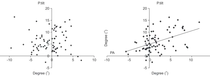
statis-tically significant relationship between the femoral component rotation angle and patellar tilt (p=0.749) (Fig. 2); however, the patellar resection angle showed a significant correlation with pa-tellar tilt (p<0.001, r=0.289) (Fig. 3). The Cronbach’ alpha value was ≥0.7, indicating good intraobserver reliability.
Discussion
In this study, we analyzed the influence of the rotational align-ment of the femoral component and patellar resection angle on patellar tilt using plain radiographs (Merchant view) in patients who received the same knee implant in TKA performed by the same surgeon to ensure consistent surgical setting. Based on the radiographic assessment, we believe that patellar resection angle has more significant influence than the rotational alignment of the femoral component on patellar tilt.
Independent influence of factors associated with patellar track-ing can be assessed through simulation in cadaveric studies, and there are in vivo studies reporting on the possible
contribut-ing factors; however, their results have failed to generate broad consensus due to the lack of standardization. In this study, we attempted to investigate the influence of factors that have been related to patellar tilt, such as the rotational alignment of the femoral component10-12) and patellar resection angle4,6), in patients
who underwent TKA according to a standardized surgical proce-dure at our institution, and compare our results with those in the previous literature.
Kanekasu et al.15) compared the preoperative axial radiographs
and computed tomography (CT) scans in the assessment of the rotational alignment of the femoral component: there was no re-markable discrepancy with regard to the posterior condylar axis of the femur and clinical transepicondylar axis (a line connecting the prominences of the medial and lateral epicondyles) between the two imaging modalities. Thus, they concluded that axial radiography could be useful for the assessment of the rotational alignment of the femoral component in TKA patients. In the cur-rent study, under the hypothesis that the anterior condylar axis and the posterior condylar axis form a constant angle, we used the surgical transepicondylar axis (a line connecting the sulcus of the medial epicondyle and the lateral epicondylar prominence) and the anterior condylar axis of the femoral component to assess the rotational alignment of the femoral component (Fig. 1).
The mean femoral component rotation angle was 0.42o±3.18o of
internal rotation, which may be attributable to the design char-acteristics of the Advance Medial Pivot Knee system where the lateral margin of the patellar groove is elevated.
Chan and Gill7) reported that over-resection of the patella is a
common occurrence because the flat or slightly convex medial patellar articular surface is thicker than the concave lateral sur-face. On the other hand, Baldini et al.17) suggested that misuse of
Fig. 2. The graph shows no positive correlation between patella tilt (P.tilt) and femoral component rotation angle (FR) (p=0.749).
P.tilt 20 15 10 5 0 -5 -15 -10 -5 0 5 10 FR ( Degree ( ( Degree (
Fig. 3. The graph shows a positive correlation between patella tilt (P.tilt) and patellar resection angle (PA) (R=0.289, p=0.019).
P.tilt -10 -5 0 5 10 15 PA 20 15 10 5 0 -5 ( Degree ( ( Degree (
instruments and errors of free-handle technique often result in medial over-resection. In the current study, we aimed to perform symmetrical patellar resection with reference to the anterior patellar cortex; however, the mean patellar resection angle was 1.82o±3.44o, indicating medial over-resection, which could be
explained by the observations described in the abovementioned studies.
The relationship between the rotational alignment of the femo-ral component and patellar latefemo-ral tilt has been addressed in various studies. Matsuda et al.9) reported that the mean femoral
component rotation angle was 6.1o of internal rotation (range, 2.7o
to 11.2o) and the mean tibial component rotation was 16.7o of
internal rotation (range, 2.4o to 27.7o) on CT scans taken with the
knee in 30o flexion. The study also showed a significant positive
correlation between patellar tilt and the sum of the internal rota-tion angles. Berger et al.2) observed significantly greater incidence
of patellofemoral complications, such as excessive patellar lateral tilt, in the group with excessive combined internal rotation of the femoral and tibial components after TKA (n=30; range, 1o of
ex-ternal rotation to 15o of internal rotation) than the control group
(range, 0o to 10o of external rotation). However, in the current
study, no statistically significant correlation was found between the femoral component rotation angle and patellar tilt (p=0.749, Fig. 2). This may be attributable to the fact that the degree of femoral component internal rotation was relatively small in the patients compared to that in previous studies where greater inter-nal rotations of the femoral and tibial components were present, and we attempted to have the tibial component placed in a neu-tral position.
On the relationship between the patellar resection angle and patellar tilt, Chan and Gill7) reported that an extensive medial
patellar resection would result in a great lateral patellar tilt. Their result was in agreement with the study of Kawano et al.6).
Ap-proximately 5o of lateral patellar tilt is normal in a healthy knee
in 60o flexion18); therefore, 5o of patellar tilt would improve the
congruence between the femoral and patellar components. On the other hand, Baldini et al.17) reported that anterior pain was
more prominent in the group with asymmetric patellar resec-tion than in the group with symmetric resecresec-tion. Anglin et al.4)
advised against excessively asymmetric patellar resection because improper patellar resection results in patellar shift. In the current study, we observed a positive correlation between the patellar resection angle and patellar tilt (p<0.001, r=0.289; Fig. 3), which also implies a positive correlation between medial over-resection and lateral tilt as shown in previous studies.
The limitations of our study include 1) the small study
popula-tion, 2) lack of clinical assessment on the postoperative outcome, 3) exclusion of CT in the assessment of femoral component rota-tion and of some factors that could influence patellar tilt in spite of our efforts to ensure standardization utilizing the same femo-ral component and to place the femofemo-ral component at a proper external rotation in the original trochlear groove and the patellar component in neutral rotation in the original position. Therefore, various factors that were not addressed in this study should be included in further research.
Patella maltracking after TKA can lead to the development of various complications including anterior knee pain, limited range of motion, asymmetrical wear and loosening, and patellar frac-ture. Therefore, proper patellar tracking is essential for successful TKA1,4,19,20). In the current study, we investigated the influence of
the rotational alignment of the femoral component and patellar resection angle on patellar tilt that could affect patellar track-ing. The results showed that patellar resection angle has greater influence on patellar tilt. For consistent patellar resection, we recommend the free-handle technique in TKA should be used 1) by referring to the preoperative radiographic findings on the shape and location of the patella, 2) taking care to avoid excessive medial resection, and 3) aiming to perform multiple piecemeal resections based on intraoperative assessment. It is also advised to assess patellar tracking during surgery with a trial patellar component placed in the knee. Ledger et al.20) suggested the
me-dial and lateral edges of the quadriceps tendon and lateral edge of the patellar tendon as anatomical landmarks for consistent patel-lar resection. Belvedere et al.21) advocated the efficacy of surgical
navigation system for patellar resection. It is our understanding that further research and efforts are needed to improve consis-tency in patellar resection.
Conclusions
The patellar resection angle had greater influence on patellar tilt than the rotational alignment of the femoral component in patients who underwent TKA performed in a consistent surgical setting. Therefore, accurate patellar resection is recommended for achieving proper patellar tracking after TKA.
Conflict of Interest
No potential conflict of interest relevant to this article was re-ported.
References
1. Bindelglass DF, Cohen JL, Dorr LD. Patellar tilt and sublux-ation in total knee arthroplasty. Relsublux-ationship to pain, fixa-tion, and design. Clin Orthop Relat Res. 1993;(286):103-9. 2. Berger RA, Crossett LS, Jacobs JJ, Rubash HE. Malrotation
causing patellofemoral complications after total knee arthro-plasty. Clin Orthop Relat Res. 1998;(356):144-53.
3. Lombardi AV Jr, Engh GA, Volz RG, Albrigo JL, Brainard BJ. Fracture/dissociation of the polyethylene in metal-backed patellar components in total knee arthroplasty. J Bone Joint Surg Am. 1988;70:675-9.
4. Anglin C, Brimacombe JM, Hodgson AJ, Masri BA, Gre-idanus NV, Tonetti J, Wilson DR. Determinants of patellar tracking in total knee arthroplasty. Clin Biomech (Bristol, Avon). 2008;23:900-10.
5. Miller MC, Berger RA, Petrella AJ, Karmas A, Rubash HE. Optimizing femoral component rotation in total knee ar-throplasty. Clin Orthop Relat Res. 2001;(392):38-45.
6. Kawano T, Miura H, Nagamine R, Urabe K, Matsuda S, Mawatari T, Moro-Oka T, Iwamoto Y. Factors affecting pa-tellar tracking after total knee arthroplasty. J Arthroplasty. 2002;17:942-7.
7. Chan KC, Gill GS. Postoperative patellar tilt in total knee arthroplasty. J Arthroplasty. 1999;14:300-4.
8. Komatsu T, Ishibashi Y, Otsuka H, Nagao A, Toh S. The ef-fect of surgical approaches and tourniquet application on patellofemoral tracking in total knee arthroplasty. J Arthro-plasty. 2003;18:308-12.
9. Matsuda S, Miura H, Nagamine R, Urabe K, Hirata G, Iwa-moto Y. Effect of femoral and tibial component position on patellar tracking following total knee arthroplasty: 10-year follow-up of Miller-Galante I knees. Am J Knee Surg. 2001; 14:152-6.
10. Akagi M, Matsusue Y, Mata T, Asada Y, Horiguchi M, Iida H, Nakamura T. Effect of rotational alignment on patellar tracking in total knee arthroplasty. Clin Orthop Relat Res. 1999;(366):155-63.
11. Jung JW, Cheon SH, Kyung HS. The correlation of
postoper-ative femoral component rotation angle and patella tilt angle with clinical results in total knee arthroplasty. J Korean Knee Soc. 2011;23:96-103.
12. Verlinden C, Uvin P, Labey L, Luyckx JP, Bellemans J, Van-denneucker H. The influence of malrotation of the femoral component in total knee replacement on the mechanics of patellofemoral contact during gait: an in vitro biomechanical study. J Bone Joint Surg Br. 2010;92:737-42.
13. Varadarajan KM, Rubash HE, Li G. Are current total knee arthroplasty implants designed to restore normal trochlear groove anatomy? J Arthroplasty. 2011;26:274-81.
14. Indelli PF, Marcucci M, Cariello D, Poli P, Innocenti M. Contemporary femoral designs in total knee arthroplasty: effects on the patello-femoral congruence. Int Orthop. 2012; 36:1167-73.
15. Kanekasu K, Kondo M, Kadoya Y. Axial radiography of the distal femur to assess rotational alignment in total knee ar-throplasty. Clin Orthop Relat Res. 2005;(434):193-7. 16. Gomes LS, Bechtold JE, Gustilo RB. Patellar prosthesis
posi-tioning in total knee arthroplasty. A roentgenographic study. Clin Orthop Relat Res. 1988;(236):72-81.
17. Baldini A, Anderson JA, Cerulli-Mariani P, Kalyvas J, Pav-lov H, Sculco TP. Patellofemoral evaluation after total knee arthroplasty: validation of a new weight-bearing axial radio-graphic view. J Bone Joint Surg Am. 2007;89:1810-7.
18. Nagamine R, Otani T, White SE, McCarthy DS, Whiteside LA. Patellar tracking measurement in the normal knee. J Orthop Res. 1995;13:115-22.
19. Barrack RL, Burak C. Patella in total knee arthroplasty. Clin Orthop Relat Res. 2001;(389):62-73.
20. Ledger M, Shakespeare D, Scaddan M. Accuracy of patellar resection in total knee replacement. A study using the me-dial pivot knee. Knee. 2005;12:13-9.
21. Belvedere C, Ensini A, Leardini A, Dedda V, Feliciangeli A, Cenni F, Timoncini A, Barbadoro P, Giannini S. Tibio-fem-oral and patello-femTibio-fem-oral joint kinematics during navigated total knee arthroplasty with patellar resurfacing. Knee Surg Sports Traumatol Arthrosc. 2014;22:1719-27.