저작자표시-비영리-변경금지 2.0 대한민국 이용자는 아래의 조건을 따르는 경우에 한하여 자유롭게 l 이 저작물을 복제, 배포, 전송, 전시, 공연 및 방송할 수 있습니다. 다음과 같은 조건을 따라야 합니다: l 귀하는, 이 저작물의 재이용이나 배포의 경우, 이 저작물에 적용된 이용허락조건 을 명확하게 나타내어야 합니다. l 저작권자로부터 별도의 허가를 받으면 이러한 조건들은 적용되지 않습니다. 저작권법에 따른 이용자의 권리는 위의 내용에 의하여 영향을 받지 않습니다. 이것은 이용허락규약(Legal Code)을 이해하기 쉽게 요약한 것입니다. Disclaimer 저작자표시. 귀하는 원저작자를 표시하여야 합니다. 비영리. 귀하는 이 저작물을 영리 목적으로 이용할 수 없습니다. 변경금지. 귀하는 이 저작물을 개작, 변형 또는 가공할 수 없습니다.
A THESIS
FOR THE DEGREE OF MASTER OF SCIENCE
Over-expression and analysis of two
endogenous UDP-glycosyltransferases genes
in Korean wild ginseng (Panax ginseng C.A.
Meyer)
Hong-Yu Li
(Supervised by Professor Hyo-Yeon Lee)
Department of Biotechnology
GRADUATE SCHOOL
JEJU NATIONAL UNIVERSITY
A THESIS
FOR THE DEGREE OF MASTER OF SCIENCE
Over-expression and analysis of two
endogenous UDP-glycosyltransferases genes
in Korean wild ginseng (Panax ginseng C.A.
Meyer)
Hong-Yu Li
(Supervised by Professor Hyo-Yeon Lee)
Department of Biotechnology
GRADUATE SCHOOL
JEJU NATIONAL UNIVERSITY
CONTENTS
CONTENTS ... I
ABBREVIATIONS ... III
LIST OF FIGURES ... V
LIST OF TABLES ... VII
SUMMARY ... 1
INTRODUCTION ... 3
MATERIALS AND METHODS ... 10
Callus induction and selection ... 10
Optimization of adventitious roots induction procedure... 10
Morphologic and histological analyses of CR and AR... 11
Gene cloning and vectors construction ... 12
Agrobacterium-mediated transformation ... 13
Production of transgenic roots ... 13
Genotype analysis of transgenic candidates ... 14
Extraction and determination of crude saponin ... 15
HPLC analysis of wild-type and transgenic lines ... 15
Statistical analysis ... 16
RESULTS ... 23
Optimization of roots induction procedure and comparison between AR and CR . 23
Production of transgenic roots and genotype analysis ... 24
HPLC analysis of wild-type and transgenic lines ... 25
DISCUSSION ... 47
CONCLUSION ... 50
REFERANCES ... 51
ABBREVIATIONS
NAA Naphthalene acetic acid IAA IBA GA3 2,4-D Indole-3-acetic acid Indole-3-butyric acid Gibberellic acid 2,4-Dichlorophenoxy-acetic acid
AR Adventitious root derived from root itself
CR Adventitious root derived from callus which induced from AR NI Root induction medium including NAA and IAA
ARIBA AR roots induced by IBA root induction medium CRIBA CR roots induced by IBA root induction medium ARNI AR roots induced by NI root induction medium CRNI CR roots induced by NI root induction medium HPLC
PEMs
High performance liquid chromatography Proembryogenic mass ACN Acetonitrile MS FW DW PPD PPT
Murashige & Skoog Fresh weight Dry weight Protopanaxadiol Protopanaxatriol
UV PAT PPT LB RB P35S RT-PCR SPSS Ultraviolet Phosphinothricin acetyltransferase Phosphinothricin Left border Right border CaMV 35S promoter
Reverse transcriptase polymerase chain reaction Statistical Package for the Social Sciences
LIST OF FIGURES
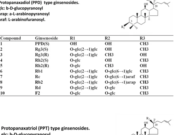
Figure 1. Structures and accurate masses of ginsenosides.
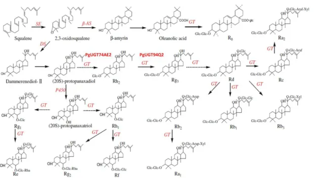
Figure 2. Putative ginsenosides biosynthesis pathway in P. ginseng.
Figure 3. Schematic extraction procedure of crude saponin.
Figure 4. Morphological comparison between CR and AR cultured on different rooting
medium.
Figure 5. Effects of plant hormones on growth of ginseng adventitious roots.
Figure 6. Southern blot analysis of PgUGT94Q2 transgenic lines.
Figure 7. RT-PCR analysis of PgUGT94Q2 over-expression lines.
Figure 8. Southern blot analysis of PgUGT74AE2 transgenic lines.
Figure 9. RT-PCR analysis of PgUGT74AE2 over-expression lines.
Figure10. Comparison of ginsenoside contents of AR and CR that induced by NI (NAA and
IAA) and IBA.
Figure 11. Effects of plant hormones on ginsenoside content in AR and CR roots.
Figure 12. Analysis of ginsenoside contents of PgUGT74AE2 transgenic lines by HPLC.
Figure 13. Ginsenosides concentration of PgUGT74AE2 transgenic lines.
Figure 14. Analysis of ginsenoside contents of PgUGT94Q2 transgenic lines by HPLC.
Figure 15. Ginsenosides concentration of PgUGT94Q2 transgenic lines.
Figure 16. PgUGT74AE2 and PgUGT94Q2 involved ginsenosides biosynthesis pathway
demonstrated in yeast system.
LIST OF TABLES
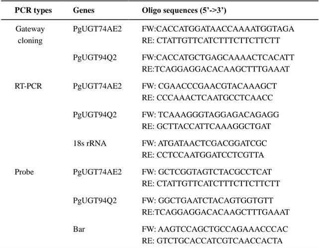
Table 1. Primers sets used in experiments.
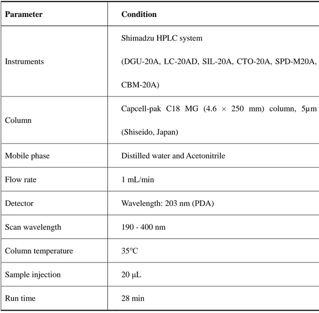
Table 2. HPLC conditions for ginsenoside analysis.
Table 3. Mobile phase of HPLC gradient conditions for ginsenoside analysis.
Table 4. Media composition for ginseng tissue culture.
Table 5. Media compositions used for transformation.
Table 6. Comparison of different medium for ginseng adventitious roots induction from
calluses and adventitious roots.
Table 7. Frequency of callus formation of ginseng adventitious roots from AR and CR
groups.
Table 8. Ginsenoside concentrations of AR and CR roots cultured on NI and IBA medium.
Table 9. Ginsenosides concentration of PgUGT74AE2 transgenic lines.
Table 10. Ginsenosides concentration of PgUGT94Q2 transgenic lines.
SUMMARY
Panax ginseng C.A Meyer (Korean wild ginseng) is an ancient and medicinal plant that
wildly used all over the world. The most beneficial compound involved inside of ginseng plants is ginsenoside, with the high attentions have been paid to ginseng researches, more and more ginsenosides had been extracted and identified from ginseng plants. Up to now, more than 70 ginsenosides have been reported from natural and processed ginseng. In the meantime, various functional genes which are engaged in ginsenosides biosynthesis pathway come to be known, including UDP-glycosyltransferases (UGTs). UGTs play roles in the final step of ginsenoside synthesis, after glycosylate reactions catalyzed by UGTs result in variety of ginsenosides. In order to explore the function of UGTs in ginenosides biosynthesis pathway and increase the content of ginsenosides in ginseng adventitious roots, two endogenous UGTs genes (PgUGT74AE2 and PgUGT94Q2) were transformed to Korean wild ginseng by
Agrobacterium-mediated transformation. The adventitious roots induction procedure was
optimized by applying different rooting hormones, and induced adventitious roots directly from callus. Because different phenotypes were observed in different hormone rooting medium during culturing, morphological and histological analysis were applied for recording. In case of hormones and different resources give rise to the concentration of ginsenoside, high performance liquid chromatography (HPLC) was supposed for ginsenoside contents analysis for both wild-type and transgenic lines. Callus derived and adventitious roots derived roots rooting by IBA was observed increasing of total content of main ginsenosides (Re, Rg1, Rc, Rb1, Rb2, Rd), especially ginsenoside Rb2. As callus acting as the most suitable host for plant
genetic engineering, transformed callus derived roots rooting by IBA was supposed to produce transgenic roots in brief period. PgUGT74AE2 and PgUGT94Q2 transgenic lines generated within 6 month, including callus induction, transformation, roots induction from transformed callus. Transgenic candidates of PgUGT74AE2 and PgUGT94Q2 were selected by Immuno strip test (Agrastrip GMO, LL test Strips) as first confirmation. 9 lines of PgUGT74AE2 (74 L2, 74 L4, 74 3L2, 74 L2-2, 74 T2, 74 T3, 74 T5, 74 T6, 74 T7) and 7 lines of PgUGT94Q2 (94 L2, 94 T2, 94 T4, 94 T5, 94 T8, 94 T11, 94 T12) were screened from transgenic candidates. Total RNA was extracted from all transgenic lines, and cDNA synthesized from total RNA, cDNA used as template for RT-PCR analysis and bar gene confirmation. Genomic DNA extracted from all transgenic lines to performed southern blotting to analyze T-DNA region insertion copies. Incorporation with HPLC results of transgenic lines, PgUGT74AE2 transgenic lines have 4 lines (74 L2-2, 74 L2, 74 L4, 74 T2) and PgUGT94Q2 transgenic lines have 4 lines (94 L2, 94 T4, 94 T2, 94 T5) gained ginsenoside contents increasing compared with wild-type ginseng. And one unknown compound among these 8 transgenic lines showed content enhancement. Further analysis of unknown compound structure is in the management.
INTRODUCTION
Ginseng (Panax ginseng C.A. Meyer) is an important medicinal plant that is widely cultivated in eastern countries. The roots have been used as drugs for over 2000 years in oriental countries and also rapidly expanding in western countries as complementary and alternative medicine (Shim et al. 2009). The beneficial effects of ginseng are attributed to glycosylated tetracyclic triterpene compounds, also known as ginsenosides, are mainly produced in ginseng roots and reported mostly concentrated in adventitious roots (Han et al. 2013). Ginsenosides are mainly consisted of two parts named oleanane- and dammarene-type ginsenosides that generated at first cyclization of 2,3-oxidosqualene. And dammarene-type triterpenes are classified into two groups, protopanaxadiol- (PPD) and protopanaxatriol-type (PPT) ginsenosides, continuously with different glycosylation and result in various ginsenosides. More than 40 different ginsenosides producing various pharmacological effects have been identified in ginseng roots (Jung et al. 2014), and more than 70 ginsenosides have been investigated from ginseng plants including these involved in fresh ginseng (FG), suncured ginseng (SG) and red ginseng (RG) (Guo et al. 2014). Ginseng is a perennial plant that has a long production cycle (4-6 years) and greater than 3 years of juvenile period are required for producing seeds (Ahn et al. 1996; Choi et al. 1998), which has made the generation of superior genotypes by conventional breeding toughly to handle. Therefore, many attempts have been carried out to achieve more rapid ginseng plant regeneration procedure and increasing production of the ginsenosides. Biotechnological techniques have been applied in previous researches, such as classical tissue culture, bioreactor culture,
breeding by γ-irradiation (Yoshikawa et al. 1987; Bae et al. 2006; Kim et al. 2013).
P. ginseng is a difficult species to manipulate in vitro, however, the regeneration of
ginseng has generally been accomplished by using somatic embryogenesis in callus derived from mature root tissues, callus derived from zygotic embryo (Lee et al. 1990), protoplast derived from callus, and cotyledons, aging callus produced numerous embryoids (Arya et al. 1991). Re-cultured of embryoids in MS solid medium supplemented with benzyladenine (6-BA) and gibberellic acid (GA3) resulted in profuse plantlet regeneration (Chang et al.
1980). Metabolic engineering of P. ginseng thorough the transgenic adventitious root culture can be an important technique to upgrade medicinal value and produce efficient production of secondary metabolites from roots. Cotyledonary explants of ginseng zygotic embryos were co-cultured with Agrobacterium tumefaciens strain LBA4404 harboring binary vector carried with β-glucuronidase (GUS) gene, and transgenic plants were obtained by somatic embryogenesis (Lee et al. 1994). A rapid and efficient genetic transformation of P. ginseng by a plasmolyzing pretreatment of cotyledon explants was reported by Choi et al. (2001). However, more than 1 year is required to induce transgenic adventitious roots from regenerated transgenic plantlets via somatic embryogenesis. We are looking forward to find a new process to generate adventitious roots directly from calluses in brief period. Various hormone combinations were used to optimized ginseng adventitious roots induction and growth on the basis of previous researches.
At present, many novel UDP-glycosyltransferases (UGTs) genes which involved inside of ginsenosides biosynthesis have been reported by using advanced sequencing tools. It is reported that putative UDP-glycosyltransferases play a complex and important role in the
synthesis and metabolism of ginsenosides (Figure 2.)(Wang et al. 2011). Since the first UGT (UGTPg1) were reported by Yan et al. (2014), soon afterwards, consecutive UGTs and encoded genes were been coherented in intricate ginsenosides biosynthesis pathway. UGTPg1 from P. ginseng was characterized as a regio-specific enzyme which glycosylate the C20-OH position of PPD and also its derived ginsenosides (Yan et al. 2014). More currently, two UGTs named PgUGT74AE2 and PgUGT94Q2 from P. ginseng were characterized to catalyze the glycosylation of C3-OH of PPD to yield Rh2 and to elongate a glucose moiety of Rh2 and F2 to form Rg3 and Rd respectively (Jung et al. 2014; Wang et al. 2015; Wei et al. 2015). Not yet, no information is available about the UGTs transgenic lines by over-expressing of UGTs genes in P. ginseng plants. In this paper, we report an efficient procedure for producing transgenic lines of P. ginseng adventitious roots based on Agrobacterium-mediated transformation and root induction directly from wild-type ginseng somatic embryos. Two UDP-glycosyltransferases (UGTs) were supposed to be transferred into and over-expressed in Korean wild ginseng to enhance concentration of ginsenosides Rh2 and Rg3 or other possible related ginsenosides.
Ginsenosides:
Triterpenoid saponins are secondary metabolites of isoprenoid compoundsand mostly exist in higher plants. Ginsenosides are considered to be the primary components of the ginseng and have been verified mostly contained in adventitious roots. P. ginseng roots contain at least 4% ginsenosides by dry weight (Shibata et al. 2001). Six dammarane-type tetracyclic triterpenes (ginsenosides Rb1, Rb2, Rc, Rd, Re and Rg1) are reported as the major ginsenoside constituents (Figure 1.). Protopanaxadiol (PPD) is the reasonable substrate lied upstream of PgUGT74AE2 and PgUGT94Q2 encoded pathway. F2, S-Rh2, R-Rh2 and S-Rg3
are the possible products after PgUGT74AE2 and PgUGT94Q2 encoded glycosylation according to published paper (Jung et al. 2014). It has been reported that ginsenosides Rh2 and Rg3 are potent medicinal agents for inducing tumor cell apoptosis (Min et al. 2006; Park et al. 1997), inhibiting tumor cell proliferation (Kim et al. 2004). Rh2 and Rg3 are also potentially effective candidates for preventing metabolic disorders of human beings, such as diabetes and obesity, via activating the AMPK signaling pathway (Hwang et al. 2007; Park et al. 2008). Indeed, as ginsenosides Rh2 and Rg3 are the versatile compound value of infrequent bioactivities, a huge demand has been existed to manufacturing them for commercial use. Whereas, their contents inside of ginseng are extremely low in natural ginseng, the contents of Rh2 and Rg3 can be increased markedly to 0.001% and 0.015%, respectively, in red ginseng, which the production process performed by steaming and drying harvested ginseng root (Shibata et al. 2001).
UDP-glycosyltransferases: Glycosylation of natural products in plants is catalyzed by
glycosyltransferases (GTs), leading to changes in hydrophilicity, stability, and subcellular localization, and thus the chemical properties and bioactivity of the natural products (Bowles et al. 2006). Enzymes belonging to the multigene families of oxidosqualenecyclases, and UDP-glycosyltransferases are key players in biosynthesis of plant triterpenoid saponins. (Augustin et al. 2011). GTs are members of a multigene superfamily that catalyzes the transfer of sugar moieties to specific acceptors. The GTs that take usage of uridine diphosphate (UDP)-activated sugar molecules as donors are referred as UDP-glycosyltransferases (UGTs) (Barvkar et al. 2012; Ross et al. 2001). Nevertheless, only a limited number of UGTs characterized to glycosylate triterpenoid aglycones have been
exposed from plants, such as Medicago truncatula (Achnine et al. 2005), Saponaria vaccaria (Meesapyodsuk et al. 2007), Barbarea vulgaris (Augustin et al. 2012), Glycine max (Shibuya et al. 2010), and P. ginseng (Yan et al. 2014; Wang et al. 2015). Two endogenous genes PgUGT74AE2 and PgUGT94Q2 from P. ginseng were characterized to catalyze the glycosylation of C3-OH of PPD to yield Rh2 and to elongate a glucose moiety of Rh2 and F2 to form Rg3 and Rd respectively, and convert reactions have been demonstrated in yeast system (Jung et al. 2014).
Figure 2. Putative ginsenosides biosynthesis pathway in P. ginseng. (Wang et al. 2011)
IPP, isopentenyl diphosphate; β-AS, β-amyrin synthase; DS, dammarenediol-II synthase; GT, UDP-glycosyltransferases.
MATERIALS AND METHODS
Callus induction and selection
Adventitious roots derived from Korean wild ginseng were provided by Sunchon National University, Sunchon, Korea. The adventitious roots were generated as described previously and have been maintained in our laboratory for over 10 years (Zhang et al. 2011). Calluses were induced from fresh adventitious roots remained on MS solid medium with 2mg/L NAA and 0.25mg/L IAA in plates culture. Wild-type adventitious roots were sectioned into 10 mm in length and were placed on Murashige and Skoog (MS) solid medium supplemented with 0.5mg/L 2,4-dichlorophenoxyacetic acid (2,4-D), 0.3mg/L kinetin, and 3% sucrose and media were solidified with 0.3% Gelrite (Zhang et al. 2014). All media were adjusted to pH 5.8 prior to autoclaving. Ten pieces of adventitious roots were placed on each petri dish. Callus formation was observed after 4 weeks of culture. The induced callus was sub-cultured at 3-wk intervals on the same medium for induction of embryogenic callus and maintenance. After 2 months of culture, we selected the compact and strong pro-embryogenic masses and used for somatic embryos induction. Somatic embryos induction medium was MS medium supplemented with 0.5mg/L 2,4-D. Induced somatic embryos were inoculated on MS solid medium for enlarge cultivation with 0.5mg/L 2,4-D every 3 weeks. All media were adjusted to pH 5.8 prior to autoclaving. All cultures were incubated at 23 ± 2 ℃ in the dark.
Optimization of adventitious roots induction procedure
Wild-type ginseng adventitious roots were used to induce calluses, from calluses to form somatic embryos and then induce roots from somatic embryos directly. We applied two
different roots induction medium according to previous research paper (Zhang et al. 2014 and Huang et al. 2010), and measured the root induction efficiency of both roots and calluses while the culturing procedure. Somatic embryos and adventitious roots were inoculated onto MS solid media containing 5.0 mg/L indole-3-butric acid (IBA), 30 g/L sucrose and 0.3% gelrite ( IBA medium) , and MS solid medium containing 2 mg/L NAA, 0.25mg/L IAA and 0.3% gelrite (NI medium). Also hormone-free MS solid medium was used for control. Three replicates were prepared for each treatment. Roots performed further development in conical flask containing 50ml liquid MS medium supplemented with 5.0 mg/L IBA or 2 mg/L NAA, 0.25mg/L IAA, respectively, on a rotary shaker (120 rpm) at 23 ± 2 ℃ and sub-cultured every 4 weeks. Plates culturing of ginseng roots were implemented in dark, then further culturing in flasks were maintained under a 16-h (light)/8-h (dark) photoperiod with light supplied by white fluorescent tubes at an intensity of 30 µmol m–2 s–1.
Morphologic and histological analyses of CR and AR
Comparison of two different roots induction medium was showed in morphological differences, such as growth ratio, secondary root number, length and diameter. Fresh weight (FW) and dry weight (DW) were measured after two months culture either on plates or in flasks culture. Harvest roots was rinsed by tap water once and then put on tissue paper for 10 minutes and blotting with tissue paper to remove residual water, straight after, measure about FW of roots. And DW was recorded after fresh roots were dried to constant weight. Root growth ratio was calculated as following formula (Yu et al. 2002).
The length of secondary roots was measured by calipers. The number of occurred secondary roots was counted from main roots body. The diameters of secondary roots were measured from microscope and Nikon NIS-Elements system by using measure scale. Newly developed adventitious roots of all treatments were selected for checking callus formation ability. Callus induction medium referred to callus induction and selection section.
Gene cloning and vectors construction
Total RNA was extracted from wild-type ginseng adventitious roots by using Trizol (Invitrogen, Carlsbad, CA, USA) reagent, and cDNA synthesized from total RNA by using M-MLV Reverse Transcriptase kit (Promega, Seoul, Korea). Synthesized cDNA used as template and gateway cloning method used for PgUGT74AE2 (JX898529) and PgUGT94Q2 (JX898530) genes cloning. Specific primers were designed according to registered sequences on NCBI (Table 1.). Pfu DNA Polymerase (Promega, Seoul, Korea) is supposed to generate blunt ends and yield high fidelity PCR products. Then PCR products ligated into pENTR vector according to pENTR™/D-TOPO® cloning kit protocol (Invitrogen, Carlsbad, CA, USA). Ligated pENTR vector was transformed to competent TOP10 E. coli cells. Positive transformant was selected and isolated plasmid DNA, and then confirm the sequence. pENTR vector contains target genes sub-cloned into destination vectors (pB2GW7,0) by LR recombination (Invitrogen, Carlsbad, CA, USA). LR reaction mixture transformed to competent TOP 10 E. coli cells, spread on LB solid medium contained 50mg/L spectinomycin. Harvested positive transformant and extracted plasmid DNA then transferred to
mg/L rifampicin and 50mg/L spectinomycin. Harvested positive transformant make
Agrobacterium cell stocks which contained transgene and used for plant transformation.
Agrobacterium-mediated transformation
Selected calluses were pre-treated in hormone-free MS liquid medium containing 30g/L sucrose for 1 day. A. tumefaciens EHA105 cells cultured in YEP medium with 25mg/L rifampicin and 50mg/L spectinomycin for 24h at 28℃, 180rpm. The pre-treated calluses were immersed in the suspension of A. tumefaciens EHA105 carried with destination vectors in hormone-free MS liquid medium containing 100 mg/L acetosyringone. After 24h cultured in dark on a gyratory shaker, the calluses were placed on sterilized filter paper for 10 min to remove extra Agrobacterium cells, and then cultured on hormone-free MS solid medium containing 100 mg/L acetosyringone, 3% sucrose, 0.3% gelrite at 23±2°C in the dark 3 days for co-culture. Thereafter, the calluses were cultured on MS medium with 3% sucrose and 600 mg/L cefotaxime 3 weeks for elimination, and then transferred to selection MS solid medium with 600 mg/L cefotaxime, 1mg/L PPT,5mg/L IBA and 0.3% gelrite. All media were adjusted to pH 5.8 prior to being autoclaved at 120°C for 15 min.
Production of transgenic roots
Transformed calluses were inoculated onto MS solid media containing 5.0 mg/L indole-3-butric acid (IBA), 600mg/L cefotaxime, 1mg/L PPT, 30 g/L sucrose and 0.3% gelrite, sub-cultured every 3 weeks. Newly developed roots from transformed calluses were selected and cut from resourced calluses to perform further development on new solid root induction
with 5.0 mg/L IBA, 600mg/L cefotaxime, 1mg/L PPT on a rotary shaker (120 rpm) at 23 ± 2 ℃ and sub-cultured every 4 weeks.
Genotype analysis of transgenic candidates
Newly developed roots from transfected calluses were supposed as transgenic candidates. Immuno strip test (Agrastrip GMO, LL test Strips) used for the most convenient method to confirmed genetically modified organism (GMO). For the first confirmation of transgenic lines, we chose the transgenic candidates length reached about 2cm, half kept for strip test, and another half remained on root induction medium for further development. Transgenic lines would get two bands on test strips, while wild-type lines would gain only one band, and under bands indicated on strips are the indication of bar gene encoded protein, PAT (phosphinothricin acetyltransferase) protein. With the first step confirmation, PgUGT74AE2 transgenic lines and PgUGT94Q2 transgenic lines were selected and amplified in conical flask containing 50ml liquid MS medium supplemented with 5.0 mg/L IBA, 600mg/L cefotaxime, 1mg/L PPT on a rotary shaker (120 rpm) at 23 ± 2 ℃ and sub-cultured every 6 weeks. Each line sampled with liquid nitrogen grounding in mortar, and collected samples divided to extract total RNA and genomic DNA. Total RNA then performed to do cDNA synthesis, cDNA of each lines was used as template to do RT-PCR (Reverse transcript-polymerase chain reaction) to check relative expression of transgene (Table 1), genomic DNA from transgenic lines was used for Southern blotting to analyze T-DNA integration and to confirm the copy number.
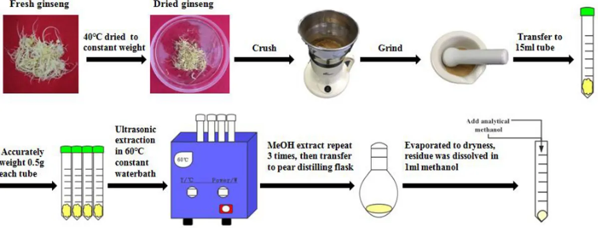
Extraction and determination of crude saponin
Extraction method of ginsenosides from ginseng roots followed and modified from the method of Jin et al. (2012) and Su et al. (2016). Ginseng samples of wild-type and transgenic lines were accurately weighed (approximately 0.5 g) and ultrasonic-extracted with 2.0 ml of methanol in an ultrasonic water bath for 15 min at the temperature of 40℃, each sample repeats ultrasonic extraction for 3 times and eventually get 6 ml volume in total. The extracts were transferred into pear-shaped flasks and evaporated to dryness at 40℃ with a vacuum rotary evaporator. Then, the residue was dissolved in 1.0 ml of methanol and filtered by a 0.22 μm PTFE syringe filter before HPLC analysis (Figure 3).
HPLC analysis of wild-type and transgenic lines
For ginsenosides assay of wild-type and transgenic lines, the HPLC (High Performance
Liquid Chromatography) conditions followed Jung et al. (2014) and made some modifications (Table 2). HPLC running conditions showed as following table (Table 3.). All the solvents were belonged to HPLC grades. Ginsenoside standards (Re, Rg1, Rb1, Rc, Rb2, Rd, Rh2 and Rg3) were purchased from SHANGHAI ZZBIO CO., LTD. An ODS C18 column (Shiseido, Japan) was selected as stationary phase, mobile phase including Solvent (A): Acetonitrile, Solvent (B): Distilled water. Ginsenoside standard solutions were prepared in 100% HPLC grade methanol. A series gradient concentration (50ppm, 100ppm, 400ppm) of each standard were tested for calibration curve.
Analysis of ginsenoside contents was performed according to Son et al. (1999a) and Yu et al. (2000). The total ginsenoside content was calculated as the sum of individual ginsenoside
fractions.
The ginsenoside content of ginseng adventitious roots was calculated as:
(GC: ginsenoside content; SGC: sample ginsenoside concentration from HPLC; SV: sample volume; AR: adventitious root derived from root itself; CR: adventitious root derived from callus which induced from AR)
Statistical analysis
Statistical analysis achieved by using SPSS (Statistical Product and Service Solutions) system, IBM SPSS Statistics version 22.0. Mean and standard errors were measured throughout datas and statistical significance between the mean values was assessed by Duncan’s multiple range tests. A probability of P < 0.05 was considered as significantly.
) ( C or AR ) ( ) )( ( ) ( 1 1 g R l SV X g mg fromHPLC SGC g mg GC
Table 1. Primers sets used in experiments.
PCR types Genes Oligo sequences (5’->3’)
Gateway cloning PgUGT74AE2 FW:CACCATGGATAACCAAAATGGTAGA RE: CTATTGTTCATCTTTCTTCTTCTT PgUGT94Q2 FW:CACCATGCTGAGCAAAACTCACATT RE:TCAGGAGGACACAAGCTTTGAAAT RT-PCR PgUGT74AE2 FW: CGAACCCGAACGTACAAAGCT RE: CCCAAACTCAATGCCTCAACC PgUGT94Q2 FW: TCAAAGGGTAGGAGACAGAGG RE: GCTTACCATTCAAAGGCTGAT 18s rRNA FW: ATGATAACTCGACGGATCGC RE: CCTCCAATGGATCCTCGTTA Probe PgUGT74AE2 FW: GCTCGGTAGTCTACGCCTCAT
RE: CTATTGTTCATCTTTCTTCTTCTT PgUGT94Q2 FW: GGCTGAATCTACAGTGGTGTT
RE:TCAGGAGGACACAAGCTTTGAAAT Bar FW: AAGTCCAGCTGCCAGAAACCCAC
Table 2. HPLC conditions for ginsenoside analysis.
Parameter Condition
Instruments
Shimadzu HPLC system
(DGU-20A, LC-20AD, SIL-20A, CTO-20A, SPD-M20A, CBM-20A)
Column
Capcell-pak C18 MG (4.6 × 250 mm) column, 5µm (Shiseido, Japan)
Mobile phase Distilled water and Acetonitrile
Flow rate 1 mL/min
Detector Wavelength: 203 nm (PDA) Scan wavelength 190 - 400 nm
Column temperature 35℃ Sample injection 20 μL
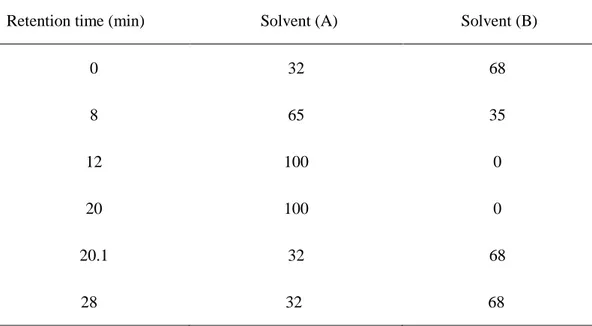
Table 3. Mobile phase of HPLC gradient conditions for ginsenoside analysis.
Solvent (A): Acetonitrile, Solvent (B): Distilled water.
Retention time (min) Solvent (A) Solvent (B) 0 8 12 20 20.1 28 32 65 100 100 32 32 68 35 0 0 68 68
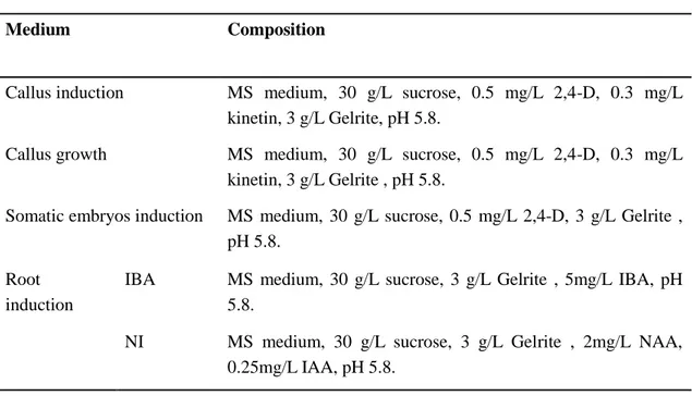
Table 4. Media composition for ginseng tissue culture.
Medium Composition
Callus induction MS medium, 30 g/L sucrose, 0.5 mg/L 2,4-D, 0.3 mg/L kinetin, 3 g/L Gelrite, pH 5.8.
Callus growth MS medium, 30 g/L sucrose, 0.5 mg/L 2,4-D, 0.3 mg/L kinetin, 3 g/L Gelrite , pH 5.8.
Somatic embryos induction MS medium, 30 g/L sucrose, 0.5 mg/L 2,4-D, 3 g/L Gelrite , pH 5.8.
Root induction
IBA MS medium, 30 g/L sucrose, 3 g/L Gelrite , 5mg/L IBA, pH 5.8.
NI MS medium, 30 g/L sucrose, 3 g/L Gelrite , 2mg/L NAA, 0.25mg/L IAA, pH 5.8.
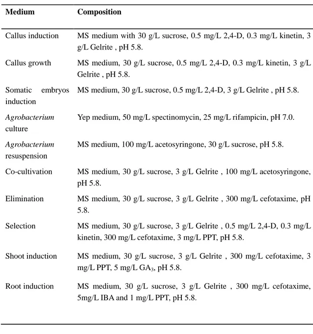
Table 5. Media compositions used for transformation.
Medium Composition
Callus induction MS medium with 30 g/L sucrose, 0.5 mg/L 2,4-D, 0.3 mg/L kinetin, 3 g/L Gelrite , pH 5.8.
Callus growth MS medium, 30 g/L sucrose, 0.5 mg/L 2,4-D, 0.3 mg/L kinetin, 3 g/L Gelrite , pH 5.8.
Somatic embryos induction
MS medium, 30 g/L sucrose, 0.5 mg/L 2,4-D, 3 g/L Gelrite , pH 5.8.
Agrobacterium
culture
Yep medium, 50 mg/L spectinomycin, 25 mg/L rifampicin, pH 7.0.
Agrobacterium
resuspension
MS medium, 100 mg/L acetosyringone, 30 g/L sucrose, pH 5.8. Co-cultivation MS medium, 30 g/L sucrose, 3 g/L Gelrite , 100 mg/L acetosyringone,
pH 5.8.
Elimination MS medium, 30 g/L sucrose, 3 g/L Gelrite , 300 mg/L cefotaxime, pH 5.8.
Selection MS medium, 30 g/L sucrose, 3 g/L Gelrite , 0.5 mg/L 2,4-D, 0.3 mg/L kinetin, 300 mg/L cefotaxime, 3 mg/L PPT, pH 5.8.
Shoot induction MS medium, 30 g/L sucrose, 3 g/L Gelrite , 300 mg/L cefotaxime, 3 mg/L PPT, 5 mg/L GA3, pH 5.8.
Root induction MS medium, 30 g/L sucrose, 3 g/L Gelrite , 300 mg/L cefotaxime, 5mg/L IBA and 1 mg/L PPT, pH 5.8.
RESULTS
Callus induction and selection
During the culture procedure, root segments started swelling after 2 weeks culture, proembryogenic mass (PEMs) produced firstly on the MS solid medium with 2,4-D and kinetin after 1 month culturing. Then PEMs were divided to pieces, around 0.5mm in diameter for following somatic embryos induction, kinetin was removed and white and strong somatic embryos gained on somatic embryos induction medium, somatic embryo calluses were used for Agrobacterium-mediated transformation and adventitious roots induction host.
Optimization of roots induction procedure and comparison between AR and CR
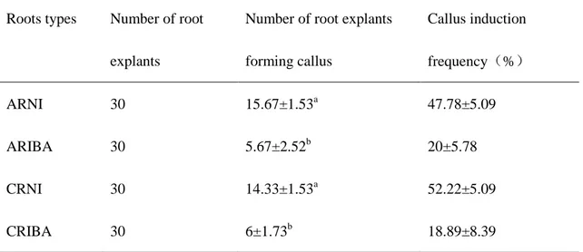
Callus-derived roots (CR) induced from compact and strong callus masses within 1 month, identical phenomenon were discovered in both roots derived adventitious roots (AR) and callus derived adventitious roots (CR) in roots induction procedure though incubating with different rooting hormone (Figure 4). Measurements of newly developed adventitious roots inducing efficiency on different rooting medium were recorded after 6 weeks of rooting culture, also first derived roots segments from callus were tested for rooting ability. IBA rooting medium gained significant enhancements for callus, CR and AR in secondary roots inducing number (Table 6), behaved similar performances in diameter of secondary roots among each treatment, but uncontemplated with extraordinary shorter length of secondary roots than NI rooting medium (Figure 4). As for growth ratio of adventitious roots development, IBA treatments behaved exemplary both in flask shaking liquid culture and
by NAA and IAA medium presented higher callus formation frequency than IBA rooting medium (Table 7).
Production of transgenic roots and genotype analysis
Transgenic candidates generated from somatic embryos after 2 month sub-culturing on roots induction medium, MS solid medium with 5mg/L IBA, PPT 1mg/L and 300mg/L cefotaxime.
Bar gene confirmation and RT-PCR (Reverse transcript-polymerase chain reaction) analysis
of and PgUGT74AE2 and PgUGT94Q2 transgenic lines (CRIBA is supposed as wild-type control) were shown in Figure 7 and Figure 9. There had 5 lines (T2, T4, T5, T8, L2) of PgUGT94Q2 transgenic lines and 8 (T2, T6, L2-2, T7, L2, L4, T5, 3L2) of PgUGT74AE2 transgenic lines gained higher relative expression level while compared with wild-type control. Incorporation with southern blot results, 7 lines (T2, T4, T5, T8, T11, T12, L2) of PgUGT94Q2 transgenic lines appeared 4, 2, 1, 1, 3, 1 and 2 bands respectively, 9 lines (L2, L4, T2, T7, T3, T5, T6, L2-2, 3L2) of PgUGT74AE2 transgenic lines appeared 1, 3, 3, 1, 7, 2, 1, 1, 2 bands respectively. Single copy of transgenic lines (PgUGT94Q2 T5, PgUGT74AE2 L2-2, PgUGT74AE2 L2) exerted high relative expression, two copies (PgUGT94Q2 T4, PgUGT94Q2 L2), triple copies (PgUGT74AE2 L4, PgUGT74AE2 T2) and quadruple copies (PgUGT94Q2 T2) also showed up high relative expression, while single copy of PgUGT94Q2 T12 and triple copies of PgUGT94Q2 T11 acted out low relative expression, 7 copies of PgUGT74AE2 acted out low relative expression (Figure 7-10). HPLC analysis of all transgenic lines was carried out to check T-DNA insert effect and ginsenosides variation.
HPLC analysis of wild-type and transgenic lines
CRIBA treatment showed highest concentration of ginsenosides among all 4 treatments (ARIBA, CRIBA, ARNI and CRNI) (Figure 10-11). CRIBA was used as wild-type control and contrasted with transgenic lines, PgUGT74AE2 transgenic groups have 4 lines (74 L2-2, 74 L2, 74 L4, 74 T2) and PgUGT94Q2 transgenic groups have 4 lines (94 L2, 94 T4, 94 T2, 94 T5) gained ginsenosides contents increasing. And the content of one unknown compound increased in these 8 transgenic lines (Figure 12-15). Transgenic line PgUGT74AE2 T2 showed the highest ginsenosides concentration among wild-type control and other PgUGT74AE2 transgenic lines, and 1.4-fold higher than that of wild-type ginseng. Transgenic line PgUGT94Q2 T2 showed the highest ginsenosides concentration among wild-type control and other PgUGT94Q2 transgenic line, and 1.3-fold higher than that of wild-type ginseng.
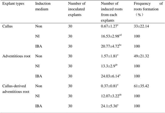
Table 6. Comparison of different medium for ginseng adventitious roots induction from
calluses and adventitious roots1)
1) Data were collected after 6 weeks of culture. The results represent the means standard error obtained from three repeats. Different corresponding letters within a column are significant different at p < 0.05 by Duncan’s multiple range test.
Non: Hormone-free.
NI: MS solid medium supplemented with 2mg/L NAA, 0.25mg/L IAA and 0.3% Gelrite. IBA: MS solid medium supplemented with 5mg/L IBA and 0.3% Gelrite.
Explant types Induction medium Number of inoculated explants Number of induced roots from each explants Frequency of roots formation (%) Callus Non 30 0.67±1.27e 33±22.14 NI 30 16.53±2.98cd 100 IBA 30 20.77±4.72bc 100
Adventitious root Non 30 1.57±1.81e 49±21.32
NI 30 13.3±2.9de 100 IBA 30 24.03±6.14a 100 Callus-derived adventitious root Non 30 0.37±0.81e 61±35.42 NI 30 12.07±3.22de 100 IBA 30 24.1±5.36a 100
Table 7. Frequency of callus formation of ginseng adventitious roots from AR and CR
groups1)
1) Data were collected after 6 weeks of culture. The results represent the means standard error obtained from three repeats. Different corresponding letters within a column are significant different at p < 0.05 by Duncan’s multiple range tests.
ARNI: Adventitious roots induced by NI medium and induced callus on callus induction medium.
ARIBA: Adventitious roots induced by IBA medium and induced callus on callus induction medium.
CRNI: Callus-derived adventitious roots induced by NI medium and induced callus on callus induction medium
CRIBA: Callus-derived adventitious roots induced by IBA medium and induced callus on callus induction medium.
Callus induction medium: MS solid medium supplemented with 0.5mg/L 2,4-D, 0.3mg/L kinetin, 30g/L sucrose and 0.3% Gelrite.
Roots types Number of root explants
Number of root explants forming callus Callus induction frequency(%) ARNI 30 15.67±1.53a 47.78±5.09 ARIBA 30 5.67±2.52b 20±5.78 CRNI 30 14.33±1.53a 52.22±5.09 CRIBA 30 6±1.73b 18.89±8.39
Figure 4. Morphological comparison between CR and AR cultured on different rooting
medium. A~G, incubated on MS solid medium, solidified with 0.3% Gelrite. H~K, incubated in MS liquid medium with rotary shaking (120 rpm). A, Calluses cultured on hormone-free medium; B, Calluses cultured on NI medium; C, Calluses cultured on IBA medium; D, H, Callus-derived roots induced by NI medium; E, J, Callus-derived roots induced by IBA medium; F, I, Adventitious roots induced by NI medium; G, K, Adventitious roots induced by IBA medium; L, Comparison of length, incidence and diameter of secondary roots in flask shaking culture among each treatment, data analyzed by R studio version 1.0.44. ARNI, Adventitious roots cultured in NI medium; ARIBA, Adventitious roots cultured in IBA medium; CRNI, Callus-derived roots cultured in NI medium; CRIBA, Callus-derived roots cultured in IBA medium. Bar 1cm.
Figure 5. Effect of plant hormones on growth of ginseng adventitious roots. A, Growth ratio
of adventitious roots in the flask culture. Original weight is 1g for all types of adventitious roots. B, Fresh weight of adventitious roots in solid root induction medium. Each plate incubated with 10 root segments originally. ARNI: Adventitious roots cultured on NI medium; ARIBA: Adventitious roots cultured on IBA medium; CRNI: Callus-derived adventitious roots cultured on NI medium; CRIBA: Callus-derived adventitious roots cultured on IBA medium.
Figure10. Comparison of ginsenoside contents of AR and CR that induced by NI (NAA and
Figure 11. Effects of plant hormones on ginsenoside content in AR and CR roots. Data are
shown as means ± standard deviation of values obtained from three experiments.
0
1
2
3
4
5
6
7
8
9
10
ARIBA
CRIBA
ARNI
CRNI
Gi
ns
eno
si
de
co
ntent
(
m
g
/g
D
W
)
PPD
S-Rh2
Rd
Rb2
Rc
Rb1
Rg1
Re
T ab le 8 . G in se nos ide con ce nt ra tion s of A R and CR r oo ts cu ltur ed on N I and IB A m ed iu m . The r esul ts rep res ent the m ea ns ± st and ar d d evi at ion o b tai ned fr o m thr ee r epe at s.
Figure 13. Ginsenosides concentration of PgUGT74AE2 transgenic lines.
*
*
*
*
0
1
2
3
4
5
6
7
8
WT 74 T3 74 3L2 74 L2-2 74 T6 74 T5 74 L2 74 L4 74 T2 74 T7Gi
ns
eno
si
de
co
ntent
(m
g
/g
D
W)
PPD
S-Rh2
Rd
Rb2
Rc
Rb1
Rg1
Re
T able 9 . G in se nos ide s c o nc ent rat io n o f PgU G T74A E2 tr an sge n ic line s. Th e r esul ts rep res ent the m ea ns ± st and ar d d evi at ion o b tai ned fr o m thr ee r epe at s.
Figure 15. Ginsenosides concentration of PgUGT94Q2 transgenic lines.
*
*
*
*
0
1
2
3
4
5
6
7
8
WT
94 L2 94 T4 94 T12 94 T2 94 T11 94 T5 94 T8
Gi
ns
eno
si
de
co
ntent
(m
g
/g
D
W)
PPD
S-Rh2
Rd
Rb2
Rc
Rb1
Rg1
Re
T able 10 . G in se no si d es c on ce nt ra tion of PgU G T94Q 2 tr an sge n ic lines . The r esul ts rep res ent the m ea ns ± st and ar d d evi at ion o b tai ned fr o m thr ee r epe at s.
DISCUSSION
Over-expression of endogenous UDP-glycosyltransferases genes in Korean wild ginseng results in a reasonable improvement of ginsenoside Rb2 and Rd, but Rg1 glycosylated by unknown UDP-glycosyltransferases also increased concentration. Transgenic lines with relative high expression (PgUGT74AE2 T5, 94 T4, 94 L2, 94 T2; PgUGT74AE2 L2-2, 74 L2, 74 L4, 74 T2) showed content enhancement of one unknown compound, that need to be confirmed in the future (LC-MS is on the progress to analyze unknown compound).
PgUGT74AE2 and PgUGT74AE2 encoded UDP-glycosyltransferases reported in yeast system that corresponding to Rh2 and Rg3 synthesis, PgUGT74AE2 add 1 glucose at C3 and PgUGT94Q2 add 1 glucose at glucose side chain of C3, PgUGT71A27 add 1 glucose at C20. PgUGT74AE2 and PgUGT94Q2 worked same function in synthesis of F2 and Rd (Jung et al. 2014) (Figure 16). But over-expression of these two UGTs in wild-type ginseng didn’t show contents improvement of Rh2 and Rg3. In the protopanaxtriol (PPT) type ginsenosides biosynthesis pathway, UGTPg 101 performed multiple functions that could add 1 glucose at both C20 and C6 site, while UGTPg100 is not specific of substrate that could add 1 glucose at C6 site of ginsenosides F1 and PPT (Wei et al. 2015) (Figure 17). Based on the reported researches, we could Figureure out that UDP-glycosyltransferases work very complicatedly involved of ginsenosides biosynthesis pathway inside of ginseng plants. As Rh2 and Rg3 are the specific ginsenosides which have been reported existence in red and processed ginseng, PgUGT74AE2 and PgUGT94Q2 encoded UDP-glycosyltransferases corresponding biosynthesis pathway inside of natural ginseng plants deserved to be verified.
Figure 16. PgUGT74AE2 and PgUGT94Q2 involved ginsenosides biosynthesis pathway
Figure 17. UDP-glycosyltransferases genes involved in ginsenosides biosynthesis pathway
CONCLUSION
Adventitious roots induction procedure of ginseng has been optimized, callus-derived adventitious roots induced by IBA performed enhancement of total 12 selected ginsenoside. Ginseng transgenic lines could be screened from transgenic candidate adventitious roots which induced from transfected callus directly.
Over-expression of endogenous UDP-glycosyltransferases genes in Korean wild ginseng couldn’t increase the contents of ginsenoside Rg3 and Rh2 directly, but enhanced the contents of possible down-stream ginsenoside Rb2, Rd and Rg1. Rb2 and Rd have been reported lied on the down-stream synthesis pathway behind Rh2 and glycosylated by unknown UGTs. And Rg1 lied on the branch pathway divided from PPD, then glycosylated by other unknown UGTs.
Through over-expression of PgUGT74AE2 and PgUGT94Q2 respectively in Korean wild ginseng, we produced transgenic ginseng lines with enrichment in Rg1, Rb2 and Rd. Ginsenoside Rg1 has the effcacy of improving cerebral and liver functions, adjusting blood pressure and anti-fatigue and anti-stress activities. Ginsenoside Rd has efficacy of relieving pain. Ginsenoside Rb2 has efficacy of relieving pain, inhibiting the metastasis of cancer cells and anti-diabetic efficacy (Choi et al. 2008)
REFERANCES
Ahn IO, Le BV, Gendy C, Van KTT (1996) Direct somatic embryogenesis through thin layer culture in Panax ginseng. Plant Cell Tiss Organ Cult 45: 237–243
Angelova N, Kong HW, Heijden RVD, Yang SY, Choi YH, Kim HK, Wang M, Hankemeier T, Greef JVD, Xu GW, Verpoorte R (2008) Recent Methodology in the Phytochemical Analysis of Ginseng. Phytochem. Anal. 19: 2–16
Arya S, Liu JR, Eriksson T (1991) Plant regeneration from protoplasts of Panax ginseng (C.A. Meyer) through somatic embryogenesis. Plant Cell Rep, 10: 277–281
Augustina JM, Kuzinaa V, Andersenb SB, Baka S (2011) Molecular activities, biosynthesis and evolution of triterpenoid saponins. Phytochemistry 72(6): 435–457
Bae KH, Choi YE, Shin CG, Kim YY, Kim YS (2006) Enhanced ginsenoside productivity by combination of ethephon and methyl jasmonate in ginseng (Panax ginseng C.A. Meyer) adventitious root cultures. Biotechnol Lett 28:1163–1166
Barvkar VT, Pardeshi VC, Kale SM, Kadoo NY, Gupta VS (2012) Phylogenomic analysis of UDP-glycosyltransferase 1 multigene family in Linum usitatissimum identified genes with varied expression patterns. BMC Genomics 13: 175.
Bowles D, Lim EK, Poppenberger B, Vaistij FE (2006) Glycosyltransferases of lipophilic small molecules. Annu. Rev. Plant Biol. 57:567–597.
Chang WC, Hsing YI (1980) Plant regeneration through somatic embryogenesis in root-derived callus of ginseng (Panax ginseng C. A. Meyer) Appl. Genetics 57: 133
Choi YE, Yang DC, Park JC, Soh WY, Choi KT (1998) Regenerative ability of somatic single and multiple embryos from cotyledons of Korean ginseng on hormone-free medium Plant Cell Rep 17: 544–551
Choi YE, Yang DC, Kusano T, Sano H (2001) Rapid and efficient Agrobacterium-mediated genetic transformation by plasmolyzing pretreatment of cotyledons in Panax ginseng. Plant Cell Rep 20: 616–621
Choi KT (2008) Botanical characteristics, pharmacological effects and medicinal components of Korean Panax ginseng C A Meyer. Acta Pharmacol Sin Sep; 29 (9): 1109–1118
Guo C, Gao YG, Zang P, He ZM, Zhao Y, Zhu HY, Yang H, Dong W, Zhang LX (2014) Simultaneous determination of sixteen ginsenosides in Panax ginseng and its preparation by HPLC. Chinese Traditional and Herbal Drugs 45(14): 2009-2013
28-Oxidase (CYP716A52v2) in Oleanane-Type Ginsenoside Biosynthesis in Panax
ginseng. Plant Cell Physiol 54(12): 2034–2046
Hwang JT, Kim SH, Lee MS, Kim SH, Yang HJ, Kim MJ, Kim HS, Ha J, Kim MS, Kwon DY (2007) Anti-obesity effects of ginsenoside Rh2 are associated with the activation of AMPK signaling pathway in 3T3-L1 adipocyte. Biochem. Biophys.Res.Commun.364: 1002–1008.
Jin X, Zhu LY, Shen H, Xu J, Li SL, Jia XB, Cai H, Cai BC, Yan R (2012) Influence of sulphur-fumigation on the quality of white ginseng: A quantitative evaluation of major ginsenosides by high performance liquid chromatography Food Chemistry 135: 1141–1147
Jung SC, Kim WH, Park SC, Jeong JK, Park MK, Lim SH, Lee Y, Im WT, Lee JH, Choi GS, Kim SC (2014) Two Ginseng UDP-Glycosyltransferases Synthesize Ginsenoside Rg3 and Rd. Plant Cell Physiol 55(12): 2177–2188
Kim DS, Song M, Kim SH, Jang DS, Kim JB, Ha BK, Kim SH, Lee KJ, Kang SY, Jeong IY (2013) The improvement of ginsenoside accumulation on Panax ginseng as a result of γ-irradiation. J Ginseng Res 37: 332–340
Rh2 on the proliferation of prostate cancer cells. Arch. Pharm. Res.27: 429–435
Kim YS, Hahn EJ, Yeung EC, Paek KY (2003) Lateral root development and saponin accumulation as affected by IBA or NAA in adventitious root cultures of Panax ginseng C.A. Meyer. In Vitro Cell. Dev. Biol.Plant 39:245–249
Lee HS, Kim SW, Lee KW, Eriksson T, Liu JR (1995) Agrobacterium-mediated transformation of ginseng (Panax ginseng) and mitotic stability of the inserted β-glucuronidase gene in regenerants from isolated protoplasts. Plant Cell Reports 14: 545
Lee HS, Liu JR, Yang SG, Lee YH, Lee KW (1990) In vitro flowering of plantlets regenerated from zygotic embryo-derived somatic embryos of ginseng. Hort Science 25: 1652–1654
Min JK, Kim JH, Cho YL, Maeng YS, Lee SJ, Pyun BJ, Kim YM, Park JH, Kwon YG (2006) 20(S)-Ginsenoside Rg3 prevents endothelial cell apoptosis via inhibition of a mitochondrial caspase pathway. Biochem Biophys Res Commun. 349: 987–994
Park MW, Ha J, Chung SH (2008) 20(S)-ginsenoside Rg3 enhances glucose-stimulated insulin secretion and activates AMPK. Biol Pharm Bull.31: 748–751
Ross J, Li Y, Lim E, Bowles DJ (2001) Higher plant glycosyltransferases. Genome Biol. 2: 3004.1–3004.6 (reviews)
Shibata S (2001) Chemistry and cancer preventing activities of ginseng saponins and some related triterpenoid compounds. J. Korean Med. 16: 28-37
Shim M, Lee YJ (2009) Ginseng as a complementary and alternative medicine for postmenopausal symptoms. J Ginseng Res 33: 89–92
Su R, Li D, Wang XH, Yang HM, Shi XY, Liu SY (2016) Determination of organophosphorus pesticides in ginseng by carbon nanotube envelope-based solvent extraction combined with ultrahigh-performance liquid chromatography mass spectrometry. Journal of Chromatography B 1022: 141–152
Wang J, Gao WY, Zhang J, Zuo BM, Zhang LM, Huang LQ (2011) Advances in study of ginsenoside biosynthesis pathway in Panax ginseng C. A. Meyer. Acta Physiol Plant 34: 397-403
Wang P, Wei Y, Fan Y, Liu Q, Wei W, Yang CS, Zhang L, Zhao GP, Yue JM, Yan X (2015) Production of bioactive ginsenosides Rh2 and Rg3 by metabolically engineered yeasts. Metab Eng 29:97–105
Wei W, Wang PP, Wei YJ, Liu QF, Yang CH, Zhao GP, Yue JM, Yan X, Zhou ZH (2015) Characterization of Panax ginseng UDP-Glycosyltransferases Catalyzing Protopanaxatriol and Biosyntheses of Bioactive Ginsenosides F1 and Rh1 in Metabolically Engineered Yeasts. Mol. Plant: 1–13
Xu RQ, Li QQ (2008) Protocol: Streamline cloning of genes into binary vectors in
Agrobacterium via the Gateway® TOPO vector system. Plant Methods. 4:4
Yan X, Fan Y, Wei W, Wang PP, Liu QF, Wei YJ, Zhang L, Zhao GP, Yue JM, Zhou ZH (2014) Production of bioactive ginsenoside compound K in metabolically engineered yeast. Cell Research 24:770-773
Yoshikawa T, Furuya T (1987) Saponin production by cultures of Panax ginseng transformed with Agrobacterium rhizogenes. Plant Cell Rep 6: 449–453
Zimmerman JL (1993) Somatic Embryogenesis: A Model for Early Development in Higher Plants. The Plant Cell 5: 1411-1423
ACKNOWLEDGMENT
First of all, I feel very grateful of Professor Hyo-Yeon Lee to give me the opportunity to study in Jeju National University and give me guidance on research, encouragement on study and tender solicitude on my living life during my master study here. I have been affected by his religious attitude on research by now and forever.
I wish to express my gratitude to Professor Hyeon-Jin Sun for advising my experiments and amendment my master manuscript. With his helping, the process of experiments had been speed up and the quality of the thesis had been improved.
I want to give my appreciation to Professor Hong-Gyu Kang, In-Ja Song, Yong-Ik Kwon, Suk-Min Ko, Dea-Hwa Yang and Doctor Kyung-Hwan Boo for their advices on my thesis and practical suggestions on my experiments.
I want to give ‘thank you’ to my lab mates for their helps. And I honor my acknowledgment to the assistant teachers of our laboratory for their helping in my daily life.
I give my special gratitude to Professor Quanchun Hong for his teaching, recommending me here to explore further degree of intellectual.
Last, but not least, I give my appreciation to my parents, sisters and brothers for their loves, supports, helps and understandings during my studies and backed behind me by giving faith and perseverance.