저작자표시-비영리-변경금지 2.0 대한민국 이용자는 아래의 조건을 따르는 경우에 한하여 자유롭게 l 이 저작물을 복제, 배포, 전송, 전시, 공연 및 방송할 수 있습니다. 다음과 같은 조건을 따라야 합니다: l 귀하는, 이 저작물의 재이용이나 배포의 경우, 이 저작물에 적용된 이용허락조건 을 명확하게 나타내어야 합니다. l 저작권자로부터 별도의 허가를 받으면 이러한 조건들은 적용되지 않습니다. 저작권법에 따른 이용자의 권리는 위의 내용에 의하여 영향을 받지 않습니다. 이것은 이용허락규약(Legal Code)을 이해하기 쉽게 요약한 것입니다. Disclaimer 저작자표시. 귀하는 원저작자를 표시하여야 합니다. 비영리. 귀하는 이 저작물을 영리 목적으로 이용할 수 없습니다. 변경금지. 귀하는 이 저작물을 개작, 변형 또는 가공할 수 없습니다.
Comparison of clinical outcome after
revascularization versus conservative
treatment in patients with borderline
fractional flow reserve measurements
by
Kyoung Woo Seo
Major in Medicine
Department of Medical Science
The Graduate School, Ajou University
Comparison of clinical outcome after revascularization
versus conservative treatment in patients with borderline
fractional flow reserve measurements
by
Kyoung Woo Seo
A Dissertation Submitted to The Graduate School of Ajou
University in Partial fulfillment of The Requirements for
The Degree of Master of Medicine
Supervised by
Seung Jea Tahk, M.D., Ph.D.
Major in Medicine
Department of Medical Science
The Graduate School, Ajou University
This certifies that the dissertation
of Kyoung Woo Seo is approved
.
SUPERVISORY COMMITTEE
Seung Jea Tahk
Seung Jea Tahk
Joon Han Shin
Joon Han Shin
Hong Seok Lim
Hong Seok Lim
The Graduate School, Ajou University
December 23rd, 2013
i
- ABSTRACT -
Comparison of clinical outcome after revascularization versus
conservative treatment in patients with borderline fractional flow
reserve measurements
Background: Measurement of fractional flow reserve (FFR) is useful tool for assessing
the functional severity of coronary artery stenosis and for clinical decision of treatment strategy. Many studies have shown that FFR measurement <0.75 is specific for ischemia, but there is a controversy about whether we need to intervene the lesion of FFR
measurement 0.75-0.80 or not. The objective of this study is to compare the clinical outcomes of revascularization versus conservative treatment in the borderline FFR measurement lesions.
Methods: We used the FFR-Registry database out of 4 centers in Korea. In 267 patients
(mean age 62 ± 10 years, male 69%), 277 lesions (LAD, 213; LCX, 40; RCA, 24) with FFR measurement between 0.75 and 0.80 (mean 0.77±0.02) were included in this study. The rate of major adverse cardiac events (MACE; death, myocardial infarction, target lesion revascularization) and target lesion related events (TLRE; FFR-evaluated lesion revascularization, FFR-evaluated lesion-related myocardial infarction) were evaluated at 1 year follow up. Sixty-seven lesions from 66 patients were deferred from
revascularization (Conservative group) and 210 lesions from 201 patients were treated with percutaneous coronary intervention (PCI group).
Results: For 1 year follow-up, 4 cases of TLRE (4 cases of FFR-evaluated lesion
revascularization and no case of FFR-evaluated lesion-related myocardial infarction) from 4 patients occurred in the Conservative group and 8 cases of TLRE (8 cases of FFR-evaluated lesion revascularization and 1 case of FFR-evaluated lesion-related myocardial infarction) from 8 patients occurred in the PCI group.
ii
Five cases of MACE (1 case of death, no case of myocardial infarction and 5 cases of target lesion revascularization) occurred in the Conservative group and 13 cases of MACE (4 cases of death, 2 cases of myocardial infarction and 9 cases of target lesion revascularization) occurred in the PCI group.
Using Cox proportional hazard model, there was no difference in lesion-related events between Conservative-group and PCI-group (hazard ratio 0.303, 95% CI 0.5-2.025, P = 0.218).
Conclusions: In coronary lesions with borderline FFR, revascularization did not show
the better clinical outcome compared to medical treatment. Therefore, lesions with borderline FFR measurement can be deferred from revascularization without an increased risk for lesion-related outcomes.
Keyword: fractional flow reserve, coronary artery disease, percutaneous coronary
iii
TABLE OF CONTENTS
ABSTRACT ··· i
TABLE OF CONTENTS ··· iii
LIST OF TABLES ··· iv
I. INTRODUCTION ··· 1
II. METHODS ··· 2
A. PATIENT POPULATION ··· 2
B. STUDY PROTOCOL ··· 2
C. FOLLOW UP AND CLINICAL OUTCOME ··· 2
D. STATISTICAL ANALYSIS ··· 3 III. RESULTS ··· 4 A. BASELINE CHARACTERISTICS ··· 4 B. CLINICAL OUTCOME ··· 7 IV. DISCUSSION ··· 11 V. CONCLUSION ··· 15 REFERENCES ··· 16 국문요약 ··· 19
iv
LIST OF TABLES
Table 1.
Baseline clinical characteristics of the patients
··· 5Table 2.
Angiographic and procedural results
··· 6Table 3.
Lesion specific outcomes as TLRE at 1 year
··· 8Table 4.
Patient specific outcomes as TLF and MACE at 1 year
··· 91
I. INTRODUCTION
Coronary artery disease can be treated with balloon or coronary stent for improvement of symptom and clinical outcome. For these purposes, we need to find the lesion inducing ischemia and to intervene only these lesions. Measurement of fractional flow reserve (FFR) is useful tool for assessing the functional severity of coronary artery stenosis and for clinical decision of treatment strategy (Kern et al., 2006). Using FFR seems to be necessary during coronary angiography in multivessel coronary artery disease patients (Tonino et al., 2009). Many studies have shown that FFR measurement <0.75 is specific for ischemia and lesions with FFR measurement > 0.8 is not inducing ischemia (Pijls et al., 1995; Pijls et al., 1996; Abe et al., 2000; Kern, 2000; Chamuleau et al., 2001; Tonino et al., 2009). There are several studies used the cutoff value as FFR measurement of 0.8, but there still is a controversy about whether we need to intervene the lesion of FFR measurement 0.75-0.80 or not. Further studies are needed for making decision for the lesions with ‘gray zone’ FFR measurements. The objective of this study is to compare the clinical outcomes of revascularization versus conservative treatment in the borderline FFR measurement lesions.
2
II. Method
A. Patient populationWe used the multicenter registry database out of 4 centers in Korea. In each hospital, patients who had at least one lesion underwent an FFR measurement during coronary angiography were enrolled. We excluded patients with significant left main disease, in-stent restenosis, acute myocardial infarction and graft vessel disease.
B. Procedure and treatment
Coronary angiography was performed following technique after intracoronary administration of 100 to 200μg nitroglycerin (NTG). Quantitative coronary angiography (QCA) analysis was performed using the Cardiovascular Angiography Analysis System II (CAAS II, Pie Medical, Maastricht, Netherlands).
FFR was measured with a 0.014-in pressure wire (St. Jude Medical System, Uppsala, Sweden). After passing the pressure wire through the lesion, distal coronary pressure (Pd) and proximal coronary pressure (Pa) were measured at baseline and maximal hyperemia. FFR was calculated by dividing mean Pd by mean Pa during maximal hyperemia. Maximal hyperemia was induced with an intracoronary bolus administration (80μg in left coronary artery, 40μg in right coronary artery), intracoronary (240-420μg/min) or intravenous
continuous infusion (140μg/kg/min) of adenosine (De Bruyne et al., 2003; Yoon et al., 2009; Seo et al., 2012). The route of adenosine administration was depended on the operator’s discretion.
The Patients who had borderline FFR > 0.75 and < 0.80 were categorized into the Conservative group (medical treatment) and the percutaneous coronary intervention (PCI) group (intervention with balloon or stent) according to the treatment strategy.
C. Follow up and endpoints
In all patients, clinical follow-up data were obtained from clinic visit or phone contact and collected through a web case reporting form. Clinical outcomes included target lesion related events (TLRE), target lesion failures (TLF) and major adverse cardiac events (MACE) at 1
3
year. Primary endpoint was TLRF at 1 year and secondary endpoints were TLF and MACE at 1 year.
Target lesion related events were defined as FFR-evaluated lesion revascularization and FFR-evaluated lesion related myocardial infarction. Target lesion failures were defined as cardiac death, FFR-evaluated vessel myocardial infarction, FFR-evaluated lesion
revascularization. And MACE included death, myocardial infarction, FFR-evaluated lesion revascularization. Through these three outcome categories, we had intended to evaluate both lesion specific outcome and patient specific outcome. The study protocol was approved by the Institutional Review Board at each participating center.
D. Statistical analysis
Categorical variables are presented as frequencies and percentages. Categorical variables were compared using chi-square tests or Fisher’s exact tests. Continuous variables are presented as mean ± SD. Independent t test or paired t test were used to compare the continuous variables between the 2 groups. A p value of < 0.05 was considered statistically significant for the comparison between the two groups. SPSS version 18.0 was used for data analysis. The times to event data were presented as Cox proportional hazard model was used to compare the clinical outcome of both groups.
4
III. Results
A. Baseline characteristics
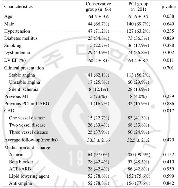
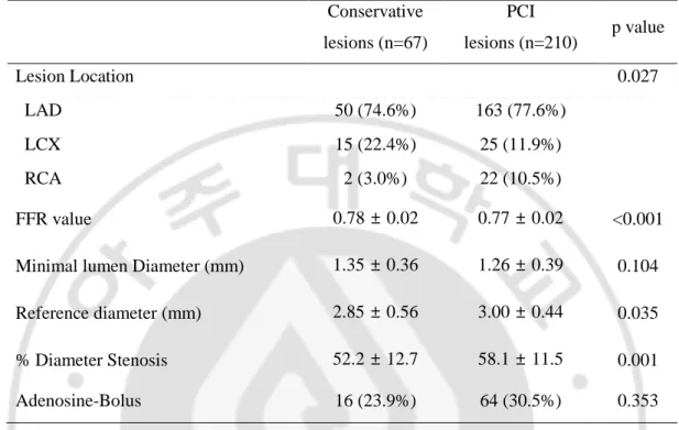
A total of 267patients and their 277 lesions were eligible and enrolled to the study.
Baseline characteristics of patients and lesions are shown in Table 1 and 2. Sixty-seven lesions from 66 patients were deferred from revascularization (Conservative group) and 210 lesions from 201 patients were treated with PCI (PCI group).
Patients in the conservative group were more likely to be older (64.5±9.6 years old vs. 61.6±9.7 years old, p=0.038), to have higher LV ejection fraction (63.4±8.2% vs. 60.2±8.0%, p=0.011) and to have more multi-vessel disease (77.3% vs. 58.7%, p=0.007) compared with those in the PCI group. There was inhomogeneous distribution of the target vessels (p=0.027) with more non-LAD lesions in the conservative group.
In patients treated with PCI, the mean FFR was lower (0.78±0.02 vs. 0.77±0.02, p<0.001) with more severe diameter stenosis by angiograph (58.1±11.5% vs. 52.2±12.7%, p=0.001) and reference diameter was bigger (3.00±0.44mm vs. 2.85±0.56, p=0.035) compared to the conservative group.
5
Table 1. Baseline clinical characteristics of the patients (n=267)
Characteristics Conservative group (n=66) PCI group (n=201) p value Age 64.5 ± 9.6 61.6 ± 9.7 0.038 Male 44 (66.7%) 140 (69.7%) 0.649 Hypertension 47 (71.2%) 127 (63.2%) 0.235 Diabetes mellitus 23 (34.8%) 73 (36.3%) 0.829 Smoking 15 (22.7%) 36 (17.9%) 0.388 Dyslipidemia 29 (43.9%) 74 (36.8%) 0.302 LV EF (%) 60.2 ± 8.0 63.4 ± 8.2 0.011 Clinical presentation 0.701 Stable angina 41 (62.1%) 113 (56.2%) Unstable angina 17 (25.8%) 60 (29.9%) Silent ischemia 8 (12.1%) 28 (13.9%) Previous MI 5 (7.6%) 8 (4.0%) 0.239 Previous PCI or CABG 11 (16.7%) 32 (15.9%) 0.886
CAD 0.017
One vessel disease 15 (22.7%) 83 (41.3%) Two vessel disease 26 (39.4%) 68 (33.8%) Three vessel disease 25 (37.9%) 50 (24.9%)
Average follow-up(months) 30.3 ± 21.6 32.5 ± 21.2 0.470 Medication at discharge
Aspirin 64 (97.0%) 200 (99.5%) 0.152 Beta blocker 28 (42.4%) 97 (48.5%) 0.410 ACEi/ARB 28 (42.4%) 86 (42.8%) 0.959 Lipid lowering agent 52 (78.8%) 152 (75.6%) 0.599 Anti-angina 52 (78.8%) 156 (77.6%) 0.842 Values are mean ± SD or n (%); PCI, percutaneous coronary intervention; LV, left
ventricular; EF, ejection fraction; MI, myocardial infarction; PCI, percutaneous coronary intervention; CABG, coronary artery bypass surgery; CAD, coronary artery disease; ACEi, angiotensin converting enzyme inhibitor; ARB, angiotensin receptor blocker
6
Table 2. Angiographic and procedural results
Conservative lesions (n=67) PCI lesions (n=210) p value Lesion Location 0.027 LAD 50 (74.6%) 163 (77.6%) LCX 15 (22.4%) 25 (11.9%) RCA 2 (3.0%) 22 (10.5%) FFR value 0.78 ± 0.02 0.77 ± 0.02 <0.001 Minimal lumen Diameter (mm) 1.35 ± 0.36 1.26 ± 0.39 0.104 Reference diameter (mm) 2.85 ± 0.56 3.00 ± 0.44 0.035 % Diameter Stenosis 52.2 ± 12.7 58.1 ± 11.5 0.001 Adenosine-Bolus 16 (23.9%) 64 (30.5%) 0.353 Values are mean ± SD or n (%); PCI, percutaneous coronary intervention; LAD, left anterior descending artery; LCX, left circumflex artery; RCA, right coronary artery; FFR, fractional flow reserve
7
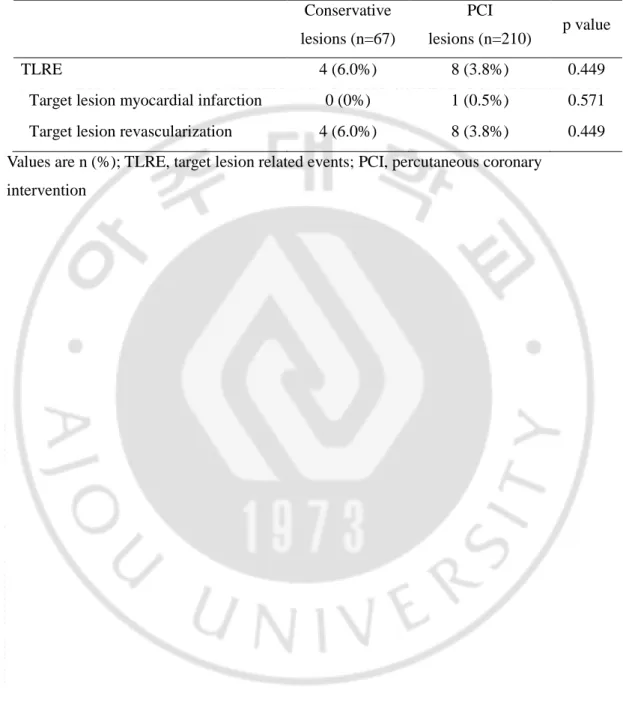
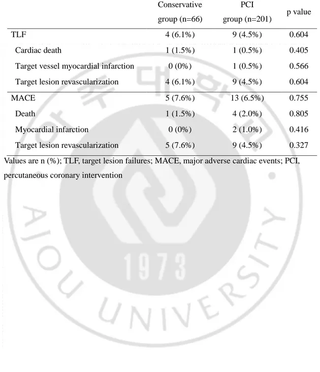
B. Clinical outcomes at 1 year
For the lesion specific outcome, at 1 year follow-up 4 cases of TLRE (4 cases of FFR-evaluated lesion revascularization and no FFR-FFR-evaluated lesion-related myocardial
infarction) from 4 patients occurred in the Conservative group. In the PCI group, eight cases of TLRE (8 cases of FFR-evaluated lesion revascularization and 1 case of FFR-evaluated lesion-related myocardial infarction) from 8 patients occurred (Table 3).
For the patient specific clinical outcome, four cases of TLF (1 case of cardiac death, 0 case of FFR-evaluated vessel myocardial infarction and 4 cases of FFR-evaluated lesion revascularization) from 4 patients occurred in the Conservative group and 9 cases of TLF (1 case of cardiac death, 1 case of FFR-evaluated vessel myocardial infarction and 9 cases of FFR-evaluated lesion revascularization) from 9 patients occurred in the PCI group (Table 4).
Five cases of MACE (1 case of death, 0 case of myocardial infarction and 5 cases of target lesion revascularization) occurred in the Conservative group and 13 cases of MACE (4 cases of death, 2 cases of myocardial infarction and 9 cases of target lesion
revascularization) occurred in the PCI group (Table 4).
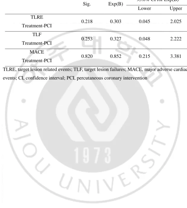
Using Cox proportional hazard model, there was no difference in lesion-related events between Conservative-group and PCI-group (hazard ratio 0.303, 95% CI 0.045-2.025, P = 0.218) (Table 5).
8
Table 3. Lesion specific outcomes as TLRE at 1 year
Conservative lesions (n=67)
PCI
lesions (n=210) p value TLRE 4 (6.0%) 8 (3.8%) 0.449
Target lesion myocardial infarction 0 (0%) 1 (0.5%) 0.571 Target lesion revascularization 4 (6.0%) 8 (3.8%) 0.449 Values are n (%); TLRE, target lesion related events; PCI, percutaneous coronary
9
Table 4. Patient specific outcomes as TLF and MACE at 1 year
Conservative group (n=66) PCI group (n=201) p value TLF 4 (6.1%) 9 (4.5%) 0.604 Cardiac death 1 (1.5%) 1 (0.5%) 0.405 Target vessel myocardial infarction 0 (0%) 1 (0.5%) 0.566 Target lesion revascularization 4 (6.1%) 9 (4.5%) 0.604 MACE 5 (7.6%) 13 (6.5%) 0.755 Death 1 (1.5%) 4 (2.0%) 0.805 Myocardial infarction 0 (0%) 2 (1.0%) 0.416 Target lesion revascularization 5 (7.6%) 9 (4.5%) 0.327 Values are n (%); TLF, target lesion failures; MACE, major adverse cardiac events; PCI, percutaneous coronary intervention
10
Table 5. Cox Proportional Hazard model for TLRE, TLF and MACE at 1 year
Sig. Exp(B) 95.0% CI for Exp(B) Lower Upper TLRE 0.218 0.303 0.045 2.025 Treatment-PCI TLF 0.253 0.327 0.048 2.222 Treatment-PCI MACE 0.820 0.852 0.215 3.381 Treatment-PCI
TLRE, target lesion related events; TLF, target lesion failures; MACE, major adverse cardiac events; CI, confidence interval; PCI, percutaneous coronary intervention
11
IV. Discussion
Because measuring FFR evaluates the target lesion functional severity, we intended to evaluate the outcome from that target lesion. Additionally we investigated the clinical outcome affected by the target lesion. So we classified the outcome results into 3 categories. TLRE was lesion specific outcome from the target lesion and MACE was patient specific outcome from the patients. TLF was middle level between TLRE and MACE. The result showed that the difference of outcome between the conservative group and the PCI group was not significant in all types of outcome categories.
The PCI group consisted of more severe stenotic lesions than the conservative group, but showed good trend of lesion events and clinical outcome. The incidence rate of lesion events and clinical outcomes were lower in the PCI group and Cox proportional hazard model revealed hazard ratio of 0.303 for PCI group. But these good trends were not significant.
There are 3 previous similar studies for the patients with gray zone FFR lesions (Lavi et al., 2007; Courtis et al., 2008; Lindstaedt et al., 2010). But, the results were not similar within those studies. One study concluded that deferral of revascularization in patients with gray zone FFR was associated with a higher rate of cardiac events after 1 year although those cardiac events consisted of revascularization (Courtis et al., 2008). The other 2 studies revealed similar findings with this study. The cardiac event rates were similar between the deferred group and the revascularization group (Lavi et al., 2007; Lindstaedt et al., 2010). Interestingly one study revealed that there was no difference in outcome between the Drug eluting stent (DES)-treated group and the deferred group. However, the Bare metal stent (BMS)-treated group showed poor outcome result (Lavi et al., 2007). So in this DES era, that study favor revascularization with DES in lower boundary in gray zone FFR. All 3 studies were not randomized study and not controlled to assign equally in two comparing group like this study.
We could consider several concerns about the result. First, for achievement of maximal hyperemia, there were 3 heterogeneous method of adenosine injection in this study; intravenous adenosine, intracoronary bolus and intracoronary continuous infusion.
12
Intracoronary adenosine has been adopted by many catheterization laboratories for achievement of maximal hyperemia (Jeremias et al., 2000). Compared with intravenous adenosine, the intracoronary route allows easier administration with fewer systemic adverse effects. But there were some concerns about the suboptimal hyperemia associated with intracoronary adenosine (Jeremias et al., 2000; Casella et al., 2004). But there are studies on higher dose of intracoronary adenosine bolus (Murtagh et al., 2003; Casella et al., 2004) or intracoronary continuous adenosine infusion by using microcatheter which could be useful and safe for inducing optimal hyperemia (Yoon et al., 2009). Due to the difference of FFR measuring protocol between the 4 centers, that could influence the FFR value. Second, the rate of ‘hard events’ such as cardiac death and myocardial infarction was very low
consistently in previous studies about the gray zone FFR lesion as well as this study (Lavi et al., 2007; Courtis et al., 2008; Lindstaedt et al., 2010). Because almost events consisted of revascularization due to angina symptoms in these studies, that suggested the conservative treatment for the gray zone FFR lesion would not make patients increased risk for serious condition. Due to the subjective aspect of symptom, revascularization rate could be variable according to patients group. Third, there was significant difference of baseline characteristics in age, FFR value, location and lesion severity between both groups. The PCI group
consisted of more severe stenotic lesions. Considering the good trend of events rate in the PCI group, if the baseline characteristics were similar between the groups, the PCI group would get a significant better event rate and hazard ratio.
From FAME-2 study (De Bruyne et al., 2012), patients in whom at least one stenotic lesion with FFR ≤0.80 were randomly assigned to FFR-guided PCI plus the best available medical therapy (PCI group) or the best available medical therapy alone (medical-therapy group). Recruitment was halted prematurely after enrollment of 1220 patients because of a significant between-group difference in the percentage of patients who had a primary endpoint event: 4.3% in the PCI group and 12.7% in the medical-therapy group (hazard ratio with PCI, 0.32; P<0.001). FFR-guided PCI plus the best available medical therapy, as compared with the best available medical therapy alone, decreased the need for urgent revascularization. Difference of primary outcome between the two groups was mainly due to the need for urgent revascularization in the medical-therapy group. And the ‘hard events’
13
such as myocardial infarction, cardiac death occurred similarly. Seeing that the mean FFR measurement in the medical-therapy group was 0.68, the lesions might be causing significant ischemia and therefore the urgent revascularization occurred more frequently during medical therapy.
In DEFER 5-year follow-up study with BMS stent (Pijls et al., 2007), in the lesion with FFR > 0.75, there were similar Cardiac death, AMI and symptom relief between the defer group and the PCI group after 5-year follow up (p=0.52). So the result showed that the lesions with FFR > 0.75 would be defer asymptomatically and safely. The lesions with FFR > 0.75 regardless of treatment type showed better clinical outcome compared to the lesions with FFR > 0.75 (p=0.03). And this study also concluded that the lesions at greatest risk of causing cardiac death or AMI are those that are functionally significant as identified by an FFR < 0.75. Again in this study, mean FFR in the defer group and the PCI group were 0.87 and these group consisted of mainly not-ischemic lesions.
Maybe the patients with gray zone FFR lesions from FAME1/2 and DEFER study would be good candidate for study and would be helpful to comprehend the prognosis of the gray FFR zone lesion.
Because this study was based on retrospective and multi-center registry analysis, it had several inherent limitations. At that time of coronary angiography, decision of whether or not to intervene the lesions with gray zone FFR measurement entirely depended on the operator. Enrolled patients were underwent coronary angiography by many operators in multi-center with various clinical techniques, devices and medication. Therefore, variety of symptoms of patients, angiographic finding and financial problem made the strategies very heterogeneous. We couldn’t standardize the reason. One year follow-up period was somewhat short to determine the difference in benefit of strategy, there were a few events from patients and lesions. If the duration was longer enough, the difference of events rate might be significant. And the various routes of adenosine injection could be another limitation influenced the FFR.
Previous 3 studies (Lavi et al., 2007; Courtis et al., 2008; Lindstaedt et al., 2010) regarding clinical outcome of lesions with gray zone FFR didn’t show a consistent result about
14
FFR is not the best decision. Nevertheless, large randomized control study may be necessary to investigate guidance of cutoff value or angiographic finding or image finding for
treatment strategy of lesions with gray zone FFR. It is possible that strategy for lesions with gray zone FFR should be made on a case-by-case basis according to lesion characteristics individually.
15
V. Conclusion
In coronary lesions with borderline FFR, revascularization did not show the better clinical outcome compared to medical treatment. Therefore, lesions with borderline FFR
measurement can be deferred from revascularization without an increased risk for target lesion-related events or MACE, especially for ‘hard events’. But, obviously some patients who have typical angina symptoms refractory to maximal antianginal medical treatment would be considered to take a revascularization.
16
REFERENCES
1. Abe M, Tomiyama H, Yoshida H, Doba N: Diastolic fractional flow reserve to assess the functional severity of moderate coronary artery stenoses: comparison with fractional flow reserve and coronary flow velocity reserve. Circulation 102: 2365-2370, 2000
2. Casella G, Leibig M, Schiele TM, Schrepf R, Seelig V, Stempfle HU, Erdin P, Rieber J, Konig A, Siebert U, Klauss V: Are high doses of intracoronary adenosine an alternative to standard intravenous adenosine for the assessment of fractional flow reserve? Am Heart J 148: 590-595, 2004
3. Chamuleau SA, Meuwissen M, van Eck-Smit BL, Koch KT, de Jong A, de Winter RJ, Schotborgh CE, Bax M, Verberne HJ, Tijssen JG, Piek JJ: Fractional flow reserve, absolute and relative coronary blood flow velocity reserve in relation to the results of technetium-99m sestamibi single-photon emission computed tomography in patients with two-vessel coronary artery disease. J Am Coll Cardiol 37: 1316-1322, 2001
4. Courtis J, Rodes-Cabau J, Larose E, Dery JP, Nguyen CM, Proulx G, Gleeton O, Roy L, Barbeau G, Noel B, DeLarochelliere R, Bertrand OF: Comparison of medical treatment and coronary revascularization in patients with moderate coronary lesions and borderline fractional flow reserve measurements. Catheter Cardiovasc Interv 71: 541-548, 2008
5. De Bruyne B, Pijls NH, Barbato E, Bartunek J, Bech JW, Wijns W, Heyndrickx GR: Intracoronary and intravenous adenosine 5'-triphosphate, adenosine, papaverine, and contrast medium to assess fractional flow reserve in humans. Circulation 107: 1877-1883, 2003
6. De Bruyne B, Pijls NH, Kalesan B, Barbato E, Tonino PA, Piroth Z, Jagic N, Mobius-Winkler S, Rioufol G, Witt N, Kala P, MacCarthy P, Engstrom T, Oldroyd KG, Mavromatis K, Manoharan G, Verlee P, Frobert O, Curzen N, Johnson JB, Juni
17
P, Fearon WF, Investigators FT: Fractional flow reserve-guided PCI versus medical therapy in stable coronary disease. N Engl J Med 367: 991-1001, 2012
7. Jeremias A, Whitbourn RJ, Filardo SD, Fitzgerald PJ, Cohen DJ, Tuzcu EM,
Anderson WD, Abizaid AA, Mintz GS, Yeung AC, Kern MJ, Yock PG: Adequacy of intracoronary versus intravenous adenosine-induced maximal coronary hyperemia for fractional flow reserve measurements. Am Heart J 140: 651-657, 2000
8. Kern MJ: Coronary physiology revisited : practical insights from the cardiac catheterization laboratory. Circulation 101: 1344-1351, 2000
9. Kern MJ, Lerman A, Bech JW, De Bruyne B, Eeckhout E, Fearon WF, Higano ST, Lim MJ, Meuwissen M, Piek JJ, Pijls NH, Siebes M, Spaan JA, American Heart Association Committee on D, Interventional Cardiac Catheterization CoCC: Physiological assessment of coronary artery disease in the cardiac catheterization laboratory: a scientific statement from the American Heart Association Committee on Diagnostic and Interventional Cardiac Catheterization, Council on Clinical Cardiology. Circulation 114: 1321-1341, 2006
10. Lavi S, Rihal CS, Yang EH, Fassa AA, Elesber A, Lennon RJ, Mathew V, David HR, Jr., Lerman A: The effect of drug eluting stents on cardiovascular events in patients with intermediate lesions and borderline fractional flow reserve. Catheter
Cardiovasc Interv 70: 525-531, 2007
11. Lindstaedt M, Halilcavusogullari Y, Yazar A, Holland-Letz T, Bojara W, Mugge A, Germing A: Clinical outcome following conservative vs revascularization therapy in patients with stable coronary artery disease and borderline fractional flow reserve measurements. Clin Cardiol 33: 77-83, 2010
12. Murtagh B, Higano S, Lennon R, Mathew V, Holmes DR, Jr., Lerman A: Role of incremental doses of intracoronary adenosine for fractional flow reserve assessment.
Am Heart J 146: 99-105, 2003
13. Pijls NH, De Bruyne B, Peels K, Van Der Voort PH, Bonnier HJ, Bartunek JKJJ, Koolen JJ: Measurement of fractional flow reserve to assess the functional severity of coronary-artery stenoses. N Engl J Med 334: 1703-1708, 1996
18
14. Pijls NH, Van Gelder B, Van der Voort P, Peels K, Bracke FA, Bonnier HJ, el Gamal MI: Fractional flow reserve. A useful index to evaluate the influence of an epicardial coronary stenosis on myocardial blood flow. Circulation 92: 3183-3193, 1995 15. Pijls NH, van Schaardenburgh P, Manoharan G, Boersma E, Bech JW, van't Veer M,
Bar F, Hoorntje J, Koolen J, Wijns W, de Bruyne B: Percutaneous coronary
intervention of functionally nonsignificant stenosis: 5-year follow-up of the DEFER Study. J Am Coll Cardiol 49: 2105-2111, 2007
16. Seo MK, Koo BK, Kim JH, Shin DH, Yang HM, Park KW, Lee HY, Kang HJ, Kim HS, Oh BH, Park YB: Comparison of hyperemic efficacy between central and peripheral venous adenosine infusion for fractional flow reserve measurement. Circ
Cardiovasc Interv 5: 401-405, 2012
17. Tonino PA, De Bruyne B, Pijls NH, Siebert U, Ikeno F, van' t Veer M, Klauss V, Manoharan G, Engstrom T, Oldroyd KG, Ver Lee PN, MacCarthy PA, Fearon WF, Investigators FS: Fractional flow reserve versus angiography for guiding
percutaneous coronary intervention. N Engl J Med 360: 213-224, 2009
18. Yoon MH, Tahk SJ, Yang HM, Park JS, Zheng M, Lim HS, Choi BJ, Choi SY, Choi UJ, Hwang JW, Kang SJ, Hwang GS, Shin JH: Comparison of the intracoronary continuous infusion method using a microcatheter and the intravenous continuous adenosine infusion method for inducing maximal hyperemia for fractional flow reserve measurement. Am Heart J 157: 1050-1056, 2009
19 - 국문요약 –
경계의 분획혈류 예비력을 가진 환자들에서 재관류술군과
보존적 치료군의 임상경과 비교
목적: 분획혈류 예비력 (FFR)을 재는 것은 심혈관의 협착 정도를 기능적으로 평가하고 치료 계획을 정하는 데에 도움이 되는 방법이다. 여러 연구를 통하여 FFR < 0.75 는 허혈의 유발에 특이적이라고 알려져 있으나 FFR 값이 0.75-0.80 인 병변에 재관류술을 할 것인지 하지 않을 것인지에 대해서는 아직 논란의 여지가 있다. 이 연구의 목적은 경계의 분획혈류 예비력을 가진 환자들에서 재관류술 군과 보존적 치료 군의 임상경과 비교하는 것이다. 방법: 이 연구는 한국의 4 개 병원에서 등록한 FFR 데이터를 이용하여 분석하였다. 총 267 명(평균 62 ± 10 세, 남성 69%)에서 FFR 값이 0.75 에서 0.80 사이인 277 개의 병변(평균 FFR 0.77±0.02 )을 분석하였다. 1 년 추적관찰에서의 주요 심장 사건과 FFR 측정 병변 관련 사건의 발생빈도를 분석하였다. 66 명 환자들의 67 개 병변들이 보존적 치료를 받았고 (보존적 치료군) 201 명 환자들의 210 개 병변들이 재관류술을 받았다 (재관류군). 결과: 1 년의 추적 기간 동안 보존적 치료군에서는 4 명의 환자에서 4 건의 FFR 측정 병변 관련 사건이 있었고 (4 건의 FFR 측정 병변 재관류술, 0 건의 FFR 측정 병변 심근경색), 재관류술군에서는 8 명의 환자에서 8 건의 FFR 측정 병변 관련 사건이 있었다 (8 건의 FFR 측정 병변 재관류술, 1 건의 FFR 측정 병변 심근경색). 또한, 보존적 치료군에서 5 건의 주요 심장 사건이 발생 (1 건의 사망, 0 건의 심근경색, 5 건의 FFR 측정 병변 재관류술) 하였고 보존적 치료군에서 13 건의 주요 심장 사건이 발생하였다 (4 건의 사망, 2 건의 심근경색, 9 건의 측정 병변 재관류술).20 Cox 비례위험모형을 이용하였을 때 FFR 측정 병변 관련 사건의 발생빈도는 재관류술군과 보존적 치료군 사이에 차이가 없었다 (위험도 0.303, 95% CI 0.045-2.025, P = 0.218). 결론: 경계의 분획혈류 예비력을 가진 병변 들에서 재관류술은 보존적 치료에 비해 좋은 임상경과를 보여주지 못하였다. 따라서 경계의 분획혈류 예비력을 가진 병변들은 FFR 측정 병변 관련 사건의 위험을 증가시키지 않으면서 재관류술을 미룰 수 있겠다. 핵심어: 분획혈류 예비력, 심혈관 협착질환, 재관류술, 보존적 약물치료