R E S E A R C H
Open Access
Effect of arthrocentesis on the clinical
outcome of various treatment methods for
temporomandibular joint disorders
Chang-Woo Kim, Sung-Jae Lee, Euy-Hyun Kim, Dong-Keon Lee, Mong-Hun Kang, In-Seok Song and Sang-Ho Jun
*Abstract
Background: We evaluated the improvement of pain and the increase in mouth opening after temporomandibular joint arthrocentesis and the possible association with various factors such as previous splint treatment, medication, and diagnosis.
Results: We studied 57 temporomandibular joint disorder patients who underwent arthrocentesis at Korea University Anam Hospital. These patients (24 males and 33 females, aged between 15 and 76 years) underwent arthrocentesis that was performed by one surgeon. The degree of mouth opening (assessed using the maximum mouth opening: MMO) and pain (assessed using the visual analog scale: VAS) were assessed pre- and post-arthrocentesis. The study also investigated whether treatment modalities other than arthrocentesis (medication and appliance therapy) were performed. Statistical analysis revealed that there was a significant difference in mouth opening and pain after temporomandibular joint arthrocentesis. Preoperative appliance therapy affected the results of arthrocentesis, but it was not statistically significant. With regard to pain relief, preoperative diagnosis did not show a significant difference. However, with regard to maximum mouth opening, patients with disc displacement without reduction with limited mouth opening (closed lock) showed the highest recovery (11.13 mm).
Conclusion: The average of MMO increase after arthrocentesis was 9.10 mm, and patients with disc displacement without reduction with locking (closed lock) showed most recovery in maximum mouth opening and it was statistically significant. The average pain relief of patients after arthrocentesis was 3.03 in the VAS scale, and patients using anterior repositioning splint (ARS) preoperatively showed the most pain relief.
Keywords: Temporomandibular joint arthrocentesis, Temporomandibular disorders Background
Temporomandibular joint (TMJ) disorder (TMD) is a term used to define disorders occurring in the mastica-tory muscles, TMJ, and surrounding tissues [1]. Regard-less of the tissue affected, TMDs show similar signs and symptoms such as pain around the ear and masticatory muscles [2]. The initial stage of TMD is characterized by normal maximum mouth opening with joint sounds, which gradually decreases with limitation of mouth opening. The advanced stage of TMD is characterized by dislocation of the articular disc and restricted sliding movements [3].
Patients with intra-articular TMDs present with vari-ous symptoms. An imbalance in the physiological rela-tionship between the articular disc and condylar head
causes various symptoms [4]. Intra-articular TMDs are
classified into six types according to the Research Diag-nostic Criteria for Temporomandibular Disorders (RDC/
TMD): “disc displacement with reduction”; “disc
dis-placement with reduction with intermittent locking”; “disc displacement without reduction with limited open-ing”; “disc displacement without reduction, without
lim-ited opening”; “degenerative joint disease”; and
“subluxation” [5]. Al-Khotani et al. [6] show that disc displacement with reduction is the most common TMJ problem.
TMJ arthrocentesis and lavage originated from the successful results of TMJ arthroscopy. Since the first
© The Author(s). 2019 Open Access This article is distributed under the terms of the Creative Commons Attribution 4.0 International License (http://creativecommons.org/licenses/by/4.0/), which permits unrestricted use, distribution, and reproduction in any medium, provided you give appropriate credit to the original author(s) and the source, provide a link to the Creative Commons license, and indicate if changes were made.
* Correspondence:[email protected]
Department of Oral and Maxillofacial Surgery, Korea University Anam Hospital, 73, Inchon-ro, Seongbuk-gu, Seoul 02841, Republic of Korea
publication on arthrocentesis by Nitzan and Dolwick in
1991 [7], TMJ arthrocentesis received widespread
ac-ceptance, as a minimally invasive surgical procedure for TMDs refractory to other conservative treatments. In order to increase mouth opening and reduce pain, arthrocentesis was performed by inserting two cathe-ters into the upper joint space under local anesthesia and irrigation with saline. This treatment proved to be very effective for disc displacement without
reduc-tion with limited mouth opening (closed lock) [8].
Compared with other surgical procedures, TMJ
arthrocentesis has been reported to be effective in re-ducing pain and increase in mouth opening with low incidence of complications [9]. Nitzan et al. reported that patients showed a significant increase in mouth opening (P < 0.001). Before the procedure, they showed a mean mouth opening of 24.1 ± 5.6 mm that increased to 42.7 ± 4 mm after arthrocentesis. The mean preoperative VAS (visual analog scale) score was 8.75 ± 2.82, within the scale of 0 to 15, and it decreased significantly (P < 0.001) to 2.31 ± 2.55 after the procedure [8].
Appliance therapy was revealed effective in TMD pa-tients with arthrogenous pain, with the purpose of
redu-cing inflammation and joint load [10]. Zhang et al.
reported in their systematic review that splint therapy can increase the MMO and reduce the frequency of pain in TMD patients [11]. Centric relation splint (CRS) and anterior repositioning splints (ARS) are the most com-mon splint used in TMD patients. CRS is the most widely accepted nonsurgical treatment of TMDs which can improve many of the clinical symptoms of TMDs
[12]. ARS can be used when the symptoms persist after
the CRS application. Many articles have reported the ef-ficacy of ARS for increase mouth opening in the disc displacement patients [4,13,14]. However, there is only scarce study about the relationship between splint ther-apy and arthrocentesis results and they only studied about one type of splint [15,16].
A muscle relaxant affects skeletal muscle function and decreases muscle tones. The decreased muscle tone can prevent the unnecessary forces that affect the TMJ, thus reducing the pain and giving stability to surrounding tis-sues. The muscle relaxants are believed to action by treating either spasticity secondary to upper motor neuron syndromes or muscular pain and spasms
sec-ondary to peripheral musculoskeletal conditions [17].
There are only few articles studied about the relation-ship between muscle relaxant and the outcome of TMJ arthrocentesis.
In this article, we evaluated the relationship between the decrease in pain and increase in mouth opening after TMJ arthrocentesis and the various factors such as pre-vious splint treatment, medication, and diagnosis.
Materials and methods Population
This study was conducted on patients who visited Korea University Anam Hospital between January 2016 and June 2018 and were diagnosed with degenerative joint disease and disc displacement with and without reduc-tion on radiographic and clinical evaluareduc-tion. TMJ arthro-centesis was performed by one surgeon, and mouth opening and the degree of pain before and after the op-eration were examined. We also investigated whether other treatments (e.g., device therapy) were performed before arthrocentesis. This study was approved by Korea University Anam Hospital in Seoul, South Korea: IRB
number 2018AN0427, and informed consent was
exempted because this study was a retrospective study. Inclusion and exclusion criteria
The sample was composed of individuals of both gen-ders older than 15 years. Patients with signs and symp-toms of intra-articular TMDs and degenerative joint disease and refractory to conservative treatment were in-cluded. They were diagnosed by the Research Diagnostic Criteria for Temporomandibular Disorders (RDC/TMD). Patients with previous surgery, hypoplasia and/or malig-nant neoplasm of the mandibular condyle, bone anky-losis, drug allergy, history of psychosomatic illness, or pregnant and lactating patients were excluded.
Procedure
The mouth opening and degree of pain were measured before TMJ lavage. The first step of TMJ arthrocentesis was extensive disinfection of the surgical site; a line was then drawn connecting the middle portion of the tragus and the outer canthus of the eye.
Articular fossa and eminence were marked at 10 mm and 20 mm respectively, in front of the tragus, along the canthal-tragus line, and vertically bellow at 2 mm and 10 mm respectively. Through palpation of the two refer-ence points, the condyle and articular eminrefer-ence were marked with methylene blue solution.
To reduce pain when inserting the needle, subcutane-ous infiltration anesthesia was performed with 0.5 ml of 2% lidocaine using a dental anesthesia syringe.
After opening the mouth of the patient, a 2-mL saline syringe was inserted with a 26G needle into the upper joint space, and saline was inserted with a gentle force. When 2 mL saline was aspirated again, it indicates that the needle is inserted into the proper place. The second needle was inserted in the same way in the same direc-tion. After the needle was inserted, irrigation was per-formed for 30 min with 500 mL saline. At this time, the fluid was located at 1.5 m above the patient who is in su-pine position.
Immediately after surgery, the surgeon injected hyalur-onic acid into the operative site of the patient, and the distance between the maxillary and mandibular central incisors was measured to determine the MMO. Pain was recorded using the VAS, which is a subjective assess-ment. Postoperative MMO and VAS was measured 4 days after the procedure when patients came to the clinic for follow-up. The splint therapy was continued after 4-day follow-up.
Splint therapy
CRS and ARS were used to some patients in this study. CRS was used to reduce the bite force and joint load. ARS was used in patients with disc displacement, pa-tients whose symptom persists after physical therapy or joint inflammation, and patients with severe inflamma-tion to reduce the occlusal load. Splints were used prior to arthrocentesis. The period and duration of the appli-ance use varied depending on each patient’s symptom. Patients with severe symptoms wore the splint for 24 h (less than 1 week), and when the symptoms subsided, they were told to wear the splint during nighttime only. Muscle relaxant
Muscle relaxant was prescribed to patients with muscle tenderness or pain. Thiosina Tab. (thiocolchicoside 4 mg, aescin 20 mg) was used to patients three times a day. The medicine was prescribed more than 7 days de-pending on the symptom of the patients. Muscle relax-ant was prescribed with painkiller (Somalgen Tab, talniflumate 370 mg) for the same days.
Statistical analysis
SPSS for Windows® (version 12.0, SPSS Inc., Chicago, IL, USA) was used for statistical analysis. To evaluate the result of arthrocentesis, pairedt test was conducted. To compare the result of arthrocentesis categorized by vari-ous factors,t test and ANOVA were used. P < 0.05 was considered statistically significant in this study.
Results
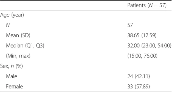
The 57 patients with TMD met the eligibility criteria and underwent arthrocentesis (24 male patients and 33 female patients). Of the 57 cases with TMJ lavage, 6 cases were performed on both sides and 51 cases were unilateral; 27 cases were of the right side and 24 were left.
The average age of the patients was 38.65 ± 17.59 years old (Table1). As a result of arthrocentesis, the preopera-tive pain score of 4.77 ± 1.98 decreased to 1.74 ± 1.89, and this difference was statistically significant (P < 0.05)
(Table 2). The MMO increased from 37.25 ±7.40 to
46.35 ± 7.39 after arthrocentesis, and this change was statistically significant (P < 0.05) (Table2). Patients were
sorted into two groups based on whether they under-went appliance therapy before arthrocentesis. Tunder-wenty- Twenty-five out of 57 patients underwent splint therapy before arthrocentesis. Patients who used TMJ splint before arthrocentesis showed slightly better improvement in MMO (9.76) and a better decrease in VAS score (3.42) than the other group (8.59, 2.73), but this change was not statistically significant (Tables3and4).
Patients treated with ARS showed most pain relief
(3.67 ± 2.50) after arthrocentesis (Table 3). However,
there was no statistical significance (P = 0.400). With re-gard to MMO, the patients who were treated by both ARS and CRS showed more improvement (10.53 ± 5.94) than other groups (treated without appliance and treated with ARS) (Table4). However, it was not statistically sig-nificant (P = 0.599). One patient was excluded in statis-tics because only one patient was treated with CRS.
Patients were diagnosed by the RDC/TMD criteria. Pa-tients were divided into the following three groups: disc displacement with locking group (RDC/TMD criteria; disc displacement without reduction with locking-closed lock), disc displacement without locking group (RDC/ TMD criteria; disc displacement with reduction, disc dis-placement with reduction with intermittent locking, disc displacement without reduction without locking), and degenerative joint disease group. There was no statisti-cally significant association between preoperative diag-nosis and pain relief (Table 3). However, patients with disc displacement without reduction with limited mouth opening (closed lock) showed the highest improvement in mouth opening (11.13 ± 6.73) and it was significantly Table 1 Baseline characteristics
Patients (N = 57) Age (year) N 57 Mean (SD) 38.65 (17.59) Median (Q1, Q3) 32.00 (23.00, 54.00) (Min, max) (15.00, 76.00) Sex,n (%) Male 24 (42.11) Female 33 (57.89)
Table 2 Result of arthrocentesis
N Pre Post P
value* Mean (SD) Mean (SD)
VAS 57 4.77 (1.98) 1.74 (1.89) < .0001 MMO 57 37.25 (7.40) 46.35 (7.39) < .0001
P < 0.05 was considered statistically significant
Pre preoperative, post postoperative, VAS visual analog scaled, MMO maximum mouth opening
higher (P < 0.05) than those with degenerative joint dis-ease and those with disc displacement without limited mouth opening (Table4).
Forty-six out of 57 patients took a muscle relaxant to reduce the TMJ symptoms before arthrocentesis (Tables
3and4). These patients showed slightly better improve-ments in MMO and VAS scores, but it was not signifi-cant (Tables3and4).
Patients were also classified by the locking period. Locking was defined as subjective mouth opening limita-tion that patients say. To evaluate the efficacy of arthro-centesis on acute and chronic locking state, patients were divided into two groups depending on the locking period. Twenty-three out of 57 patients suffered from
TMJ locking for more than 6 months (Tables 3 and 4).
Patients with TMJ locking more than 6 months showed better results after arthrocentesis. However, this was not statistically significant when compared with the patients with TMJ locking period less than 6 months.
Discussion
TMDs originate from the joint itself or arise due to any
pathology of the muscles [18]. The TMJ can adjust to
the function of the jaws continuously by remodeling
[19]. However, when the functional load exceeds the
regenerative capacity of the joints, the remodeling be-comes insufficient and results in structural change. These changes cause deformities in the TMJ, resulting in clinical symptoms.
Arthrocentesis has been demonstrated to be effective
in treating disc displacement without reduction [20].
Arthrocentesis and lavage have been suggested to be useful for the management of other TMDs. Murakami et al. [21] reported that arthrocentesis showed favorable results in mitigating the symptoms of the advanced stage of internal derangement. It should be pointed out that arthrocentesis is effective in treating
symp-tomatic TMDs [9]. Patients with intra-articular TMDs
were divided into three groups in this study. Patients with disc displacement were sorted by whether they have limited mouth opening (MMO < 40) or not. Pa-tients with degenerative joint disease (DJD) were the third group. Late-stage DJD with acquired mandibular retrognathia requires surgical management; however, early-stage DJD without significant bony changes should be treated with nonsurgical methods or min-imally invasive surgical methods, such as arthrocent-esis and arthroscopy [22]. In this study, arthrocentesis was effective in the early stage of DJD patient in im-proving range of MMO and reducing pain just as the Table 3 Improvement of VAS score due to various factor
VAS
N Pre Post P value* Difference†of VAS P value
Mean (SD) Mean (SD) Mean (SD)
Appliance treatment 0.1768a No splint 32 4.48 (1.94) 1.75 (1.92) < .0001 − 2.73 (1.88) Splint tx 25 5.14 (2.02) 1.72 (1.88) < .0001 − 3.42 (1.87) Type of appliance tx 0.4002b No splint 32 4.48 (1.94) 1.75 (1.92) < .0001 − 2.73 (1.88) ARS 9 5.00 (2.87) 1.33 (2.18) 0.0023 − 3.67 (2.50) ARS and CRS 15 5.23 (1.50) 2.07 (1.71) < .0001 − 3.17 (1.46) Dx 0.9568b DD with locking 32 4.81 (2.30) 1.84 (1.92) < .0001 − 2.97 (1.84) DD without locking 15 4.80 (1.74) 1.67 (1.72) < .0001 − 3.13 (2.07) DJD 10 4.60 (1.26) 1.50 (2.17) 0.0008 − 3.10 (1.97) Muscle relaxants 0.6760a No 11 5.00 (2.41) 2.18 (1.78) 0.0004 − 2.82 (1.78) Yes 46 4.72 (1.90) 1.63 (1.91) < .0001 − 3.09 (1.93) Locking period 0.0697a < 6 months 34 4.46 (2.24) 1.79 (1.97) < .0001 − 2.66 (1.96) ≥ 6 months 23 5.24 (1.44) 1.65 (1.80) < .0001 − 3.59 (1.68)
P < 0.05 considered statistically significant
DD disc displacement, DJD degenerative joint disease, ARS anterior repositioning splint, CRS centric relation splint *P value calculated by paired t test
†Difference = post− pre a
P value calculated by t-test
b
formal study by Nitzan et al. [9]. When comparing the re-sult of arthrocentesis in the three groups, the disc dis-placement with locking group showed significant MMO recovery than the other two groups (Table4). The mech-anism of MMO recovery in closed lock patients is uncer-tain, because the cause of the limited motion and pathology is still unknown [23]. The relieve of negative pressure on the disc, adhesion, and surface friction might be the reason for the significant MMO recovery of the closed lock group.
TMJ splints such as ARS and CRS are most widely used conservative treatment that relieve the joint and
muscle pain based on TMD education [14]. It was
dem-onstrated that stabilization splints reduce the number of painful muscles and the degree of the pain after short use [24]. ARS was used for patients with anterior disc displacement and patients with onset of limited ability to mouth opening. To reduce the occlusal instability, ARS was used for only a short time and the use of stabilization splint was followed. CRS was used for pa-tients with muscle hyperactivity and parafunctional ac-tivity in this study. There was no significant additional pain relief and MMO recovery by applying the occlusal splint before arthrocentesis, as shown in Tables3 and4.
The result does not concur with the formal study that concluded arthrocentesis is more effective when used in conjunction with splint therapy [18, 25]. In this study, patients underwent appliance treatment in various pe-riods and duration. Also, only short-term results were analyzed. In a long-term study by Lee et al, simultaneous wearing of splint after arthrocentesis showed a better re-sult than preoperative splint treatment [16]. So further long-term study is needed for determining the relation-ship between the splint use and arthrocentesis.
There were studies about arthrocentesis that pre-scribed muscle relaxant after the procedure [26, 27]. However, there were scarce studies comparing patients with or without muscle relaxant. The results of arthro-centesis with or without muscle relaxant were statisti-cally analyzed in our study. There was no statistical difference between the two groups in short-term evaluation.
Locking period was one of the factors that were ana-lyzed in this study. The result was that patient with lock-ing more than 6 months showed slightly better results, but it was not statistically significant (Tables 3 and 4). However, the study was conducted in a short period to evaluate the relationship between the efficacy of Table 4 Improvement in MMO due to various factor
MMO
N Pre Post P value* Difference†of MMO P value
Mean (SD) Mean (SD) Mean (SD)
Appliance treatment 0.4792a No splint 32 36.84 (8.33) 45.44 (8.64) < .0001 8.59 (6.61) Splint tx 25 37.76 (6.12) 47.52 (5.33) < .0001 9.76 (5.46) Type of appliance tx 0.5993b No splint 32 36.84 (8.33) 45.44 (8.64) < .0001 8.59 (6.61) ARS 9 38.11 (5.58) 46.89 (5.80) 0.0007 8.78 (4.94) ARS and CRS 15 37.27 (6.69) 47.80 (5.39) < .0001 10.53 (5.94) Dx 0.0156b DD with locking 32 33.38 (6.97) 44.50 (8.14) < .0001 11.13 (6.73) DD without locking 15 42.40 (4.40) 48.80 (5.56) < .0001 6.40 (4.27) DJD 10 41.90 (4.61) 48.60 (5.99) 0.0003 6.70 (3.80) Muscle relaxants 0.6190a No 11 38.09 (6.47) 46.36 (5.18) 0.0005 8.27 (5.37) Yes 46 37.04 (7.65) 46.35 (7.87) < .0001 9.30 (6.31) Locking period 0.6441a < 6 months 34 37.26 (8.06) 46.06 (8.37) 0.0005 8.79 (5.80) ≥ 6 months 23 37.22 (6.46) 46.78 (5.78) < .0001 9.57 (6.64)
P < 0.05 was considered statistically significant
DD disc displacement, DJD degenerative joint disease, ARS anterior repositioning splint, CRS centric relation splint *p value calculated by paired t-test
†Difference = post− pre a
P value calculated by t test
b
arthrocentesis and locking period. Thus, further study is needed to evaluate the relationship between locking period and TMJ arthrocentesis.
We investigated the MMO to evaluate the function of the TMJ. However, this study lacks information about the lateral movements of the jaws and TMJ. Hence, fur-ther study is needed to evaluate the improvement in TMJ function after arthrocentesis. The study was con-ducted to short-term follow-up, so it could not evaluate the long-term results.
In this study, there were no complications such as fa-cial nerve injury, pre-auricular hematoma, superfifa-cial temporal artery injury, and needle breakage in the joint [28] after TMJ arthrocentesis.
Conclusion
The average MMO increase after arthrocentesis was 9.10 mm, and patients with disc displacement without reduction with locking (closed lock) showed most recov-ery in MMO and showed statistically significant results. Among other factors, patients who used ARS and CRS preoperatively showed the second-best results in MMO.
The average pain relief of patients after arthrocentesis was 3.03 in VAS scale, and patients using ARS preopera-tively showed the most pain relief.
Abbreviations
MMO:Maximum mouth opening; TMDs: Temporomandibular disorders; TMJ: Temporomandibular joint; VAS: Visual analog scale
Acknowledgements
This work was supported by the Basic Science Research Program through the National Research Foundation funded by the Ministry of Education (NRF-2017R1D1A1B03028418).
Authors’ contributions
CWK is the first author. SHJ is the corresponding author. Each author took part in the design of the study, the clinical data collection, and writing of the manuscript, and all agreed with the accuracy of the content of the paper. This work has not been published elsewhere in any form and any language. All authors read and approved the final manuscript.
Funding
The authors declare that they have no funding.
Availability of data and materials Please contact the author for data requests.
Ethics approval and consent to participate
Approved (Korea University Anam Hospital in Seoul, South Korea: IRB number 2018AN0427).
Consent for publication
All participants included in the paper agreed with providing information and publication of papers.
Competing interests
The authors declare that they have no competing interests.
Received: 25 June 2019 Accepted: 9 September 2019
References
1. Monje-Gil F, Nitzan D, Gonzalez-Garcia R (2012) Temporomandibular joint arthrocentesis. Review of the literature. Med Oral Patol Oral Cir Bucal 17: e575–e581
2. Larheim TA, Hol C, Ottersen MK, Mork-Knutsen BB, Arvidsson LZ (2018) The role of imaging in the diagnosis of temporomandibular joint pathology. Oral Maxillofac Surg Clin North Am.https://doi.org/10.1016/j.coms.2018.04. 001
3. Wilkes CH (1989) Internal derangements of the temporomandibular joint. Pathological variations. Arch Otolaryngol Head Neck Surg 115:469–477 4. Pihut M, Gorecka M, Ceranowicz P, Wieckiewicz M (2018) The efficiency of
anterior repositioning splints in the management of pain related to temporomandibular joint disc displacement with reduction. Pain Res Manag 2018:9089286
5. Schiffman E, Ohrbach R, Truelove E et al (2014) Diagnostic Criteria for Temporomandibular Disorders (DC/TMD) for clinical and research applications: recommendations of the International RDC/TMD Consortium Network* and Orofacial Pain Special Interest Group†. J Oral Facial Pain Headache 28:6–27
6. Al-Khotani A, Naimi-Akbar A, Albadawi E, Ernberg M, Hedenberg-Magnusson B, Christidis N (2016) Prevalence of diagnosed temporomandibular disorders among Saudi Arabian children and adolescents. J Headache Pain 17:41 7. Nitzan DW, Dolwick MF (1991) An alternative explanation for the genesis of
closed-lock symptoms in the internal derangement process. J Oral Maxillofac Surg 49:810–815 discussion 815-816
8. Nitzan DW, Dolwick MF, Martinez GA (1991) Temporomandibular joint arthrocentesis: a simplified treatment for severe, limited mouth opening. J Oral Maxillofac Surg 49:1163–1167 discussion 1168-1170
9. Nitzan DW, Price A (2001) The use of arthrocentesis for the treatment of osteoarthritic temporomandibular joints. J Oral Maxillofac Surg 59:1154– 1159 discussion 1160
10. Greene CS, Menchel HF (2018) The use of oral appliances in the management of temporomandibular disorders. Oral Maxillofac Surg Clin North Am 30:265
11. Zhang C, Wu JY, Deng DL et al (2016) Efficacy of splint therapy for the management of temporomandibular disorders: a meta-analysis. Oncotarget 7:84043–84053
12. Al-Ani Z, Gray RJ, Davies SJ, Sloan P, Glenny AM (2005) Stabilization splint therapy for the treatment of temporomandibular myofascial pain: a systematic review. J Dent Educ 69:1242–1250
13. Okeson JP (1988) Long-term treatment of disk-interference disorders of the temporomandibular joint with anterior repositioning occlusal splints. J Prosthet Dent 60:611–616
14. Carraro JJ, Caffesse RG (1978) Effect of occlusal splints on TMJ symptomatology. J Prosthet Dent 40:563–566
15. Alpaslan C, Kahraman S, Guner B, Cula S (2008) Does the use of soft or hard splints affect the short-term outcome of temporomandibular joint arthrocentesis? Int J Oral Maxillofac Surg 37:424–427
16. Lee HS, Baek HS, Song DS et al (2013) Effect of simultaneous therapy of arthrocentesis and occlusal splints on temporomandibular disorders: anterior disc displacement without reduction. J Korean Assoc Oral Maxillofac Surg 39:14–20
17. Chou R, Peterson K, Helfand M (2004) Comparative efficacy and safety of skeletal muscle relaxants for spasticity and musculoskeletal conditions: a systematic review. J Pain Symptom Manage 28:140–175
18. Bilici IS, Emes Y, Aybar B, Yalcin S (2018) Evaluation of the effects of occlusal splint, trigger point injection and arthrocentesis in the treatment of internal derangement patients with myofascial pain disorders. J Craniomaxillofac Surg 46:916–922
19. Milam SB, Schmitz JP (1995) Molecular biology of temporomandibular joint disorders: proposed mechanisms of disease. J Oral Maxillofac Surg 53:1448– 1454
20. Okeson JP, Hayes DK (1986) Long-term results of treatment for
temporomandibular disorders: an evaluation by patients. J Am Dent Assoc 112:473–478
21. Murakami K, Moriya Y, Goto K, Segami N (1996) Four-year follow-up study of temporomandibular joint arthroscopic surgery for advanced stage internal derangements. J Oral Maxillofac Surg 54:285–290 discussion 291
22. Mercuri LG (2008) Osteoarthritis, osteoarthrosis, and idiopathic condylar resorption. Oral Maxillofac Surg Clin North Am 20:169–183 v-vi 23. Frost DE, Kendell BD (1999) The use of arthrocentesis for treatment of
temporomandibular joint disorders. Journal of Oral and Maxillofacial Surgery 57:583–587
24. Turk DC, Zaki HS, Rudy TE (1993) Effects of intraoral appliance and biofeedback/stress management alone and in combination in treating pain and depression in patients with temporomandibular disorders. J Prosthet Dent 70:158–164
25. Tvrdy P, Heinz P, Zapletalova J, Pink R, Michl P (2015) Effect of combination therapy of arthrocentesis and occlusal splint on nonreducing
temporomandibular joint disk displacement. Biomed Pap Med Fac Univ Palacky Olomouc Czech Repub 159:677–680
26. De Riu G, Stimolo M, Meloni SM et al (2013) Arthrocentesis and temporomandibular joint disorders: clinical and radiological results of a prospective study. Int J Dent 2013:790648
27. Thomas H, Neelakantan RS, Thomas TK (2012) Role of arthrocentesis in the management of acute closed lock of TM joint: a pilot study. J Maxillofac Oral Surg 11:390–393
28. Vaira LA, Raho MT, Soma D et al (2018) Complications and post-operative sequelae of temporomandibular joint arthrocentesis. Cranio 36:264–267
Publisher’s Note
Springer Nature remains neutral with regard to jurisdictional claims in published maps and institutional affiliations.