Original Paper
Cell Physiol Biochem 2010;25:233-240 Accepted: October 09, 2009
Cellular Physiology
Cellular Physiology
Cellular Physiology
Cellular Physiology
Cellular Physiology
and Biochemistr
and Biochemistr
and Biochemistr
and Biochemistr
and Biochemistryyyyy
Copyright © 2010 S. Karger AG, Basel
Superoxide Generated by
Lysophosphatidyl-choline Induces Endothelial Nitric Oxide Synthase
Downregulation in Human Endothelial Cells
Shinkyu Choi
1,*, Seonghee Park*, Guo Hua Liang, Ji Aee Kim and
Suk Hyo Suh
Department of Physiology and Medical Research Center, School of Medicine, Ewha Womans University, Seoul, 1Current address: Department of Microbiology and Immunology, Innovative Research Institute for Cell Therapy, Medical Research Center, Seoul National University Hospital, Seoul, *S Choi and S Park contributed equally to this publication and therefore share the first authorship
Key Words
Superoxide • eNOS • Endothelial cell • Lysophosphatidylcholine • Vascular dysfunction
Abstract
We examined the mechanism through which lysophosphatidylcholine (LPC) induces endothelial nitric oxide (eNOS) downregulation. Human umbilical vein endothelial cells (HUVECs) were treated with LPC (50-150 µM) for 0.5-2 h or the reactive oxygen spe-cies (ROS) donors, xanthine/xanthine oxidase (X/XO), 1,4-hydroquinone (HQ) or tert-butylhydroperoxide (TBHP) for 2 h. Protein levels of eNOS, superoxide dismutase1 (SOD1), catalase, and phospho-extracel-lular signal regulated kinase 1/2 (pERK 1/2) were assessed using immunoblotting. LPC treatment re-duced SOD1 levels but increased catalase levels. The superoxide donors X/XO and HQ showed similar ef-fects. The hydroperoxide donor TBHP increased SOD1 levels but did not change catalase levels. LPC concentration- and time-dependently decreased eNOS levels, but this effect was blocked by antioxi-dants and SOD and potentiated by the SOD1 inhibi-tor, ammonium tetrathiomolybdate. LPC and X/XO inhibited ERK1/2 phosphorylation, whereas TBHP stimulated phosphorylation. Taken together, these
data indicate that LPC induces superoxide overload in HUVECs via SOD1 inhibition and downregulates phospho-ERK1/2 and eNOS levels.
Introduction
Endothelial dysfunction refers to the perturbation of
anticoagulant, anti-inflammatory properties, and impaired
vascular reorganization against several
pathophysiologi-cal conditions [1]. Endothelial dysfunction is the primary
step in the initiation and development of atherosclerosis
and promotes the ingress of lipoproteins from plasma into
the intima [2, 3]. The endothelium, which is the lining
between tissues and the blood, responds to external
fac-tors such as shear stress and blood-borne signals to
main-tain homeostasis. One diffusible regulator, nitric oxide
(NO), an endothelium-derived relaxing factor, is
benefi-cial under conditions of vessel impairment or platelet and
leukocyte adhesion [4]. It is produced by NO synthase
(NOS) as a by-product of the biochemical conversion of
the amino acid L-arginine to L-citrulline. Since NO
func-tions as quencher of reactive oxygen species (ROS),
endothelial NO synthase (eNOS) could help reduce
oxidative status caused by risk factors such as
hyper-cholesterolemia.
ROS are pleiotropic factors that play important roles
in a wide variety of cellular events, but excess
produc-tion causes cell damage through lipid peroxidaproduc-tion and
the disruption of structural proteins, enzymes, or DNA.
Low-density lipoprotein (LDL) and its oxidative
modifi-cation (ox-LDL) cause extreme oxidative stress in
en-dothelial cells, which affects cell signaling, differentiation
and apoptosis [5, 6]. Lysophosphatidylcholine (LPC) is
an atherogenic phospholipid generated during LDL
oxi-dation [7]. LPC leads to ROS-induced endothelial
dys-function via impairing endothelium-dependent
vasorelaxation caused by loss of NO [8, 9]. Although
LPC modulates NO production by upregulating eNOS in
endothelial cells [10], the regulatory mechanism is
un-clear.
In the present study, we investigated the
mecha-nism by which LPC-induced oxidative injury impacts
eNOS regulation in human endothelial cells. Our results
suggest that the loss of superoxide dismutase1 (SOD1)
caused by LPC increases the production of intracellular
superoxide and suppresses phospho-extracellular
signal-regulated protein kinase 1/2 (pERK1/2) to downregulate
eNOS.
Materials and Methods
Materials
N-acetyl-L-cysteine (NAC), Tempol, Tiron, 1,4-hydroquinone (HQ), tert-butyl hydroperoxide (TBHP), ammo-nium tetrathiomolybdate (TM), SOD, catalase, dihydroethidine (DHE), 3-(4,5-dimethylthiazol-2-yl)-2,5-diphenyltetrazolium bromide (MTT) and L-α-lysophosphatidyl choline (LPC) from egg yolk were purchased from Sigma (St. Louis, MO); 4',6-Diamidino-2-phenylindole dihydrochloride (DAPI) and 2’,7’-dichlorofluorescin diacetate (DCFH-DA) were from Molecular Probes (Eugene, OR), xanthine and xanthine oxidase (X/XO) were from Calbiochem. Mouse anti-eNOS monoclonal antibody was obtained from BD Bioscience (Rockville, MD). Rabbit anti-superoxide dismutase 1, rabbit anti-catalase, goat anti-extra-cellular signal-regulated protein kinase (ERK), and mouse anti-phospho-ERK (pERK) were purchased from Santa Cruz Biotechnology (Santa Cruz, CA).
Mammalian Cell Cultures
Human umbilical vein endothelial cells (HUVECs, CRL-1730) were purchased from the American Type Culture Collection (Manassas, VA) and cultured as a monolayer in Medium 199 (M199, Hyclone, Logan, UT) supplemented with 10% fetal bovine serum (FBS), 100 units/ml penicillin, 100 µg/ ml streptomycin, and 15 µg/ml endothelial cell growth supplement, or Eagle basal medium 2 (EBM2, Clonetics, Walkersville, MD) supplemented with 2% FBS, 0.1% vascular
endothelial growth factor, 0.1% ascorbic acid, 0.1% gentamycin sulfate amphotericin-B, 0.04% hydrocortisone, and 0.1% heparin. All cells were maintained at 37 °C in humidified condi-tions under 5% CO2. Media were changed twice weekly, and cultures were split at 1:5 weekly. For experiments, HUVECs were plated in 60 mm or 100 mm plates and equilibrated with 0.3% serum containing media for 16 h. The medium was then removed and replaced with fresh medium, and the cells were maintained for the time periods indicated.
Measurement of Intracellular ROS
ROS in HUVECs were determined using DHE or DCFH-DA as described previously [11]. Cells were seeded at 2×10⁴ cells per well in 96-well plates with EBM2 media one day
before the experiment. Cells were pre-treated with various agents in EBM2 media for 30 min followed by treatmemt with LPC in EBM2 media for 120 min. Cells were then loaded with DCFH-DA in Hank’s balanced salt solution for 20 min in the dark, washed twice with phosphate-buffered saline (PBS) to eliminate contamination of diffused ROS, and treated with vehicle alone or the indicated compounds for 30 min and then stimulated with LPC for 2 h. Fluorescence was directly read at an emission wavelength of 538 nm after excitation at 485 nm. Cell numbers were normalized to detect maximum fluorescence increases in wells.
Western Blotting
HUVECs were pre-treated with various agents in 10% FBS containing media for 30 min followed by stimulation with LPC for 120 min. After drug incubation, cells were washed once in ice-cold PBS and lysed in protein extraction buffer containing a protease inhibitor cocktail. Protein concentrations in the supernatant were determined by the Bradford protein assay. For Western blot analysis, 30 µg of protein was sub-jected to SDS-PAGE gels (7.5 - 12%), and proteins were then transferred to a nitrocellulose membrane. Membranes were blocked for 1 h with TBST (10 mM Tris-HCl, 150 mM NaCl, and 1 v/v% Tween 20, pH 7.6) containing 5% bovine serum albumin at room temperature. The blots were incubated for 3 h with primary antibody followed by incubation with horseradish peroxidase-conjugated secondary antibodies for 1 h. Bands were visualized by chemiluminescence. Data collection and processing were performed using a luminescent image analyzer LAS-3000 and IMAGE GAUGE software (Fuji Film, Japan).
RT-PCR
The expression of eNOS mRNA was analyzed by the RNeasy mini kit (Qiagen, GmbH) for RNA isolation and OneStep RT-PCR kit (Qiagen, GmbH) for RT-PCR. For blocking DNA contamination in isolated RNA, DNaseI was treated before the final washing step. RNA (1 µg) was subjected to RT-PCR for 28 cycles. Primers for human eNOS were 5´-ACC CTC ACC GCT ACA ACA TC-3´ (sense) and 5´-GCT CAT TCT CCA GGT GCT TC-3´ (antisense). eNOS mRNA expression is normalized to the house-keeping gene human, GAPDH [5-GAG TCA ACG GAT TTG GTC GT-3´ (sense) and 5´-TTG ATT TTG GAG GGA TCT CG-3´(antisense)].
Nuclear Extraction
Cells were washed twice in ice-cold phosphate-buffered saline and suspended in ice-cold lysis buffer (10 mM HEPES, pH 7.9, 1.5 mM MgCl2, 10 mM KCl, 0.5 mM dithiothreitol, 0.1% Nonidet P-40, 1 mM phenylmethylsulfonyl fluoride, and 1 µg/ ml each of pepstatin A, leupeptin, and aprotinin), and incu-bated on ice for 30 min. To prepare nuclear extracts, lysed cells were centrifuged at 8,000 rpm for 5 min at 4 °C, and the pellets were resuspended in a buffer containing 20 mM HEPES, pH 7.9, 25% glycerol, 0.42 M KCl, 1.5 mM MgCl2, 0.2 mM EDTA, 0.5 mM dithiothreitol, 0.1 mM β-glycerophosphate, 0.05 mM vanadate, and the protease inhibitor mixture. After extraction on ice for 30 min, samples were centrifuged at 14,000 rpm for 30 min at 4 °C. Supernatant, containing nuclear proteins, was trans-ferred to a microcentrifuge tube, an aliquot was removed for protein determination, and samples were stored at -20 °C.
Electrophoretic Mobility Shift Assay (EMSA)
EMSAs were performed using a double-stranded oligo-nucleotide (5'-CCTGTGCTCCGGGAATTTCCCTGGCC-3') containing the NF-κB binding motif. For the binding reaction, nuclear protein extract (10 µg) was incubated in a total volume of 20 µl in binding buffer containing 10 mM HEPES, pH 7.5, 5% glycerol, 50 mM KCl, 1 mM dithiothreitol, 1 µg of poly(dI-dC), and radiolabeled (about 30,000 cpm) DNA for 30 min at room temperature. DNA-protein complexes were resolved in a pre-electrophoresed 6% nondenaturing polyacrylamide gel at 4 °C. Subsequently, the gel was dried under vacuum and exposed to film.
Flow Cytometry
HUVECs (106) were treated with LPC for 2 h. To evaluate
apoptotic cell death, cells were stained with Annexin V-FITC
(BD Bioscience, Rockville, MD) in Annexin-V staining buffer for 15 min at room temperature and counterstained with propidium iodide. Data were analyzed with a FACSCalibur flow cytometer (Becton Dickinson, San Jose, CA).
MTT assay
HUVEC cells were seeded onto 96-well plates (2 × 10⁴
cells/well) and stimulated with LPC in 2% or 10% FBS media for 120 min. MTT (0.1 mg) in phosphate-buffered saline was then added to each well, and the cells were incubated for an addi-tional 4 h at 37 °C. After removing the medium, the cells were lysed with dimethyl sulfoxide to dissolve the formazan prod-uct. The absorbance of reduced MTT at 590 nm was measured with a plate reader.
Statistical analysis
Data are expressed as means ± S.E. Statistical analysis was performed using Student’s t-test and ANOVA as appropri-ate. P<0.05 was considered statistically significant.
Results
LPC-induced ROS involve in the downregulation
of eNOS on HUVECs
To examine which kinds of cell injury were induced
by LPC treatment, we performed MTT assay and FACS
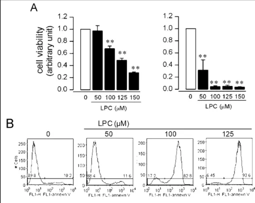
analysis. LPC was concentration-dependently cytotoxic
in 10% FBS media and concentration-independently in
2% FBS media in the MTT assay (Fig. 1A). Because
the physiological concentration of LPC in body fluids is
Fig. 1. HUVEC death induced by LPC.
(A) Cell viability was determined by MTT assay after 2 h stimulation with LPC in M199 containing 10% (left panel) or 2% (right panel) serum. Data are shown as means ± S.E. from three independent experiments. Asterisks indicate significant differences from vehicle treated cells (P < 0.01). (B) Af-ter LPC treatment in M199 containing 10% serum for 2 h, cells were analyzed by flow cytometry. The numbers show the percentages of annexin V-positive cells. Data are representative of three experiments.
between 5 and 180 µM [12, 13] and low serum itself can
damage cells, we used 10% FBS media for subsequent
experiments. FACS results showed that LPC
concentra-tion-dependently induced apoptotic changes in HUVECs
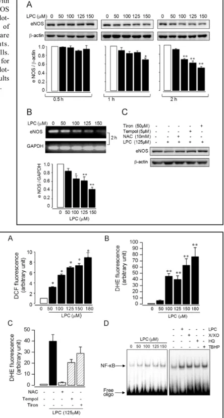
(Fig. 1B). LPC time-dependently decreased protein
lev-els of eNOS (Fig. 2A) when treated for 1 h at 150 µM or
2 h at ≥100 µM. In addition, LPC decreased eNOS mRNA
Fig. 2. Effects of LPC on eNOS. After stimulation with
vehicle or LPC for 0.5, 1, or 2 h, expression of eNOS protein (A) or mRNA (B) was measured by western blot-ting or RT-PCR analysis. Results are representative of three independent experiments. The data shown are means ± S.E. of three independent experiments. ** P < 0.01, * P < 0.05 versus vehicle-treated cells. (C) HUVECs were treated with NAC, Tempol or Tiron for 0.5 h prior to stimulation with LPC for 2 h. Western blot-ting was performed with an anti-eNOS antibody. Results are representative of three independent experiments.
levels in RT-PCR analysis (Fig. 2B). The antioxidants,
NAC, Tempol, and Tiron could block the effects of LPC
(Fig. 2C), implying a ROS-mediated mechanism. Indeed,
LPC increased levels of total ROS (Fig. 3A) as well as
superoxide (Fig. 3B), consistent with previous reports
[14], and these increases could be blocked with NAC,
Tempol, or Tiron (Fig. 3C). LPC (50 – 150 µM) also
in-Fig. 3. ROS generation induced by LPC. HUVECs
were treated with vehicle or LPC for 2 h, and the amounts of total ROS (A) or superoxide (B) were measured using DCF or DHE dye. (C) HUVECs were treated with vehicle or LPC (125 µM) for 2 h with or without pretreatment of NAC (10 mM), Tempol (5 µM) or Tiron (50 µM) for 30 min, and the amounts of superoxide were measured. Results are representative of three independent experiments. The data shown are means ± S.E. of three independent experiments. ** P < 0.01 ver-sus vehicle-treated cells. (D) HUVECs were treated with vehicle, LPC (left panel), LPC (125 µM), X/XO (100 µM/100 mU), HQ (100 µM) or TBHP (100 µM) for 2 h (right panel). EMSA was per-formed in nuclear extracts incubated with 32P-end
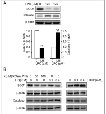
Fig. 4. Effects of LPC-induced ROS on antioxidant enzymes.
HUVECs were treated with vehicle, LPC (125 µM) (A), X/XO, HQ or TBHP (B) for 2 h. Antibodies against SOD1 or catalase were used for blots. Results are representative of three inde-pendent experiments. The data shown are means ± S.E. of three independent experiments. ** P < 0.01 versus vehicle-treated cells.
creased levels of NF-κB, a redox-sensitive transcription
factor (Fig. 3D). These results indicate that LPC-induced
ROS regulate eNOS levels in human endothelial cells.
LPC-induced ROS affect intracellular
antioxi-dant enzymes
Because intracellular oxidative stress is modulated
by antioxidant enzymes such as SOD and catalase, we
examined whether LPC affected the expression of these
enzymes. LPC (125 µM) decreased protein levels of
SOD1, which neutralizes superoxide radicals, and
in-creased catalase, which detoxifies hydroperoxide (Fig.
4A). To differentiate the effects of superoxide radicals
and hydroperoxide, we used an X/XO mixture or HQ as
superoxide radical donors [15, 16] and TBHP as a
hydroperoxide donor. X/XO and HQ decreased levels of
intracellular SOD1, but TBHP increased them. The
superoxide donors increased catalase levels as did LPC
stimulation, but TBHP did not affect catalase expression.
These results indicate that LPC changes antioxidant
en-zymes via superoxide.
LPC leads to superoxide-mediated eNOS
downregulation
As for LPC, the superoxide donors, X/XO and HQ,
diminished eNOS levels (Fig. 2 & 5), whereas TBHP
increased them, suggesting that superoxide is a major
effector in LPC-induced eNOS downregulation. To test
the role of SOD1 in LPC activity, we used TM, a SOD1
inhibitor [17]. Whereas TM alone did not affect eNOS
levels, preincubation of TM (100 µM) prior to LPC (100
µM) treatment potentiated the downregulation of eNOS
(Fig. 6A). In contrast, exogenous SOD could block LPC
downregulation of eNOS (Fig. 6B). These treatments also
changed LPC-induced NF-κB activation (Fig. 6C),
con-sistent with their effects on eNOS. Thus, superoxide
gen-erated by LPC promotes the downregulation of eNOS
via SOD1 inhibition or direct superoxide donation.
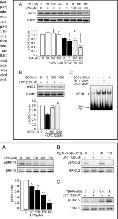
LPC leads to inactivation of ERK1/2
Although pERK1/2 can modulate eNOS [18, 19], it
is not known whether ROS affects ERK activation in
endothelial cells. Thus our study was focused on the
re-Fig. 5. Effects of ROS donors on eNOS regulation. HUVECs
were treated with vehicle, X/XO, HQ (A), or TBHP (B) for 2 h. Western blot was performed using an anti-eNOS antibody. Results are representative of three independent experiments. The data shown are means ± S.E. of three independent experi-ments. ** P < 0.01 versus vehicle-treated cells.
lationship between LPC-induced ROS and ERK1/2
acti-vation in HUVECs. LPC concentration-dependently
in-hibited ERK1/2 activation (Fig. 7A). To confirm whether
LPC effect on pERK1/2 was caused by ROS, we tested
the effect of X/XO or TBHP on pERK1/2. X/XO (100
mU/ml) inhibited ERK1/2 as did LPC (Fig. 7B),
suggest-ing that LPC evoke superoxide-mediated ERK1/2
sup-pression. In contrast, TBHP activated ERK1/2 (Fig. 7C).
These results indicate that LPC reduces
ERK1/2-medi-ated eNOS expression by inducing superoxide.
Fig. 6. The role of SOD1 on LPC-induced eNOS regulation.
(A) HUVECs were treated with vehicle or LPC (70 or 100 µM) for 2 h with or without pretreatment of TM (100 µM) for 30 min. Results are representative of three independent experiments. The data shown are means ± S.E. of three independent experi-ments. ‡ indicates significant difference between LPC (100 µM) and TM (100 µM)-pretreated LPC (100 µM) at P < 0.05 by ANOVA. *, P < 0.05 or **, P < 0.01 versus vehicle-treated cells. (B) HUVECs were pretreated with SOD for 30 min and then stimulated with LPC (125 µM) for 2 h. Results are representa-tive of three independent experiments. The data shown are means ± S.E. of three independent experiments. ** P < 0.01 versus LPC (125 µM)-stimulated cells. (C) HUVECs were treated with vehicle or LPC (125 µM) for 2 h with or without pretreat-ment of SOD (1000 U) or TM (100 µM) for 30 min. EMSA was performed in nuclear extracts incubated with 32P-end labeled
oligonucleotide. Results are representative of three independ-ent experimindepend-ents.
Discussion
We found that LPC downregulates eNOS via a
superoxide-mediated mechanism in human endothelial
cells. LPC stimulates superoxide production by
suppress-ing SOD and/or upregulatsuppress-ing catalase expression to
sup-press ERK1/2 phosphorylation and eNOS exsup-pression.
This is an important finding that differs from previous
reports that LPC promotes total ROS generation and
decreases nitric oxide diffusion [20, 21].
Fig. 7. Effects of LPC-induced superoxide on pERK1/
2. HUVECs were stimulated with vehicle or LPC for 2 h. Western blot was performed using anti-pERK antibody. Results are representative of three independent experiments. The data shown are means ± S.E. of three independent experiments ** P < 0.01, * P < 0.05 versus vehicle-treated cells. (A) HUVECs were treated with X/XO (B), TBHP (C), or LPC 125µM (B, C) for 2 h. Western blot was performed using an anti-pERK antibody. Results are representative of three independent experiments.
Our findings provide novel evidence that eNOS is
downregulated by LPC-induced ROS. LPC-induced
eNOS downregulation was ameliorated by exogenous
SOD (Fig. 6B) and superoxide donors had similar
ef-fects as LPC on eNOS (Fig. 5A), suggesting that
LPC-induced eNOS downregulation was mediated by
superoxide. LPC suppressed SOD1 and increased
cata-lase expression (Fig. 4), indicating LPC increases
superoxide but not hydroperoxide. Inducers of oxidative
stress in atherosclerotic disease like oxLDL, tumor
necro-sis factor-α, and thrombin also promote eNOS
downregulation [22-24].
External stress and physiological stimuli can signal
through the ERK cascades, the best known MAP kinase
cascade [18]. LPC or superoxide donors inhibited
ERK1/2 phosphorylation and eNOS expression, whereas
THBP stimulated them (Fig. 2 & Fig. 5). Thus, inhibition
of ERK1/2 may suppress eNOS expression, as suggested
previously [25].
The effect of LPC on eNOS in endothelial cells is
controversial. LPC induces a rapid, transient rise in
intracellular Ca
2+levels [26], a pathway that can affect
eNOS activity or expression [27]. eNOS expression and
function can be upregulated by
hydroperoxide-depend-ent CaM Kinase II [28]. Conversely, LPC inhibits
phosphorylation of eNOS-Ser1177 and modulates Ca
2+signals through Akt phosphorylation or a
PKCδ-independ-ent pathway [29]. Thus, eNOS may be differPKCδ-independ-entially
regulated by LPC in different cell types.
SOD1 is an endogenous antioxidant, but the
relationship between ROS and SOD1 and its influences
on target molecules is unclear. SOD1 is essential to
hydroperoxide-mediated oxidation and can regulate
growth factors [30, 31].
ERK can regulate the endothelial cell cycle,
proliferation, growth, and apoptosis [32] and regulates
the expression of substrates such as eNOS [33]. LPC
induces superoxide and inactivates ERK1/2 (Fig. 7).
Liu et al [34] also suggested that NADPH oxidase, a
prominent contributor of endothelial superoxide [35],
inactivates ERK and activates P-p38, which LPC
treatment also activated (data not shown).
NF-κB is sensitive to oxidative stress [36]. We
confirmed that NF-κB was activated by LPC and both
types of ROS in an ERK-independent manner (Fig. 3D),
with exogenous SOD blocking this activation and
TM potentiating it (Fig. 6C). Because NF-κB induces
overexpression of the proapoptotic protein Fas ligand
(FasL) in plaques of patients with carotid atherosclerosis
[37], NF-κB may be involved in LPC-induce apoptosis
in HUVECs.
eNOS and ROS are important in endothelial
func-tion in cardiovascular disease. Here, LPC induced
superoxide levels and decreased SOD and eNOS,
producing oxidative stress and causing cytotoxicity.
Physiological LPC levels (5 to 180 µM) [12, 13] could
induce similar toxicity. Lower levels of LPC (20 µM) can
inhibit Ser
1177-eNOS phosphorylation but not protein
lev-els [29, 38], indicating an alternative mechanism for LPC
regulation: lower LPC (≤ 20 µM) decreases
phosphor-ylation but higher levels (≥ 100 µM) induce eNOS
downregulation.
In conclusion, our study highlights a mechanism for
LPC-induced eNOS downregulation. Inhibition of eNOS,
SOD1, and pERK mediated by LPC-induced superoxide
suggests that superoxide can influence eNOS regulation.
Pharmacological modulation of eNOS expression in the
vascular endothelium may be key for the treatment of
atherosclerosis and endothelial damage.
Acknowledgements
This work was supported by the Korea Research
Foundation Grant funded by the Korean Government
(MOEHRD) (KRF-2007-313-E00032).
References
1 Cai H, Harrison DG: Endothelial dysfunc-tion in cardiovascular diseases: The role of oxidant stress. Circ Res 2000;87:840-844.
2 Holvoet P: Endothelial dysfunction, oxi-dation of low-density lipoprotein, and cardiovascular disease. Ther Apher 1999;3:287-293.
3 Mehta JL, Chen J, Hermonat PL, Romeo F, Novelli G: Lectin-like, oxidized low-density lipoprotein receptor-1 (lox-1): A critical player in the development of atherosclerosis and related disorders. Cardiovasc Res 2006;69:36-45. 4 Shah AM, MacCarthy PA: Paracrine and
autocrine effects of nitric oxide on myo-cardial function. Pharmacol Ther 2000;86:49-86.
5 Droge W: Free radicals in the physiologi-cal control of cell function. Physiol Rev 2002;82:47-95.
6 Gutierrez J, Ballinger SW, Darley-Usmar VM, Landar A: Free radicals, mitochon-dria, and oxidized lipids: The emerging role in signal transduction in vascular cells. Circ Res 2006;99:924-932.
7 Matsumoto T, Kobayashi T, Kamata K: Role of lysophosphatidylcholine (lpc) in atherosclerosis. Curr Med Chem 2007;14:3209-3220.
8 Gimbrone MA, Jr., Cybulsky MI, Kume N, Collins T, Resnick N: Vascular endothe-lium. An integrator of pathophysiologi-cal stimuli in atherogenesis. Ann N Y Acad Sci 1995;748:122-131; discussion 131-122.
9 O’Donnell VB, Chumley PH, Hogg N, Bloodsworth A, Darley-Usmar VM, Free-man BA: Nitric oxide inhibition of lipid peroxidation: Kinetics of reaction with lipid peroxyl radicals and comparison with alpha-tocopherol. Biochem 1997;36:15216-15223.
1 0 Searles CD: Transcriptional and posttranscriptional regulation of en-dothelial nitric oxide synthase expres-sion. Am J Physiol Cell Physiol 2006;291:C803-816.
1 1 Engelmann J, Volk J, Leyhausen G, Geurtsen W: Ros formation and glutath-ione levels in human oral fibroblasts ex-posed to tegdma and camphorquinone. J Biomed Mater Res B Appl Biomater 2005;75:272-276.
1 2 Croset M, Brossard N, Polette A, Lagarde M: Characterization of plasma unsatu-rated lysophosphatidylcholines in human and rat. Biochem J 2000;345 Pt 1:61-67.
1 3 Okita M, Gaudette DC, Mills GB, Holub BJ: Elevated levels and altered fatty acid composition of plasma lysophosphati-dylcholine(lysopc) in ovarian cancer patients. Int J Cancer 1997;71:31-34. 1 4 Zmijewski JW, Landar A, Watanabe N,
Dickinson DA, Noguchi N, Darley-Usmar VM: Cell signalling by oxidized lipids and the role of reactive oxygen species in the endothelium. Biochem Soc Trans 2005;33:1385-1389.
1 5 Dixit K, Moinuddin, Ali A: Immunologi-cal studies on peroxynitrite modified hu-man DNA. Life sciences 2005;77:2626-2642.
1 6 Duprat F, Girard C, Jarretou G, Lazdunski M: Pancreatic two p domain k+ chan-nels talk-1 and talk-2 are activated by nitric oxide and reactive oxygen species. J Physiology 2005;562:235-244. 1 7 Carpenter A, Rassam A, Jennings MH,
Robinson-Jackson S, Alexander JS, Erkuran-Yilmaz C: Effects of ammonium tetrathiomolybdate, an oncolytic/ angiolytic drug on the viability and pro-liferation of endothelial and tumor cells. Inflamm Res 2007;56:515-519. 1 8 Merla R, Ye Y, Lin Y, Manickavasagam
S, Huang MH, Perez-Polo RJ, Uretsky BF, Birnbaum Y: The central role of ad-enosine in statin-induced erk1/2, akt, and enos phosphorylation. Am J Physiol 2007;293:H1918-1928.
1 9 Vouyouka AG, Jiang Y, Rastogi R, Basson MD: Ambient pressure upregulates nitric oxide synthase in a phosphorylated-ex-tracellular regulated kinase- and protein kinase c-dependent manner. J Vasc Surg 2006;44:1076-1084.
2 0 Frank GD, Eguchi S, Motley ED: The role of reactive oxygen species in insulin signaling in the vasculature. Antioxid Redox Sig 2005;7:1053-1061. 2 1 Jia SJ, Jiang DJ, Hu CP, Zhang XH, Deng
HW, Li YJ: Lysophosphatidylcholine-induced elevation of asymmetric dimethylarginine level by the nadph oxi-dase pathway in endothelial cells. Vascul Pharmacol 2006;44:143-148. 2 2 Agnoletti L, Curello S, Bachetti T,
Malacarne F, Gaia G, Comini L, Volterrani M, Bonetti P, Parrinello G, Cadei M, Grigolato PG, Ferrari R: Serum from pa-tients with severe heart failure downregulates enos and is proapoptotic: Role of tumor necrosis factor-alpha. Cir-culation 1999;100:1983-1991. 2 3 Eto M, Barandier C, Rathgeb L, Kozai T,
Joch H, Yang Z, Luscher TF: Thrombin suppresses endothelial nitric oxide syn-thase and upregulates endothelin-con-verting enzyme-1 expression by distinct pathways: Role of rho/rock and mitogen-activated protein kinase. Circ Res 2001;89:583-590.
2 4 Liao JK, Shin WS, Lee WY, Clark SL: Oxidized low-density lipoprotein de-creases the expression of endothelial ni-tric oxide synthase. J Biol Chem 1995;270:319-324.
2 5 Ladage D, Brixius K, Steingen C, Mehlhorn U, Schwinger RH, Bloch W, Schmidt A: Mesenchymal stem cells in-duce endothelial activation via paracine mechanisms. Endothelium 2007;14:53-63.
2 6 Wolfram Kuhlmann CR, Wiebke Ludders D, Schaefer CA, Kerstin Most A, Backenkohler U, Neumann T, Tillmanns H, Erdogan A: Lysophosphatidylcholine-induced modulation of ca(2+)-activated k(+)channels contributes to ros-depend-ent proliferation of cultured human en-dothelial cells. J Mol Cell Cardiol 2004;36:675-682.
2 7 Michel JB, Feron O, Sacks D, Michel T: Reciprocal regulation of endothelial ni-tric-oxide synthase by ca2+-calmodulin and caveolin. J Biol Chem 1997;272:15583-15586.
2 8 Drummond GR, Cai H, Davis ME, Ramasamy S, Harrison DG: Transcrip-tional and posttranscripTranscrip-tional regulation of endothelial nitric oxide synthase ex-pression by hydrogen peroxide. Circ Res 2000;86:347-354.
2 9 Gousset-Dupont A, Robert V, Grynberg A, Lacour B, Tardivel S: The effect of n-3 pufa on enos activity and expression in ea hy 926 cells. Prostaglandins Leukot Essent Fatty Acids 2007;76:131-139. 3 0 Grzenkowicz-Wydra J, Cisowski J,
Nakonieczna J, Zarebski A, Udilova N, Nohl H, Jozkowicz A, Podhajska A, Dulak J: Gene transfer of cuzn superoxide dismutase enhances the synthesis of vas-cular endothelial growth factor. Mol Cell Biochem 2004;264:169-181.
3 1 Juarez JC, Manuia M, Burnett ME, Betancourt O, Boivin B, Shaw DE, Tonks NK, Mazar AP, Donate F: Superoxide dismutase 1 (sod1) is essential for h2o2-mediated oxidation and inactivation of phosphatases in growth factor signaling. PNAS U S A 2008;105:7147-7152. 3 2 Huang D, Ding Y, Luo WM, Bender S,
Qian CN, Kort E, Zhang ZF, VandenBeldt K, Duesbery NS, Resau JH, Teh BT: Inhi-bition of mapk kinase signaling pathways suppressed renal cell carcinoma growth and angiogenesis in vivo. Cancer Res 2008;68:81-88.
3 3 Yi T, Cho SG, Yi Z, Pang X, Rodriguez M, Wang Y, Sethi G, Aggarwal BB, Liu M: Thymoquinone inhibits tumor ang-iogenesis and tumor growth through sup-pressing akt and extracellular signal-regu-lated kinase signaling pathways. Mol Cancer Ther 2008;7:1789-1796. 3 4 Liu WH, Cheng YC, Chang LS:
Ros-me-diated p38alpha mapk activation and erk inactivation responsible for upregulation of fas and fasl and autocrine fas-medi-ated cell death in taiwan cobra phospholi-pase a(2)-treated u937 cells. J Cell Physiol 2009;219:642-651.
3 5 Ray R, Shah AM: Nadph oxidase and en-dothelial cell function. Clin Sci (Lond) 2005;109:217-226.
3 6 Andalibi A, Liao F, Imes S, Fogelman AM, Lusis AJ: Oxidized lipoproteins influence gene expression by causing oxidative stress and activating the transcription factor nf-kappa b. Biochem Soc Trans 1993;21:651-655.
3 7 Martin-Ventura JL, Blanco-Colio LM, Munoz-Garcia B, Gomez-Hernandez A, Arribas A, Ortega L, Tunon J, Egido J: Nf-kappab activation and fas ligand overexpression in blood and plaques of patients with carotid atherosclerosis: Potential implication in plaque instabil-ity. Stroke 2004;35:458-463.
3 8 Thors B, Halldorsson H, Clarke GD, Thorgeirsson G: Inhibition of akt phos-phorylation by thrombin, histamine and lysophosphatidylcholine in endothelial cells. Differential role of protein kinase c. Atherosclerosis 2003;168:245-253.