Biomaterials
Research
CThe Korean Society for Biomaterials
전기방사 나노섬유막을 이용한 배양 피부대체물의 제조 및 평가
Electrospun Nanofibrous Membranes for the Engineering of Cultured
Skin Substitutes
박시내·김정환·김일훈·설아람·서 활*
Si-Nae Park, Jeong Hwan Kim, Il Hoon Kim, Ah Ram Sul, and Hwal Suh*
연세대학교의과대학 의학공학교실
Department of Medical Engineering, Yonsei University College of Medicine, Seoul 120-752, Korea (Received March 7, 2006/Accepted May 17, 2006)
In this study, nanofibrous membranes from biodegradable PLGA and collagen were fabricated to mimic natural extra-cellular matrix (ECM) and investigated on the morphology, conformational stability, cytotoxicity and cell attachment. The effect of PLGA or collagen nanofibrous membranes incorporating human dermal fibroblasts on wound healing was also evaluated using an in vivo full thickness dermal defect model. The circular dichroism measurements showed that
electrospun collagen maintained its triple helix structure. In cytotoxicity test using L929 fibroblastic cells, electrospun PLGA or collagen nanofibrous membrane demonstrated no significant toxicity. It was also found that collagen and PLGA nanofibers favored cell attachment and proliferation. In vivo testing showed that the regeneration of dermis and epidermis treated with PLGA or collagen nanofibrous membrane incorporating dermal fibroblasts was accelerated. Therefore, this electrospun PLGA or collagen nanofibrous membrane might have potential efficacy in tissue engineering as skin substitutes.
Key words: Skin substitute, Collagen, PLGA, Electrospun nanofiber, Biocompatibility
서 론 년대 급속하게 발달하기 시작한 생명공학기술의 발전은 손상된인체 조직을인공적으로 수복
,
재 생하는기술로 이용되기시작하여,
최근조직공학 기술의발달 로 체외조직배양을통한 인공피부,
인공연골,
인공간,
인공혈 관 등이 실용화되고 있는 등 다양한 조직공학제품이연구 및 생산에 들어가는단계에 있다.
1-4) 특히 매년 수백만명이발생하는화상환자및 당뇨 등으로인 한 피부궤양 환자에게 적용할 수 있는 인공 피부제품은 손 상피부의 재생및 회복에매우 효과적임이밝혀짐에따라인 공피부 제품의 지속적인개발및 상용화가 촉진되고있다.
5) 인공피부를 통한손상 피부 치료법은현재 사용되고있는일 반 치료법에 비해 창상의 완전한 치유에 요구되는 공여 부위 면적의 감소,
수술procedure
및 입원 기간 감소,
흉터 감소 등 환자의 편의 및 의료비 면에 많은 이점을 가져올 수 있 다.
6,7) 특히자가 혹은 타가각질세포와 진피섬유아세포,
콜라 겐,
히알루론산과 같은생물학적매트릭스 등을이용하여 제조 되는조직공학적 인공피부는 넓은부위의 피부손상을 치료해 줄 수있는 방법으로소개되어현재 널리연구되고있다.
8) 현 재 조직공학적제품에사용되는재료로서생체 조직 가공물및 타가 세포의 사용이 빈번하다.
현재 체내사용이 허가되어 있 는 합성 고분자 재료의 안전성 뿐만 아니라 이종(xenogenic)
혹은 타가 조직으로부터의 추출된 천연 고분자의 생체 내 안 전성,
자가 혹은 세포은행으로부터 제공받은 타가세포의 배 양과정에서의 미생물오염 등에 대한 안전성 평가가 요구되고 있다.
따라서 본 연구에서는 국내·외에서 이용되고 있는 대표적인 의료용 고분자 소재인
poly (D,L-lactic-co-glycolic acid)
(PLGA)
와 제1
형아텔로콜라겐을 전기방사하여각각나노섬유 막을 제조하고 피부조직의 치환 및 재생을 유도하기 위해 진 피섬유아세포를 함께 배양한 나노섬유 기반 인공피부의 체외 및 체내안전성을 평가하기위한 실험을 수행하였다.
재료와 방법PLGA
나노섬유막의 제조PLGA (LA:GA=75:25, Mw. 90,000-126,000)
는Sigma
Chemical Co. (St. Louis, MO, USA)
로부터 구입하였으며PLGA
나노섬유막의 제조를 위하여PLGA(LA:GA=75:25)
를10 ml
의methylene chloride
에35% w/v
로 녹인 후 유기 용매 필터를 이용하여 여과한 후 총8 ml
의 양을18 gauge
*책임연락저자: [email protected]needle
을 끼운10.0 ml syringe
에 넣어 전기방사에 사용하였 다.
전기방사는 전압25 kV,
유속2.5 ml/h, metal collector
의 회전속도300 rpm, spinneret
과metal collector
와의 거리15 cm,
그리고metal collector
의 폭은2 cm
의 조건에서NanoNC (Seoul, Korea)
에서 제조한 전기방사 시스템을 이용하여시행하였다
.
제조된 나노섬유를진공오븐에서48
시간동안 건조하여 잔류 유기용매를 제거하고
ion sputter (E1010,
HITACHI, Tokyo, Japan)
내에서300
Å의 두께로gold/Pt
코팅한 후 시차주사현미경
(S-800, HITACHI, Tokyo, Japan)
을이용하여형태 및구조를 관찰하였다
.
콜라겐 나노섬유막의 제조 제1
형 아텔로콜라겐은Suh
등의방법을 이용하여소 피부로 부터추출하였다.
9) 제1
형 아텔로콜라겐용액은 건조된제1
형 아텔로콜라겐을1,1,1,3,3,3-hexafluoro-2-propanol (HFP)
에7%
의 농도로 녹인후유기용매필터로 여과하여제조하였다.
이후 콜라겐용액을
18 gauge needle
을 끼운5.0 ml syringe
에 채운 후syringe pump
를 통해PLGA
나노섬유 제조 조건과 같은 조건에서 전기방사하였다
.
제조된 콜라겐 나노섬유는12
시간 동안50 mM 1-ethyl-3-(3-dimethyl aminopropyl)
carbodiimide (EDC) solution (EtOH: H
2O=95:5)
용액에서가교시킨후 세척하였다
.
세척된 섬유 다발은 충분한 시간동 안 동결건조기를 이용하여 동결 건조 한 후, PLGA
나노섬유 와 같은방법으로시차주사현미경을 이용하여형태 및구조를 관찰하였다.
콜라겐 나노섬유의Circular Dichroism (CD)
분석 아텔로콜라겐의triple helix
구조가 전기방사 및 화학적 가 교화 이후에 유지되었는지를 확인하기 위해CD
분석을 이용 하였다.
비처리 아텔로콜라겐과 나노섬유 형태로 제조된 아텔로콜라겐의
CD spectra
는0.01 cm length thermostatized
cuvette
과Neslab RTE-111 thermostat
을 이용하여Jasco
J-715 dicrograph (Jasco Corp, Tokyo, Japan)
로 측정하였다.
각 시편을3 mg/ml
의 농도로0.001 N HCl
에 용해시킨 후,
25
oC
에서 가열해놓은CD cuvette
에 넣고30
분 경과 후 시료 에 대한CD spectra
를 기록하였다.
시편(
단백질)
의 양은Bradford method
를사용하여 결정하였다.
10) 세포독성시험PLGA
나노섬유막과가교화된 아텔로콜라겐나노섬유막의세 포독성유무를확인하기 위하여직접접촉법에의한세포독성시 험을 실시하였다.
11-13) 먼저L929
섬유아세포(ATCC CCL1,
NCTC clone 929, from mouse connective tissue)
을6-well
plate
에 농도가2
×10
5cells/well
되도록plating
하고, 600
µl
의10%
태아우혈청(JBI Inc, Daejeoun, Korea)
을 함유한Dulbecco's modified Eagle's medium (DMEM) (JBI Inc,
Daejeoun, Korea)
을 공급하면서2
일 동안37±2
oC, 5%
CO
2, 99% humidity
조건으로 배양시켰다.
그 위에1
cm
×1 cm
크기의나노섬유막을얹고,
배지를새로넣은다음,
같은조건에서24
시간 동안배양하였다. 0.2% crystal violet
으 로 염색하여 세포의 성장이 억제된zone size
를 측정하였다(Table 1).
음성대조군과 양성대조군으로서high-density
polyethylene (HDPE)
필름과Latex
필름을 각각사용하였다.
세포배양콜라겐 나노섬유막과
PLGA
나노섬유막 위에 점착 및 배양시킬 세포로서 연세대학교의
The Medical Material Control
and Management Committee
로부터 허가를 얻어 연세의료원성형외과로부터 공급받은인간섬유아세포를이용하였다
.
인간진피 섬유아세포는
175 cm
2 조직배양 플라스크(NUNC,
Roskilde, Denmark)
에서1%
페니실린/
스트렙토마이신/
암포테리 신-
비(penicillin/streptomycin/amphotericin-B), 10%
태아우혈청(JBI Inc, Daejeoun, Korea)
을 함유한DMEM (JBI Inc,
Daejeoun, Korea)
성장배지를 이용하여 세포를 배양하였다.
배양은37±2
oC, 5% CO
2, 99% humidity
환경이 유지되는 배양기에서 이루어졌다.
본 실험을 위해5
와7 passage
사이 의 섬유아세포가 사용되었다.
세포점착성 실험을 위해1 cm
×1 cm
크기의PLGA
나노 섬유막과가교화된콜라겐 나노섬유막을1
×10
7의 섬유아세포 를 함유한80 ml
의 배지가 들어있는 각각의spinner
형 플라 스크에서60 rpm
의 속도로 교반하면서8
시간동안세포점착을 유도하였다.
세포의 부착능 및 증식능은 교반 배양 이후0
시간, 4
시간 및24
시간이 지났을 때3-(4, 5-dimethylthiazolyl)-2,
5-di-phenyltetrazolium bromide (MTT) assay
14)법에 의해ELISA
reader (Spectra Max 340, Molecular Device Inc. CA,
USA)
로측정되었다.
통계학적유의성은Student's t-test
를통하여 평가하였다
.
PCR
기법을 이용한Mycoplasma
검사PLGA
나노섬유막과 콜라겐 나노섬유막에서 세포를 배양한 후PCR assay kit (MycoSensor
TMPCR Assay Kit, Stratagene,
La Jolla, CA, USA)
을 이용하여mycoplasma
오염 여부를 측 정하였다.
먼저 세포 배양액100
µl
를microcentrifuge tube
에서
5
분간 끓이고60
초간 원심분리한 후Manual
에 따라template
을 준비하였다. PCR amplification
을 위해5
µl
의sample template, kit
에서 제공하는 양성 대조군 또는 음성대 Table 1. Zone descriptionZone Index Description of Zone
0 No detectable zone around or under specimen
1 Zone limited to area under specimen
2 Zone extends less than 0.5cm beyond specimen
3 Zone extends 0.5-1.0 cm beyond specimen
4 Zone extends greater than 1.0 cm beyond specimen but does not involve entire dish
조군
(
증류수)
을MgCI
2(
최종농도20 mM), 10
× Tagreac-tion buffer 5
µl, dNTP/dUTP mix 1
µl, Mycoplasma primer
mix 2
µl, Internal control template 4
µl,
TagDNA
poly-merase 2.5 U
및 멸균수를 포함하는45
µl
의reaction
mixture
에 가하였다. PCR
프로그램의thermal profile
은94
oC
에서30
초간denaturation, 55
oC
에서1
분간primer
annea-ling, 72
oC
에서1
분간primer extension
으로 구성하여 각각35 cycle
씩 실시하였다.
이후 반응물은2% agarose gel
을 이용한 전기영동에 사용되었고
, gel
상에 나타난banding
pattern
을 분석하였다.
동물이식시험섬유아세포가 배양된
PLGA
나노섬유막과 콜라겐 나노섬유막을 각각
15
마리의athymic mice (BALB/c-nu)
에 이식 후 이식재와host cells
와의 상호작용 및 분해거동을 평가하고자 하였다.
이를 위해 약6-8
주령의athymic mice (NIH Swiss
nude) (24-32 g)
를ketamine
과xylazine (
각각200
µg/g
과10
µg/g)
로 마취시킨 후.
각 동물의 등 중앙부위에 약1
cm
2직경의full-thickness skin wound
를 만들고 제조된 나노섬유막를 손상부위에 수술실로 고정하였다
.
이후 투명 필름(Tegaderm
TM,
3M, St. Paul, MN, USA)
으로 수술 부위를감쌌다
.
모든 동물 실험은연세대학교의the Guidelines and
Regulations for Use and Care of Animals
에 따라 시행되 었다.
수술 후7, 14, 21
일째에동물들을희생시키고인접 손상시 키지않은정상피부와 근육층을 포함하여채취한 후 포르말린 고정,
수세,
파라핀 포매과정을 거쳐5
µm
두께로 박절하여hematoxylin-eosin (H-E)
염색을 통한조직학적검사를 시행하 였다.
결과 및 고찰PLGA
나노섬유막과 콜라겐 나노섬유막 지금까지 조직공학적 이식재의임상결과를향상시키기 위하여 콜라겐
, poly (l-lactic acid)
와poly (glycolic acid)
등의 재료를지지체나합성 매트릭스구조체로서 이용하는 시도가있 어왔다
.
특히최근에는 인체의세포외기질구조를 모방하기위 하여천연 고분자용액의전기방사를통해 제조된구조체를조 직공학적으로 이용하는 연구가 활발히 진행되고 있다.
15-17) 본 연구에서는 조직공학적 인공진피막의 지지체로서PLGA
나노 섬유막과콜라겐 나노섬유막을 제조하기위하여 전기방사법을 이용하였다.
예비실험을통해3%
농도 이하의 콜라겐 용액은 나노섬유를 형성하지못하는 것을확인하였으며 이에따라용 액의 농도를 결정하였다.
전기방사된 후 각각의 나노섬유막들 은 실질적구조 연결성을갖고있기 때문에metal collector
로 부터 쉽게 떼어낼 수 있었다.
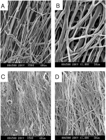
시차주사현미경을 통한 관찰을 통해전기방사된PLGA
섬유막 및콜라겐 섬유막은서로연결 되고교차하는섬유 네트워크와 공극들로이루어진 구조를나 타내고 있었으며 직경이 각각0.3-3
µm
및0.1-0.5
µm
인 것을 확인하였다(Figure 1(A-D)).
콜라겐의Triple Helix
구조 확인콜라겐의 구조는 기본적으로
triple helical structure
를 이루고 있으며 이 구조의 유지는 생체 내 세포와의 상호작용 시 매우 중요한 요소로 여겨지고 있다
. CD
는 단백질 구조에서의 형태적 변화를연구하기 위해널리사용되는 방법이며,
콜라겐 분자의 구조적folding
은 매우 독특하여CD spectra
역시 다 른 단백질과 차이를 보인다.
따라서triple helix
구조의 변화 는CD spectra
를 통해 쉽게 관찰될 수 있다.
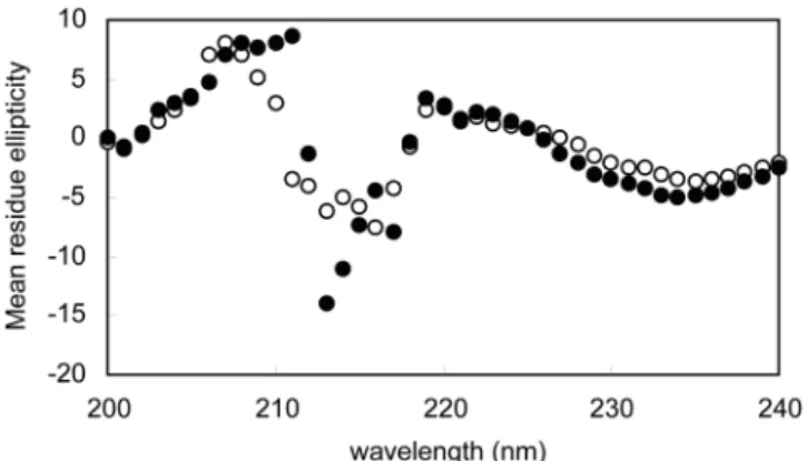
콜라겐의CD
spectrum
에 나타나는 특징은221 nm
에서 보이는 작은positive band
와197 nm
에서 보이는 보다 큰negative
band
이다.
18) 본실험결과에서 나노섬유화콜라겐은219 nm
에서
34516 deg
×cm
2×dmol
-1 크기의positive peak
와213
nm
에서140236 deg
×cm
2×dmol
-1 크기의negative peak
를 나타냈다(Figure 2).
나노섬유화 콜라겐은 비교를 위해 사용된 비처리 콜라겐의spectrum
과 비교하였을 때molar ellipticity
의 강도에서만차이를 나타냈을뿐 전체
spectrum
은 유사하였Figure 1. (A and B) Electrospun PLGA collected on a mandrel
revolv-ing at 300 RPM (Magnification 500X with the inserted scale bar at 60 microns (A)), (Magnification 100X with the inserted scale bar at 30 microns (B)), (C and D) Electrospun collagen collected on a mandrel revolving at 300 RPM (Magnification 500X with the inserted scale bar at 60 microns (C)), (Magnification 100X with the inserted scale bar at 30 microns (D)).
고이는다른연구들에서의 결과와 일치하는것이다
.
19) 따라서 본 실험을 통하여 전기방사와 화학적 가교화 과정을 거친 콜 라겐분자의triple helix
구조가 그대로 유지되었다는것을확 인하였다.
생체 외 세포독성 직접접촉법에 의거하여인공피부용 재료로 사용된PLGA
나 노섬유막과콜라겐 나노섬유막의세포독성정도를zone index
로써 살펴보았다.
양성 대조군과 음성 대조군의zone index
결과를보면,
적정시험조건에서실험이 수행되었음을알 수있 었다(Table 2).
양성대조군인Latex
필름이 놓인 아랫면과 그 주변의 세포들은cell lysis
가 일어나서culture dish
바닥면에 붙어있지 못하고 배지위로 떨어져 주변으로0.45
±0.04 cm
크기의zone
이 형성된 반면HDPE
음성대조군이나PLGA
나노섬유막혹은콜라겐나노섬유막의 경우에는배양접시에서자 란 세포들의 모양과 동일한 상태로 필름의 아랫면과 그 주변 에 그대로 붙어있으면서
zone
을 형성하지 않는 것으로 보아 세포독성이없는것으로 확인되었다.
세포 부착능 및 증식능 나노섬유 구조가세포의점착과증식을유도할수 있음이널 리 알려져 있으며 이미 많은 이들이PLGA, Polycaprolactone
(PCL))
등의합성고분자또는 천연고분자로이루어진나노섬 유상에서 섬유세포,
연골세포,
골수 유래줄기세포등을배양 하는 시도를 하였다.
20-22) 한편Schindler, M.
et al.은 실험을통해 섬유세포와
normal rat kidney
세포의 점착 및 증식에영향을 주는 것이
polyamide
나노섬유의3
차원적 지형이라고 주장하였고, J. VENUGOPAL
et al. 등은 콜라겐 나노섬유와PCL
나노섬유상에서의세포의 거동을비교하여본 결과PCL
재료 단독 보다는 생체 고분자인 콜라겐 분자가 코팅된 나노 섬유상에서 세포의 증식 및이주능이 향상되었다고보고하였 다.
22,23) 본 연구에서MTT assay
를 통한 세포배양 실험결과, dynamic seeding
이후 모든종류의나노섬유상에서 세포가 초기에 잘 점착하고7
일간 크게 증식하는 것을 관찰하였고(Figure 3),
예상한 바와 같이1
일과7
일째에 콜라겐 나노섬유 가PLGA
나노섬유와비교하였을 때더 향상된증식능을보여 주는 것을확인할 수 있었다(p<0.05).
Mycoplasma
검사 현재세포가포함된 조직공학적제품을최종 ‘멸균화’시키 기는 어렵기 때문에 공여 물질이나 제조단계에서의 첨가물로 부터의 어떠한감염도 공정 중 증식을통해 치료받는 환자에 게 치명적인 영향을 줄 수 있다.
특히mycoplasma
오염은 세포배양과 생물학적 재료에 흔히 되풀이되는 위험 요소로서Mollicutes (Mycoplasma, Ureaplasma, Acholeplasma
species
포함)
감염은 실험결과의신뢰도를 떨어뜨리고생물학 적 제품에 손상을주게된다.
24)Mollicute
감염은 박테리아수의 증가에 따라 배지 탁도가 달라지지 않으며 세포병리적 영
향이 거의 없기 때문에 검사하기가 쉽지 않다
.
25) 이에 따라이미 유럽약전
(European Pharmacopoeia)
과USP(United States
Pharmacopoeia)
에 멸균 및mycoplasma
에 대한 시험 규격이 제정되어 있지만, 1
개월 이상의시험기간이 소요되며그 동안 세포 혹은 조직공학적제품의생장능(viability)
이유지되지 못할 것이다.
26) 반면, PCR
기술은 다른 종류의primer set
의 제작을 통해 일반적인mycoplasma
감염원을 수시간 내 검색할 수 있는 장점이 있으므로이미mycoplasma
검사의 한 방법으로 받아 들여지고 있다.
27) 따라서 본 실험에서는 세포배양 중에 흔히발견되는
mycoplasma species
의16S rRNA
에 대한gene
sequence
를annealing
하는primer mixture
를 이용한PCR
분 Figure 2. CD spectra of native type I collagen (open circle) andelec-trospun type I collagen (closed circle).
Table 2. The numerical values for zone index of the materials 시험 대상 형성된 Zone 크기(cm) IndexZone
음성 대조군 - 0
양성 대조군 0.45±0.04 3
PLGA nanofibrous membrane - 0
Collagen nanofibrous membrane - 0
Figure 3. Cell attachment and proliferation assessed by MTT testing
performed 0 hr, 4 hr, 1 day and 7 day after dynamic seeding. The data reported are means±SD for n = 5.
석을 통해
1
주간 세포가 배양된PLGA
나노섬유막과 콜라겐 나노섬유막이mycoplasma
에 감염되었는지 알아보고자 하였 다.
그 결과,
진피섬유세포가부착된PLGA
나노섬유막과콜라 겐 나노섬유막의배양상층액 모두mycoplasma DNA
가함유 되어 있지 않은 것이 확인되었다(Figure 4).
본 검사를 통해mycoplasma
에 의해감염되지 않은것으로 확인된 진피섬유세 포 부착PLGA
나노섬유막과 콜라겐 나노섬유막은 다음 동물 이식시험에적용되었다.
동물이식시험 실험적으로유발한athymic mice
의창상에 각종진피섬유아 세포가 부착된 나노섬유막 인공피부를 이식한 후 창상크기의 변화를 관찰하고사진 촬영하였다(Figure 5).
또한피부생검의 조직학적소견상 인공피부이식후 급성염증반응이심하고7
일째상처 부위아래쪽에서의 혈관형성이 활발해지는 것을관 찰하였다.
초기에 염증세포들이진피조직전체에서관찰되었고Figure 4. Electrophoretic analysis of mycoplasma polymerase chain
reaction (PCR). Supernatants from cell cultures on PLGA or collagen nanofibrous membrane were subjected to the sample preparation. Lane 1: size marker (100bp DNA ladder), lane 2: collagen nanofiber with cells, lane 3: PLGA nanofiber with cells, lane 4: medium with cells, lane 5: medium without cells, lane 6: negative control (water) and lane 7: positive control.
Figure 5. Macroscopic observation of wounds treated with fibroblast seeded collagen nanofibrous or PLGA nanofibrous membrane at day 0, day
7, day 14 and day 21 after implantation.
Figure 6. Photomicrographs of biopsy specimens from a wound treated with fibroblast seeded collagen nanofibrous (A, B, E, F, I, J) or PLGA
nanofibrous membrane (C, D, G, H, K, L) on the postoperative 7th day (A-D), 14th day (E-H), and 21th day (I-L). Nanofibrous membrane implan-tion (B, D, F, H, J, L) and sham-operaimplan-tion controls (A, C, E, G, I, K) were compared (Magnificaimplan-tion 40X).
(Figure 6(A-D)), 14
일째에는섬유아세포의 뚜렷한증식과 콜라젠 생성이관찰되었으며 진피의 혈관은 감소하기시작하고표
피의 재생이 뚜렷했다
(Figure 6(E-H)). PLGA
나노섬유막 기반인공피부와 콜라겐 나노섬유막 기반 인공피부를 비교했을 때 염증 정도의 의미 있는 차이는 보이지 않았으며 콜라겐 나노 섬유막 기반인공피부가 적용된 군의경우표피재생이 대조군 에비해빨리시작되었다
(Figure 6(I-L)). 21
일째에는모든실험 군의조직에서성숙된 표피및진피 형성을관찰할 수있었다.
특히나노섬유막기반인공피부를적용한경우더 두껍게형 성된 각질층(Stratum corneum)
을 확인하였으며 잘 배열된 표 피층을 통해재생의plateau
단계에이르고 있음을 확인할 수 있었다.
결 론 본 연구에서는 대표적 의료용 고분자인PLGA
와 콜라겐을 전기방사 기법을 통해 나노섬유막 형태로 제조하였고,
콜라겐 나노섬유의경우 물성 유지를 위해화학적으로 가교화하였다.
CD
분석을 통해 콜라겐의triple helix
구조가 전기방사 및 화학적가교화 처리이후에유지되었음을 확인하였다.
또한제 조된PLGA
혹은 콜라겐나노섬유막이세포독성을나타내지않 으며콜라겐 나노섬유막의경우PLGA
나노섬유막과비교하여 세포증식능이1
일째와7
일째 유의적으로향상된것을 관찰하 였다. PCR
기법을 통해mycoplasma
감염여부를 검사한 후 세포를 포함하는PLGA
나노섬유막과 콜라겐 나노섬유막을anthimic mice
에 이식한 결과 초기 급성 염증반응이 심하고7
일째상처부위 아래쪽에서의혈관형성이 활발해지는 것을관 찰하였다. 14
일째에는 섬유아세포의뚜렷한 증식과 콜라젠 생 성이관찰되었으며콜라겐나노섬유막기반인공피부가적용된 군의 경우 표피재생이 대조군에 비해 빨리 시작되었다. 21
일 째에는 모든 실험군의 조직에서 성숙된 표피 및 진피 형성을 관찰할수 있었다.
따라서본 연구를 통해PLGA
혹은콜라겐 나노섬유막의 조직공학적 인공피부로서의 생체적합성과 효용 가능성을확인할 수 있었다.
감사의 글 본 연구는식품의약품안전청(KFDA2005-5036)
의지원으로이 루어진 것으로 이에감사드립니다.
참고문헌1. S. Kaihara and J. P. Vacanti, “Tissue engineering toward new
solutions for transplantation and reconstructive surgery,” Arch.
Surg., 134, 1184-1188 (1999).
2. R. Langer and J. P. Vacanti, “Tissue engineering,” Science, 260,
920-926 (1993).
3. T. Shinoka, C. K. Breuer, and R. E. Tanel, et al., “Tissue
engineered heart valves,” Ann. Thorac. Surg., 60, S513-S516
(1995).
4. C. A. Vacanti and J. P. Vacanti, “Bone and cartilage
recon-struction with tissue engineering approaches,” Otolaryngol. Clin.
North Am., 27, 263-276 (1994).
5. S. L. Hansen, D. W. Voigt, P. Wiebelhaus, and C. N. Paul, “Using
skin replacement products to treat burns and wounds,” Adv. Skin
Wound Care, 14, 37-46 (2001).
6. D. Heimbach, A. Luterman, and J. F Burke, “Artificial dermis for
major burns. A multi-center randomized clinical trial,” Ann.
Surg., 208, 313-320 (1988).
7. R. Stern, M. McPherson, and M Longaker, “Histologic study of
artificial skin used in the treatment of full-thickness thermal injury,” J. Burn Care Rehab., 11, 7-13 (1990).
8. R. E. Horch, J. Kopp, U. Kneser, J. Beier, and A. D. Bach. “Tissue
engineering of cultured skin substitutes,” J. Cell. Mol. Med., 9,
592-608 (2005).
9. H. Suh, S. W. Suh, and B. G. Min, “Anti-infection treatment of a
transcutaneous device by a collagen-rifampicine composite,”
ASAIO J., 40, M406- M411 (1994).
10. X. Liu, H. Wu, M. Byrne, J. Jeffrey, S. Krane, and R. Jaenisch. “A
targeted mutation at the known collagenase cleavage site in mouse type I collagen impairs tissue remodeling,” J. Cell. Bio., 130, 227-237 (1995).
11. S. N. Park, J. C. Park, H. O. Kim, M. J. Song, and H. Suh,
“Characterization of porous collagen/hyaluronic acid scaffold
modified by 1-ethyl-3-(3-dimethylaminopropyl)carbodiimide cross-linking,” Biomaterials, 23, 1205-1212 (2002).
12. “Biological evaluation of medical device, part 5. Test for
cytotoxicity: in vitro methods,” ISO 10993, 1992.
13. “Biological reactivity tests,” USP 24 <87>, 2000.
14. H. W. Sung, I. L. Liang, C. N. Chen, R. N. Huang, and H. F. Liang, “Stability of a biological tissue fixed with a naturally
occurring crosslinking agent (genipin),” J. Biomed. Mater. Res., 55, 538-546 (2001).
15. J. Matthews J, D. Simpson, G. Wnek, and G. Bowlin, “
Electro-spinning of collagen nanofibers,” Biomacromolecules, 3,
232-238 (2002).
16. W. S. Kim, J. P Vacanti, L. Cima, D. Mooney, J. Upton, W. C. Puelacher, and C. A. Vacanti, “Cartilage engineered in
predeter-mined shapes employing cell transplantation on synthetic biodegradable polymers,” Plast. Reconstr. Surg., 94, 233-237
(1994).
17. Y. Cao, J. P. Vacanti, K. T. Paige, J. Upton, and C. A. Vacanti,
“Transplantation of chondrocytes utilizing a polymer-cell
construct to produce tissue-engineered cartilage in the shape of a human ear,” Plast. Reconstr. Surg., 100, 297 -302 (1997)
18. M. H. Li, P. Fan, B. Brodsky, and J. Baum, “Two-dimensional
NMR assignments and conformation of (Pro-Hyp-Gly)10 and a designed collagen triple-helical peptide,” Biochemistry, 32,
7377-7387 (1993).
19. K. A. Piez and M. R. Sherman, “Characterization of the product
formed by renaturation of alpha 1-CB2, a small peptide from collagen,” Biochemistry, 9, 4129-4133 (1970).
20. W. J. Li, C. T. Laurencin, E. J. Caterson, R. S. Tuan, and K. K. Frank, “Electrospun nanofibrous structure: A novel scaffold for
tissue engineering,” J. Biomed. Mater. Res., 60, 613-621, (2001).
21. W. J. Li, R. Tulia, C. Okafor, A. Derfoul, K. G. Danielson, D. J. Hall, and R. S. Tuan, “A three-dimensional nanofibrous scaffold
for cartilage tissue engineering using human mesenchymal stem cells,” Biomaterials, 26, 599-609 (2005).
22. S. Melvin, A. Ijaz, K. Jabeen, N. Alam, H. Timothy, H. Grafec, C. Young, and M. Sally, “A synthetic nanofibrillar matrix promotes in
vivo-like organization and morphogenesis for cells in culture,”
Biomaterials, 26, 5624-5631 (2005).
23. J. Venugopal and S. Ramakrishna, “Biocompatible nanofiber
matrices for the engineering of a dermal substitute for skin regeneration,” Tissue eng., 11, 847-854 (2005)
24. H. G. Drexler and C. C. Uphoff, “Contamination of cell culture,
mycoplasma,” in The Encyclopedia of Cell Technology: E. Spier
(Ed.), Wiley, New York, 2000, pp. 609-627.
25. I. Bruchmuller, E. Pirkl, R. Herrmann, M. Stoermer, H. Eichler, H.
Kluter, and P. Bugert, “Introduction of a validation concept for a
PCR-based Mycoplasma detection assay,” Cytotherapy., 8, 62-69
(2006).
26. D. N. Galbraith, “Regulatory and microbiological safety issues
surrounding cell and tissue-engineering products,” Biotechnol.
Appl. Biochem. 40, 35-39 (2004).
27. C. C. Uphoff and H. G. Drexler, “Detection of mycoplasma
contaminations in cell cultures by PCR analysis,” Hum. Cell, 12,