796 Copyright © 2012 The Korean Society of Cardiology Korean Circulation Journal
Introduction
Degenerative aortic valve disease is frequently found in elderly people. However, approximately 30% of elderly patients with se-vere aortic stenosis (AS) remain unreferred and untreated because these patients are considered too high risk or inappropriate for conventional open heart surgery.1) Transcatheter aortic valve
impl-antation (TAVI) using a balloon expandable Sapien valve (Edwards Lifesciences, Irvine, CA, USA) or self-expandable CoreValve (Medtro-nic, Minneapolis, MN, USA) has demonstrated favorable outcomes in patients with high surgical risk. The most common approach for
Case Report
http://dx.doi.org/10.4070/kcj.2012.42.11.796 Print ISSN 1738-5520 • On-line ISSN 1738-5555
The First Korean Patient With Severe Aortic Stenosis and Bilateral
Iliofemoral Artery Disease Treated With Transcatheter Aortic Valve
Implantation by Transsubclavian Approach
Seung-Jun Lee, MD
1, Young-Guk Ko, MD
1, Ji-Young Shim, MD
1, Sak Lee, MD
2, Byung-Chul Chang, MD
2,
Jae-Kwang Shim, MD
3, Young-Ran Kwak, MD
3, and Myeong-Ki Hong, MD
11Division of Cardiology, 2Departments of Thoracic and Cardiovascular Surgery and 3Anesthesiology and Pain Medicine, Severance Cardiovascular Hospital,
Yonsei University Health System, Seoul, Korea
Transcatheter aortic valve implantation (TAVI) is indicated as an alternative treatment modality to surgical aortic valve replacement for high risk patients. The standard retrograde approach through the femoral artery is not feasible in the case of unfavorable iliofemoral anatomy or severe peripheral arterial disease (PAD). However, patients with aortic stenosis (AS) have a higher prevalence of for PAD because both diseases are consequences of atherosclerotic degenerative changes. Transsubclavian, transapical, and direct access to the ascending aorta by thoracotomy are alternative routes for the TAVI procedure. In this report, we present the first Korean patient with symptomatic severe AS and bilateral iliofemoral artery disease who was successfully treated with TAVI using a CoreValve (Medtronic, Minneapolis, MN, USA) by transsubclavian approach. (Korean Circ J 2012;42:796-799)
KEY WORDS: Aortic valve stenosis; Catheters; Heart valve prosthesis; Prosthesis implantation.
Received: April 11, 2012 Accepted: May 2, 2012
Correspondence: Myeong-Ki Hong, MD, Division of Cardiology, Severance Cardiovascular Hospital, Yonsei University Health System, 50 Yonsei-ro, Seodaemun-gu, Seoul 120-752, Korea
Tel: 82-2-2228-8460, Fax: 82-2-2227-7732 E-mail: [email protected]
• The authors have no financial conflicts of interest.
This is an Open Access article distributed under the terms of the Creative Commons Attribution Non-Commercial License (http://creativecommons. org/licenses/by-nc/3.0) which permits unrestricted non-commercial use, distribution, and reproduction in any medium, provided the original work is properly cited.
TAVI is the transfemoral approach because the relatively large pro-file (18 Fr) of the delivery catheter requires an access route with a dia-meter larger than 6 mm. However, peripheral arterial disease (PAD) is also relatively common in elderly patients with AS because degen-erative aortic valve disease shares many characteristics with ath-erosclerotic disease.2) When a transfemoral approach is not feasible
due to small or diseased iliofemoral arteries, alternative access rou-tes such as the transapical approach, transsubclavian approach, as well as direct aortic access via minimal thoracotomy may be consi-dered.3)4)
In this case report, we present the first Korean patient with symp-tomatic severe AS at high surgical risk accompanied by bilateral il-iofemoral artery disease that was successfully treated with TAVI us-ing a CoreValve by transsubclavian approach.
Case
An 82-year-old male patient presented with worsening symptoms of chest discomfort and exertional dyspnea {New York Heart Asso-ciation (NYHA) III} for 2 months. He had past medical history of hy-pertension and dyslipidemia. Four years ago, the patient had been treated with percutaneous coronary intervention with implantation of stents at the left main artery and the distal left circumflex artery.
797 Seung-Jun Lee, et al.
http://dx.doi.org/10.4070/kcj.2012.42.11.796
www.e-kcj.org
At that time he also underwent percutaneous transluminal angio-plasty with implantation of self-expandable nitinol stents at the bi-lateral iliac arteries. On admission, transthoracic and transesopha-geal echocardiography showed severe AS and mild aortic regurgit-ation (AR) due to degenerative change with calcificregurgit-ation (Fig. 1). The aortic valve area was 0.44 cm2. The peak and mean pressure
gradi-ents across the aortic valve were estimated to be 97 and 59 mm Hg, respectively, while the diameter of the aortic annulus by echocar-diography was 25 mm. The left ventricle (LV) showed a dilated end-diastolic dimension (64 mm) with LV hypertrophy. The LV systolic function was globally reduced with an LV ejection fraction (EF) of 39%. Based on the coronary angiography, the stent at the left main artery was patent. However, the stent implanted at the distal left
circumflex artery showed in-stent restenosis, which was subse-quently treated with a 3×30 mm paclitaxel-eluting balloon (SeQu-ent® Please, B. Braun Melsungen AG, Berlin, Germany). The previ-ously inserted stents at the right and left iliac arteries were patent, however computed tomography (CT) angiography revealed that the minimal lumen diameter of the stented right iliac artery was 5.5 mm and that of the left iliac artery was 4.5 mm. Both common femoral arteries also showed significant stenosis with a minimum lumen di-ameter of 4.0 mm (Fig. 2). The didi-ameter of the left subclavian artery was measured to be 7 mm on CT angiography. The calculated Eu-roSCORE was 44.08%.
In this patient we decided to perform TAVI by transsubclavian ap-proach. The procedure was carried out in a hybrid operating room
A B
Fig. 1. Transthoracic echocardiography showed severely narrowed aortic valve area (0.44 cm2) measured by the continuity equation (A), and color Doppler
revealed aliasing due to severe aortic stenosis (B).
Fig. 2. Angiography and computed tomography image showed iliofemoral arteries with severe peripheral arterial occlusive disease. Previous stent at Lt. common iliac artery was patent but showed a minimum diameter of 4.5 mm and both common femoral arteries showed a minimum diameter of 4.0 mm. Lt. CIA: left common iliac artery, Rt. CFA: right common femoral artery, Lt. CFA: left common femoral artery.
798 Transsubclavian Approach TAVI in Severe AS Patient With PAD
http://dx.doi.org/10.4070/kcj.2012.42.11.796 www.e-kcj.org
under general anesthesia. After surgical exposure of the left subcla-vian artery, a 0.035 inch Amplatz Super Stiff wire (Boston Scientific, Natick, MA, USA) was inserted into the LV through an 18 Fr intro-ducer sheath (Fig. 3). Balloon dilation of the stenotic aortic valve was performed with a 23 mm balloon (Z-med, NuMED Inc., Hopkinton, NY, USA) under rapid pacing using a temporary pacemaker. Under angiographic guidance, a 29 mm CoreValve was slowly deployed at the aortic annulus. An immediate post-procedural aortogram sh-owed good position of the CoreValve with mild AR (Fig. 4). The vas-cular access site at the left subclavian artery was closed surgically without any complications. The post-procedural echocardiography
demonstrated well functioning bioprosthetic aortic valve with mild paravalvular AR. The peak and mean pressure gradients across the aortic valve decreased from 97 and 59 mm Hg to 42 and 23 mm Hg, respectively. The aortic valve area increased from 0.44 cm2 to 2.86
cm2. The LVEF improved from 39% to 52%. Four days post
proce-dure, the patient was discharged with improved symptoms (NYHA I). There was no sign of stroke or any conduction abnormality as indi-cated by the electrocardiogram. The patient has been free from any major cardiovascular events or symptom aggravation for a total follow-up duration of 6 months.
Discussion
Transcatheter aortic valve implantation has emerged as a promis-ing alternative treatment modality to surgical aortic valve replace-ment for patients with severe AS at high surgical risk. Patients with an estimated mortality risk >20% by logistic EuroSCORE or >10% by the Society of Thoracic Surgeons score system are generally con-sidered candidates for the TAVI procedure. In addition, combined re-spiratory failure, pulmonary hypertension, previous cardiac surgery, right ventricular failure, hostile thorax (such as radiation, burns, pre-vious thoracic pleurodesis, or multiple thoracotomies), severe con-nective tissue disease, liver cirrhosis, cachexia, and porcelain aorta are further indications for TAVI. Currently, two types of stented val-ves, the balloon-expandable Edwards SAPIEN valve and the self-ex-pandable CoreValve system are commonly used for the percutane-ous treatment of severe AS. There has been no randomized study comparing the two systems to date. However, the procedural suc-cess rates (>95%) have been reported to be high with both valves. The mortality rates are similar at 30 days (Edwards 12% vs.
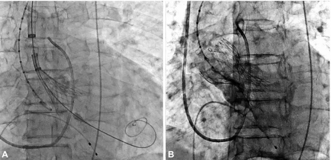
CoreV-Fig. 3. CoreValve delivery system is advanced through 18 Fr sheath insert-ed into the Lt. subclavian artery.
Fig. 4. CoreValve delivery catheter and bioprosthetic valve deployed across the aortic valve (A) and spontaneously expanded. An aortogram showed good positioning of the CoreValve with mild aortic regurgitation (B).
799 Seung-Jun Lee, et al.
http://dx.doi.org/10.4070/kcj.2012.42.11.796
www.e-kcj.org
alve 11%, p=0.99) and one year (Edwards 18% vs. CoreValve 21%) for both systems. The need for pacemaker appears to be significantly higher with the CoreValve.5)
Commonly, the transfemoral route is used for the TAVI procedure due to the vessel diameter requirement of >6 mm for the 18 Fr device introducer sheath and delivery catheter. However, when transfemo-ral approach is not amenable due to the concomitant PAD, alterna-tive routes by transapical, transsubclavian or direct aortic access through thoracotomy can be utilized for the TAVI procedure. The pa-tient we observed here is the first Korean case of TAVI by transsub-clavian approach using a CoreValve. Recent studies have shown that the safety of transsubclavian TAVI is not inferior to transfem-oral TAVI.6) Petronio et al.7) reported a 100% procedural success
rate and 0% intraprocedural mortality according to the analysis of 54 transsubclavian approach cases. Thirty-day mortality was 0% (vs. 6.1% in transfemoral approach group, p=0.13) and the six-month mortality rate was 9.4% (vs. 15.8%, p=0.44) showing no difference compared with transfemoral TAVI.
Moynagh et al.8) reported that the transsubclavian TAVI achieved
better outcomes compared with the transfemoral approach in opti-mal valve positioning (88.6% vs. 60.5%, p<0.0001) and in major ad-verse cardiovascular and cerebrovascular events (2.9% vs. 13.4%, p=0.09), even though transsubclavian TAVI patients had significantly higher EuroSCOREs because they were frequently accompanied by PAD, coronary artery disease, prior myocardial infarct, and prior per-cutaneous coronary intervention experience. CoreValve is currently not available for the transapical approach. However, patients treated by transapical approach using Edwards SAPIEN showed significantly lower one-year survival than those treated by transfemoral appro-ach. More renal failure and stroke events were observed in the tr-ansapical TAVI group compared with transfemoral TAVI group.5)
Tr-ansapical approach generally accompanies the risk from general anesthesia, thoracotomy and the incision of the LV apex. Moreover, patients with structural change of the LV due to remodeling and distorted angle, the risk of transapical TAVI may increase.9) By
con-trast, transsubclavian approach can be safely performed via local
anesthesia combined with administration of a mild systemic seda-tive and analgesic agent. Therefore, if the transfemoral approach is not applicable due combined PAD in a patient with severe AS requir-ing TAVI procedure, and if the subclavian artery has a diameter >6 mm, a transsubclavian approach should be considered as a primary alternative route.
References
1. Iung B, Cachier A, Baron G, et al. Decision-making in elderly patients with severe aortic stenosis: why are so many denied surgery? Eur Heart J 2005;26:2714-20.
2. Aronow WS, Ahn C, Kronzon I. Association of valvular aortic stenosis with symptomatic peripheral arterial disease in older persons. Am J Cardiol 2001;88:1046-7.
3. Bruschi G, De Marco F, Fratto P, et al. Direct aortic access through right minithoracotomy for implantation of self-expanding aortic biopros-thetic valves. J Thorac Cardiovasc Surg 2010;140:715-7.
4. Da Gama Ribeiro V, Vouga L, Markowitz A, et al. Vascular access in transcatheter aortic valve implantation. Int J Cardiovasc Imaging 2011;
27:1235-43.
5. Bosmans JM, Kefer J, De Bruyne B, et al. Procedural, 30-day and one year outcome following CoreValve or Edwards transcatheter aortic valve implantation: results of the Belgian National Registry. Interact Cardiovasc Thorac Surg 2011;12:762-7.
6. Witkowski A, Da˛browski M, Chmielak Z, et al. Transcatheter aortic valve implantation using transfemoral/transsubclavian or transapical ap-proach: 30-day follow-up of the initial 30 patients. Kardiol Pol 2011;
69:105-14.
7. Petronio AS, De Carlo M, Bedogni F, et al. Safety and efficacy of the subclavian approach for transcatheter aortic valve implantation with the CoreValve revalving system. Circ Cardiovasc Interv
2010;3:359-66.
8. Moynagh AM, Scott DJ, Baumbach A, et al. CoreValve transcatheter aortic valve implantation via the subclavian artery: comparison with the transfemoral approach. J Am Coll Cardiol 2011;57:634-5.
9. Fraccaro C, Napodano M, Tarantini G, et al. Expanding the eligibility for transcatheter aortic valve implantation the trans-subclavian retro-grade approach using: the III generation CoreValve revalving system.