2010;70:6352-6358.
Cancer Res
Kwang Je Kim, Je-Wook Yu, Hyun Sub Hwang, et al.
CIIA Is a Novel Regulator of Detachment-Induced Cell Death
Updated version
http://cancerres.aacrjournals.org/content/70/15/6352
Access the most recent version of this article at:
Material
Supplementary
http://cancerres.aacrjournals.org/content/suppl/2010/07/22/70.15.6352.DC1.html
Access the most recent supplemental material at:
Cited Articles
http://cancerres.aacrjournals.org/content/70/15/6352.full.html#ref-list-1
This article cites by 20 articles, 6 of which you can access for free at:
Citing articles
http://cancerres.aacrjournals.org/content/70/15/6352.full.html#related-urls
This article has been cited by 1 HighWire-hosted articles. Access the articles at:
E-mail alerts
Sign up to receive free email-alerts
related to this article or journal.
Subscriptions
Reprints and
.
pubs@aacr.org
Department at
To order reprints of this article or to subscribe to the journal, contact the AACR Publications
Permissions
.
permissions@aacr.org
Department at
Tumor and Stem Cell Biology
CIIA Is a Novel Regulator of Detachment-Induced Cell Death
Kwang Je Kim1, Je-Wook Yu2, Hyun Sub Hwang1, and Eui-Ju Choi1
Abstract
Detachment-induced cell death, or anoikis, is a type of apoptosis that occurs when epithelial cells lose their attachment to the extracellular matrix. Anoikis serves as a physiologic barrier to metastasis. Deviation from the tightly regulated mechanism of detachment-induced cell death might result in progression to metastatic cancer. Here, we investigated the function of CIIA in the regulation of anoikis. CIIA protein was upregulated in colon cancer tissue samples. Knockdown of CIIA in metastatic colorectal carcinoma SW620 and KM12SM cells promoted detachment-induced cell death through the regulation of caspase activation. Knockdown of CIIA also inhibited anchorage-independent growth in soft agar and colony formation after suspension stress. These observations suggest that CIIA is a novel negative regulator of anoikis.Cancer Res; 70(15); 6352–8. ©2010 AACR.
Introduction
Cancer is one of the most dangerous human diseases. Cell immortality, abnormal cell growth regulation, self-sufficient cell growth, cellular evasion of apoptosis, sustained angiogen-esis, cell invasion, and metastasis are well-defined hallmarks of tumors (1). Due primarily to these characteristics of tumor cells, successful cancer treatments remain elusive. Metastasis, in particular, poses one of the major difficulties in cancer treat-ment. Normal cells die when they lose attachment to the ex-tracellular matrix. However, many malignant cancer cells avoid detachment-induced cell death and acquire metastatic potential. Anoikis serves as a physiologic barrier to metastasis (2–4). Identification of the critical regulators of anoikis might provide important information leading to the development of more effective treatments for malignant metastatic cancer.
There are many signaling pathways involved in anoikis (5). Many cancer cells evade detachment-induced cell death through the amplification of survival signals or inhibition of cell death signals. One of the major apoptosis pathways, the caspase cascade, is also involved in anoikis (3, 6, 7). Caspase activation cascades are associated with two pathways in cell death, the death receptor–mediated extrinsic pathway and the mitochondria-mediated intrinsic pathway. In the extrinsic pathway, a death-inducing signaling complex is formed to activate initiator procaspase-8 (8). In the mitochondria-mediated intrinsic pathway, a signaling complex called the apoptosome is formed to activate initiator procaspase-9 (8, 9). In response to a number of diverse apoptotic stimuli,
cyto-chrome c is released from the mitochondria into the cytoplasm. Upon binding of released cytochrome c to Apaf1, Apaf1 forms heptameric oligomers through interactions of its CARD do-main. Initiator procaspase-9 is recruited to the Apaf1oligomer through the CARD domain and then activated (10, 11). Activated initiator caspases subsequently induce the activation of effector caspases that leads to apoptosis. Under detachment condi-tions, caspase activation occurs; however, in metastatic cancer cells, caspase activation is circumvented for survival (7, 12, 13). Previously, we reported that CIIA functions as an anti-apoptotic protein that blocks tumor necrosis factor-α– and H2O2–induced cell death (14). Here, we showed that CIIA
inhi-bits detachment-induced apoptosis through the regulation of caspase activation. CIIA protein was upregulated in colon cancer tissue samples, and knockdown of CIIA induced anoikis in me-tastatic colorectal carcinoma cells. These results show that CIIA is a novel negative regulator of detachment-induced cell death.
Materials and Methods
Cell culture, transfection, and induction of anoikis Colon cancer SW480, SW620, KM12C, and KM12SM cells were kindly provided by Dr. S.G. Chi (Korea University, Seoul, Korea). SW480 and SW620 cells were purchased from Amer-ican Type Culture Collection. KM12C and KM12SM cells were from the Korean Cell Line Bank in 2008. SW620 cells were derived from a lymph node metastasis of SW480 colon cancer cells (15). KM12SM cells are metastatic variants from the KM12C cells (16). Madin-Darby canine kidney (MDCK) cells were purchased from the Korean Cell Line Bank in 2002. 293T cells were provided by Dr. D.S. Lim (Korea Advanced Institute of Science and Technology, Daejeon, Korea; cells were purchased from American Type Culture Collection) in 2007. SW620 and SW480 cells were grown in RPMI medium supplemented with 10% fetal bovine serum (FBS). KM12SM, MDCK, and 293T cells were in DMEM supplemented with 10% FBS. KM12C cells were grown in MEM supplemented with 10% FBS. SW480, SW620, KM12C, and KM12SM cells were transfected by electroporation using a Microporator (Invitrogen). MDCK cells were transfected Authors' Affiliations:1Laboratory of Cell Death and Human Diseases, School
of Life Sciences and Biotechnology, Korea University, Seoul, Korea and
2Division of Biosciences and Bioinformatics, Myongji University, Yongin, Korea
Note: Supplementary data for this article are available at Cancer Research Online (http://cancerres.aacrjournals.org/).
Corresponding Author: Eui-Ju Choi, School of Life Sciences and Biotechnology, Korea University, Anam-dong, Seoul 136-701, Republic of Korea. Phone: 82-23290-4285; Fax: 82-23290-4741; E-mail: ejchoi@ korea.ac.kr.
doi: 10.1158/0008-5472.CAN-09-4394
©2010 American Association for Cancer Research.
Cancer
Research
Cancer Res; 70(15) August 1, 2010 6352
Research.
on December 22, 2013. © 2010 American Association for Cancer cancerres.aacrjournals.org
with the use of LipofectAMINE (Invitrogen). 293T cells were trans-fected with polyethylenimine (Sigma-Aldrich). For the induction of anoikis, cells were cultured in suspension on dishes coated with poly-2(hydroxyethyl methacrylate) (10 mg/mL in ethanol). RNA interference
Noble agar). SW620 cells (2 × 10⁴cells) in 1.0 mL of top agar (RPMI medium containing 10% FBS and 0.4% Noble agar) were added to each well and incubated for 20 days. Colonies were stained with 0.005% crystal violet and counted. Colony-forming assay
SW620 cells were cultured in poly-2(hydroxyethyl methac-rylate)–coated dishes for 48 hours, and then transferred to
six-well culture plates (300 cells/well). After 15 days, colonies were stained with 0.05% crystal violet.
Immunohistochemistry
To determine the levels of endogenous CIIA in normal and colon cancer tissues, premade AccuMax Array colon cancer tissues with liver metastasis slides (ISU ABXIS CO. LTD., Seoul, Korea) were immunoreacted with anti-CIIA antibody
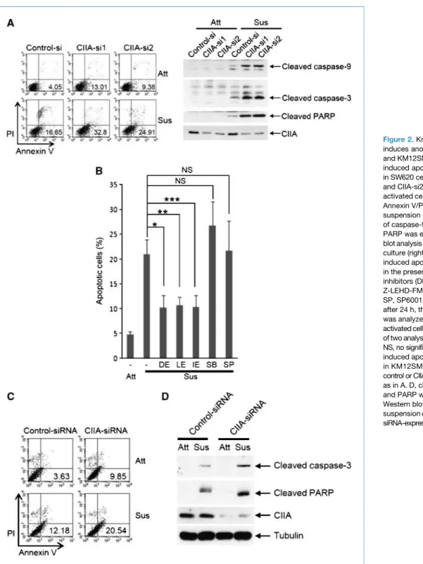
Figure 2.Knockdown of CIIA induces anoikis in SW620 and KM12SM cells. A, suspension-induced apoptosis was examined in SW620 cells expressing CIIA-si1 and CIIA-si2 by fluorescence-activated cell sorting analysis using Annexin V/PI staining after 48 h of suspension culture (left). Cleavage of caspase-9, caspase-3, and PARP was examined by Western blot analysis after 48 h of suspension culture (right). B, detachment-induced apoptosis of CIIA-si1 cells in the presence of the indicated inhibitors (DE, Z-DEVD-FMK; LE, Z-LEHD-FMK; IE, Z-IETD-FMK; SP, SP600125; SB, SB203580) after 24 h, the suspension culture was analyzed by fluorescence-activated cell sorting. Columns, mean of two analyses (*, **, and ***,P < 0.01; NS, no significance). C, suspension-induced apoptosis was examined in KM12SM cells transfected with control or CIIA siRNA oligonucleotides as in A. D, cleavage of caspase-3 and PARP was examined by Western blotting after 48 h of suspension culture in control or CIIA siRNA-expressing KM12SM cells. Kim et al.
Cancer Res; 70(15) August 1, 2010 Cancer Research
6354
Research.
on December 22, 2013. © 2010 American Association for Cancer cancerres.aacrjournals.org
(Santa Cruz Biotechnology). Immunoreactive proteins were visualized with fast 3,3′-diaminobenzidine tetrahydrochlor-ide dehydrate tablets (Sigma) after signal amplification using avidin-biotin-peroxidase complex (Vector Laboratories).
Images were acquired by Olympus DP71 microscopy using DP controller software (Olympus). Relative expression levels of CIIA were quantified by Adobe Photoshop CS-based image analysis using the equation (1− mean of the staining intensity
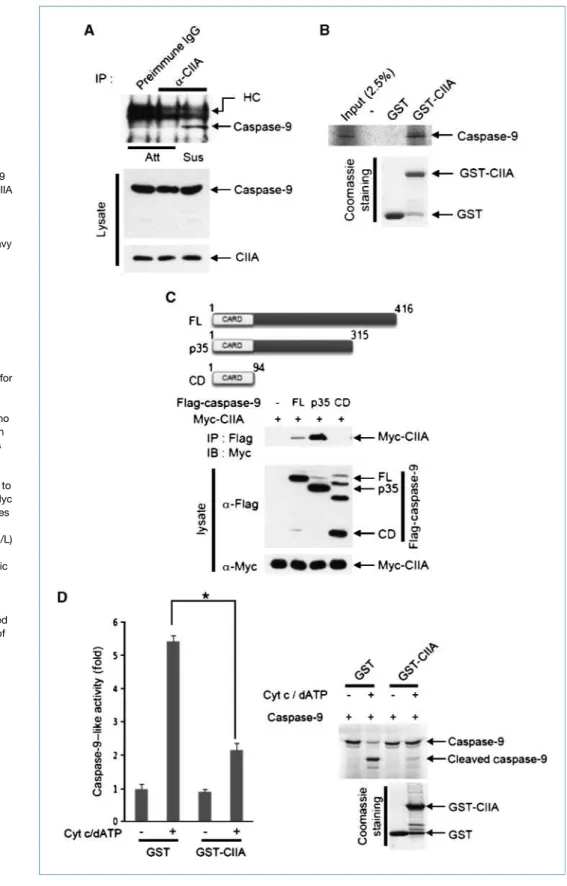
Figure 3.CIIA inhibits caspase-9 activation. A, the interaction of CIIA and caspase-9 in attached and 24-h suspension conditions was examined by
coimmunoprecipitation. HC, heavy chain. B,in vitro–translated
35
S-labeled caspase-9 was applied to GST-CIIA on glutathione-conjugated beads. Bead-bound35
S-labeled caspase-9 was collected and analyzed by autoradiography. C, 293T cells were transfected for 48 h with a vector encoding Myc-CIIA together with vectors for Flag-tagged caspase-9 variants [FL, full-length (amino acids 1–416); p35, p35 fragment (amino acids 1–315); CD, CARD domain (amino acids 1–94)]. Cell lysates were immunoprecipitated with anti-Flag antibody, and the resulting pellets were subjected to immunoblot analysis with anti-Myc antibody. D, left, 293T cell lysates were added with cytochromec (0.5μmol/L) and dATP (100 μmol/L) as indicated, and assayed for caspase-9 activity by colorimetric assay (*,P < 0.01). Right,
35
S-labeled caspase-9 was incubated for 1 h at 37°C with cytosolic S-100 solution obtained from 293 cells in the presence of cytochromec, dATP, and either GST or GST-CIIA as indicated. Cleavage of caspase-9 was analyzed by autoradiograph.
CIIA Inhibits Anoikis
Cancer Res; 70(15) August 1, 2010
of the selected region / mean of the staining intensity of background) × 100.
Results and Discussion
CIIA is upregulated in colon cancer tissues
We previously reported that CIIA is an antiapoptotic pro-tein (14). Many antiapoptotic propro-teins are frequently up-regulated in cancer cells as compared with their normal counterparts, and contribute to the ability of cancer cells to evade cell death. We investigated the protein levels of CIIA in colon cancer tissues by immunohistochemistry using premade AccuMax Array slides. CIIA was upreg-ulated in colon cancer tissues and corresponding liver metastasis colon cancer tissues as compared with normal tissues in each patient examined (P1–P9; Fig. 1). Further-more, the protein levels of CIIA were higher in metastatic colon cancer tissues than in nonmetastatic colon cancer tissues (Fig. 1B). CIIA expression was also higher in metastatic SW620 or KM12SM cells than in their non-metastatic counterpart SW480 or KM12C cells (Supple-mentary Fig. S1).
Knockdown of CIIA induces anoikis in metastatic colon cancer cells
To better understand the possible role of CIIA in cancer de-velopment, we examined the effect of CIIA on detachment-induced apoptosis in metastatic cancer SW620 cells. We first examined the effect of siRNA-mediated CIIA depletion on an-oikis resistance in SW620 cells. We generated two SW620 cell lines (CIIA-si1 and CIIA-si2) that stably expressed CIIA siRNA.
Depletion of CIIA expression by RNA interference (RNAi) pro-moted detachment-induced apoptosis and the cleavage of caspase-9, caspase-3, and PARP in SW620 cells (Fig. 2A). Next, we examined whether caspase activation might mediate an-oikis in CIIA knockdown SW620 cells. Inhibitors of caspase-3, caspase-8, and caspase-9 (Z-DEVD-FMK, Z-IETD-FMK, and Z-LEHD-FMK, respectively) suppressed detachment-induced apoptosis in CIIA knockdown SW620 cells (CIIA-si1), whereas inhibitors of c-Jun NH2-terminal kinase and p38 (SP600125
and SB203580, respectively) had no effect (Fig. 2B). We con-firmed the inhibitory effect of CIIA on detachment-induced apoptosis in SW620 cells transiently transfected with CIIA siRNA oligonucleotides (Supplementary Fig. S2A). Fur-thermore, siRNA-mediated CIIA depletion promoted the cleavage of caspase-9, caspase-3, and PARP in the cells (Sup-plementary Fig. S2A and B). These results suggested that CIIA depletion converts anoikis-resistant SW620 cells to an anoikis-susceptible phenotype through caspase activa-tion. The promoting effects of CIIA siRNA on detachment-induced apoptosis (Fig. 2C) as well as cleavage of caspase-3 and PARP (Fig. 2D) were also observed in another meta-static colon cancer cell line, KM12SM cells. Furthermore, overexpression of CIIA inhibited detachment-induced caspase activation and apoptosis in MDCK cells (Supple-mentary Fig. S3).
CIIA inhibits the activation of caspase-9
Given that RNAi-mediated CIIA depletion promoted detachment-induced caspase activation and apoptosis, we next examined a possible action of CIIA on the activation of caspase-9. We first examined the interaction of endoge-nous CIIA and caspase-9 in SW620 cells. The interaction of
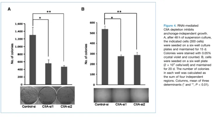
Figure 4.RNAi-mediated CIIA depletion inhibits anchorage-independent growth. A, after 48 h of suspension culture, the indicated cells (300 cells) were seeded on a six-well culture plates and maintained for 15 d. Colonies were stained with 0.05% crystal violet and counted. B, cells were seeded on a six-well plate (2 × 10⁴
cells/well) and maintained for 20 d. The number of colonies in each well was calculated as the sum of four independent regions. Columns, mean of three determinants (* and **,P < 0.01). Kim et al.
Cancer Res; 70(15) August 1, 2010 Cancer Research
6356
Research.
on December 22, 2013. © 2010 American Association for Cancer cancerres.aacrjournals.org
endogenous CIIA and caspase-9 was detected under suspen-sion growth conditions (Fig. 3A). Furthermore, ectopically expressed CIIA bound to full-length and p35 fragment of caspase-9, but not to its CARD domain (Fig. 3C). In vitro binding data also showed that caspase-9 directly bound to CIIA, as a GST-fusion protein, but not to GST (Fig. 3B). Moreover, overexpressed CIIA inhibited oligomerization of recombinant caspase-9 in transfection studies (Supplementa-ry Fig. S4). Together, these data suggest that CIIA physically interacts with caspase-9. We next examined whether CIIA af-fected the activation of caspase-9 triggered by cytochrome c and dATP. Cytochrome c/dATP-mediated caspase-9 activa-tion was significantly reduced by GST-CIIA (Fig. 3D). These results suggested that CIIA interacts with caspase-9 in cells under suspension culture and inhibits the activation of caspase-9 and downstream caspases such as caspase-3.
Given that caspase-8 as well as caspase-9 might be as-sociated with detachment-induced apoptosis in CIIA-depleted SW620 cells (Fig. 2B), we examined the cleavage of caspase-8 and caspase-9 at various times after suspen-sion culture in control and CIIA-depleted SW620 cells (Supplementary Fig. S5A). The time course of the activa-tion was similar between caspase-8 and capase-9. More-over, a caspase-8 inhibitor Z-IETD-FMK did not block the detachment-induced activation of caspase-9 in CIIA-depleted SW620 cells (Supplementary Fig. S5B). These results thus suggested that caspase-8 activation was not involved in caspase-9 activation induced by detachment in CIIA-depleted cells.
CIIA is necessary for anchorage-independent growth Next, we carried out colony-forming assays after suspen-sion culture, and examined anchorage-independent growth in soft agar (Fig. 4A and B). The colony formation assay was carried out to quantify living cells after growth in sus-pension. Anchorage-independent growth is one of the char-acteristics of transformed cells, including metastatic cancer cells. After 48 hours of suspension culture, cells were plated in fresh dishes for 15 days. CIIA knockdown cells (CIIA-si1 and CIIA-si2) formed fewer olonies than control SW620 cells (Fig. 4A). Similarly, when we examined colony formation in soft agar, CIIA knockdown cells formed fewer colonies than control cells (Fig. 4B). Thus, CIIA facilitated anchorage-independent growth.
The caspases are the major signaling players in anoikis. Activation of the caspase cascade is also one of the main tar-gets of cancer treatment (13). Many reports have shown that antiapoptotic proteins that negatively regulate the caspase cascade, such as XIAP, Bcl-2, Bcl-xL, and MCL-1 are fre-quently overexpressed in cancer cells, and loss or inhibition of these antiapoptotic proteins is involved in anoikis (12, 13, 18, 19). Our results showed that CIIA inhibits detachment-induced apoptosis through the negative regulation of cas-pase activation. CIIA was upregulated in colon cancer and corresponding liver metastasis colon cancer tissues as com-pared with normal tissues. Knockdown of CIIA promoted detachment-induced apoptosis in anoikis-resistant colon cancer cells. These results suggest that CIIA is a novel inhib-itor of anoikis. CIIA induces epithelial-mesenchymal transi-tion and cell invasion (20). Thus, it is conceivable that CIIA induces cancer cell differentiation to metastatic cancer cells through the inhibition of anoikis and induction of invasion. Because anoikis serves as a physiologic barrier that inhibits metastasis, the induction of anoikis has im-portant implications in cancer treatment. On the basis of our findings in this study, we propose that CIIA functions as a novel regulator of anoikis, and may be a possible target for metastatic cancer treatment.
Disclosure of Potential Conflicts of Interest No potential conflicts of interest were disclosed.
Acknowledgments
We thank Dr. S.G. Chi for SW480, SW620, KM12C, and KM12SM cells, and Dr. D.S. Lim for 293T cells.
Grant Support
Korea Research Foundation grant (KRF-2006-341-C00023), Basic Science Research Program (2009-0080895) through the National Research Foundation of Korea (NRF), and a NRF grant (20090081488) funded by the Ministry of Education, Science & Technology, South Korea (E-J. Choi). K.J. Kim was supported by the Korea Student Aid Foundation grant (S2-2009-000-01286-1) funded by MEST.
The costs of publication of this article were defrayed in part by the payment of page charges. This article must therefore be hereby marked advertisement in accordance with 18 U.S.C. Section 1734 solely to indicate this fact.
Received 12/02/2009; revised 05/19/2010; accepted 06/07/2010; posted online 08/01/2010.
References
1. Hanahan D, Weinberg RA. The hallmarks of cancer. Cell 2000;100:57–70. 2. Frisch SM, Francis H. Disruption of epithelial cell-matrix interactions
induces apoptosis. J Cell Biol 1994;124:619–26.
3. Mehlen P, Puisieux A. Metastasis: a question of life or death. Nat Rev Cancer 2006;6:449–58.
4. Rennebeck G, Martelli M, Kyprianou N. Anoikis and survival connec-tions in the tumor microenvironment: is there a role in prostate cancer metastasis? Cancer Res 2005;65:11230–5.
5. Frisch SM, Screaton RA. Anoikis mechanisms. Curr Opin Cell Biol 2001;13:555–62.
6. Cory S, Adams JM. The Bcl2 family: regulators of the cellular life-or-death switch. Nat Rev Cancer 2002;2:647–56.
7. Simpson CD, Anyiwe K, Schimmer AD. Anoikis resistance and tumor metastasis. Cancer Lett 2008;272:177–85.
8. Riedl SJ, Shi Y. Molecular mechanisms of caspase regulation during apoptosis. Nat Rev Mol Cell Biol 2004;5:897–907.
9. Jiang X, Wang X. Cytochrome C-mediated apoptosis. Annu Rev Biochem 2004;73:87–106.
10. Pop C, Timmer J, Sperandio S, Salvesen GS. The apoptosome activates caspase-9 by dimerization. Mol Cell 2006;22:269–75.
CIIA Inhibits Anoikis
Cancer Res; 70(15) August 1, 2010
11. Riedl SJ, Salvesen GS. The apoptosome: signalling platform of cell death. Nat Rev Mol Cell Biol 2007;8:405–13.
12. LaCasse EC, Mahoney DJ, Cheung HH, Plenchette S, Baird S, Korneluk RG. IAP-targeted therapies for cancer. Oncogene 2008; 27:6252–75.
13. MacKenzie SH, Clark AC. Targeting cell death in tumors by activating caspases. Curr Cancer Drug Targets 2008;8:98–109.
14. Cho SG, Kim JW, Lee YH, et al. Identification of a novel antiapoptotic protein that antagonizes ASK1 and CAD activities. J Cell Biol 2003; 163:71–81.
15. Leibovitz A, Stinson JC, McCombs WB III, McCoy CE, Mazur KC, Mabry ND. Classification of human colorectal adenocarcinoma cell lines. Cancer Res 1976;36:4562–9.
16. Morikawa K, Walker SM, Nakajima M, Pathak S, Jessup JM, Fidler IJ. Influence of organ environment on the growth, selection, and metastasis of human colon carcinoma cells in nude mice. Cancer Res 1988;48:6863–71.
17. Park HS, Huh SH, Kim Y, et al. Selenite negatively regulates caspase-3 through a redox mechanism. J Biol Chem 2000;275:8487–91. 18. Schimmer AD, Dalili S, Batey RA, Riedl SJ. Targeting XIAP for the
treatment of malignancy. Cell Death Differ 2006;13:179–88. 19. Wu H, Tschopp J, Lin SC. Smac mimetics and TNFα: a dangerous
liaison? Cell 2007;131:655–8.
20. Han SY, Hwang HS, Chae JS, et al. CIIA induces the epithelial-mesenchymal transition and cell invasion. Biochem Biophys Res Commun 2009;387:548–52.
Kim et al.
Cancer Res; 70(15) August 1, 2010 Cancer Research
6358
Research.
on December 22, 2013. © 2010 American Association for Cancer cancerres.aacrjournals.org