pISSN 1738-1843 © 2013 The Korean Society of Pathologists/The Korean Society for Cytopathology
Recent advances in molecular pathology and targeted thera-pies have opened a new era of personalized medicine for lung cancer treatment. Driver genetic alterations such as epidermal growth factor receptor (EGFR) mutations, as well as Kirsten rat sarcoma viral oncogene homolog (KRAS) and anaplastic lym-phoma kinase (ALK) rearrangements, have been identified and are currently used as predictive biomarkers for targeted thera-pies.1 Activating somatic mutations in the EGFR gene are known
to be major driver mutations in that they exhibit a high inci-dence in lung cancers and have played an important role in the development of targeted molecular therapies for lung cancer.2
EGFR tyrosine kinase inhibitors (TKIs), such as gefitinib and erlotinib, are associated with anti-tumor activity, inhibiting multiple downstream signaling processes that activate cell pro-liferation and other cell responses, including cell migration and angiogenesis.3 EGFR TKIs are approved in Korea as a first-line
Guideline Recommendations for
EGFR Mutation Testing in Lung Cancer:
Proposal of the Korean Cardiopulmonary Pathology Study Group
Hyo Sup Shim* · Jin-Haeng Chung1*
Lucia Kim2· Sunhee Chang3
Wan-Seop Kim4· Geon Kook Lee5
Soon-Hee Jung6· Se Jin Jang7
Department of Pathology, Yonsei University College of Medicine, Seoul; 1Department of
Pathology, Seoul National University Bundang Hospital, Seongnam; 2Department of Pathology,
Inha University School of Medicine, Incheon;
3Department of Pathology, Ilsan Paik Hospital,
Inje University College of Medicine, Goyang;
4Departments of Pathology, Konkuk University
School of Medicine, Seoul; 5Department of
Pathology, National Cancer Center, Goyang;
6Department of Pathology, Yonsei University
Wonju College of Medicine, Wonju; 7Department
of Pathology, Asan Medical Center, University of Ulsan College of Medicine, Seoul, Korea *Hyo Sup Shim and Jin-Haeng Chung contributed equally to this work.
Mutations of the epidermal growth factor receptor (EGFR) are the strongest predictive factor for response to EGFR tyrosine kinase inhibitors (TKIs), such as gefitinib and erlotinib. EGFR TKIs are approved in Korea as a first-line treatment for lung cancer patients with mutated EGFR. Rapid and accurate EGFR mutation testing is essential for patient selection and establishing targeted therapies with EGFR TKIs. Thus, a standard set of guideline recommendations for EGFR muta-tion testing suitable for the Korean medical community is necessary. In this article, we propose a set of guideline recommendations for EGFR mutation testing that was discussed and approved by the Cardiopulmonary Pathology Study Group of the Korean Society of Pathologists.
Key Words: Mutation; Receptor, epidermal growth factor; Guideline
Received: March 16, 2013 Revised: March 28, 2013 Accepted: April 1, 2013
Corresponding Author
Se Jin Jang, M.D.
Department of Pathology, University of Ulsan College of Medicine, Asan Medical Center, 88 Olympic-ro 43-gil, Songpa-gu, Seoul 138-736, Korea
Tel: +82-2-3010-5966 Fax: +82-2-472-7898 E-mail: [email protected]
treatment for advanced non-small cell lung cancer (NSCLC) with mutated EGFR (Fig. 1). In the Iressa Pan-Asia Study (IPASS) trial, tumors with mutated EGFR exhibited a 71.2% clinical response to first-line gefitinib treatment, while only 1.1% of tumors with wild-type EGFR responded to the treat-ment.4 Therefore, patient selection is critical for the clinical use
of EGFR TKIs as a first-line treatment. Clinical characteristics such as female gender, never-smoker status, and Asian ethnicity were also found to be associated with the response to EGFR
TKIs; however, the results of the IPASS study confirmed that molecular selection-based EGFR mutation testing is the stron-gest predictive factor for EGFR TKI treatment response.4,5
Thus, EGFR mutation testing is very important for lung cancer therapy. Likewise, rapid and accurate EGFR mutation testing is essential for proper patient selection when consider-ing targeted therapy with EGFR TKIs. In addition, a standard set of guidelines suitable for the Korean medical community is necessary. In this article, we propose guideline recommenda-tions for EGFR mutation testing that were discussed and ap-proved by the Cardiopulmonary Pathology Study Group of the Korean Society of Pathologists (Table 1).
PATIENT SELECTION
The most important reason for EGFR mutation testing is to select patients who might benefit from EGFR TKI therapy. Pa-tients that receive EGFR mutation testing are primarily those with advanced stage disease. EGFR mutations are more preva-lent in female patients, never-smokers, and patients of Asian ethnicity. However, clinical features alone cannot entirely pre-dict EGFR mutation status.6,7 Most of the guidelines published
thus far recommend histologic type as the most important fac-tor for determining whether EGFR mutational testing should be performed.8-10 Specifically, when patients are diagnosed with
Fig. 1. Epidermal growth factor receptor (EGFR) tyrosine kinase in-hibitors (TKIs) are approved as a first-line treatment for advanced non-small cell lung cancer harboring EGFR mutation.
Advanced non-small cell lung carcinoma
Positive
1st line: EGFR-TKI (e.g., gefitinib, erlotinib) EGFR mutation testing
Table 1. Recommendation summary for EGFR mutation testing
Recommendation
Patient selection Pathologic diagnosis is the most important factor
Patients with non-small cell carcinoma, especially adenocarcinoma componenta
Other types if clinically indicated
Sample source Primary and metastatic sites are equally suitable
Biopsy (formalin-fixed paraffin-embedded tissue) and cytology specimens are equally suitable
Sample processing Routine preparation for tissue or cytology is suitable
Tumor content The presence of tumor cells must be verified by a pathologist
High percentage (ideally more than 50%) of tumor cells for direct sequencing Lower percentage acceptable for methods with higher sensitivity
Method for mutation testing Various methods can be used for mutation testing
New techniques must be approved by the Korean government
The pathologist should consider available facilities and the pros and cons of each method
Turnaround time The entire workflow process should be supervised by the pathologist
Pathologic diagnosis: 1-2 working days Molecular diagnosis: 5-7 working days
Repeat examination The pathologist should consider repeating the examination under the following situations Poor sequence data
Cycle threshold too close to the defined cut-off limit
Result are not matched with previously well-defined clinical-pathologic characteristics
Reporting format Sample information, type of method, mutation status, comments
EGFR, epidermal growth factor receptor.
aIn this regard, poorly differentiated non-small cell carcinoma should be further classified into a more specific type whenever possible. A minimum
immunohis-tochemical panel (such as thyroid transcription factor 1/napsin A/p63 or p40) is recommended in small specimens to preserve as much tissue as possible for molecular testing.
NSCLC including an adenocarcinoma component or NSCLC-not-otherwise-specified after immunohistochemistry, EGFR
mutation testing is routinely recommended.9 Thus,
patholo-gists should try to further classify poorly differentiated NSCLC into more specific types, such as adenocarcinoma or squamous cell carcinoma, whenever possible (Fig. 2). In addition, to pre-serve as much tissue as possible for molecular testing in small specimens, a minimum immunohistochemical panel such as thyroid transcription factor 1/napsin A/p63 or p40 is recom-mended.9,11,12
EGFR mutations are detected in approximately 40% of
Ko-rean NSCLC patients with adenocarcinoma histology.13
Fur-thermore, it has been reported that EGFR mutations are more prevalent in specific subtypes of adenocarcinomas such as lepid-ic, papillary, or micropapillary, although it should be noted that these subtypes are not fully predictive of EGFR mutation sta-tus.14,15 Although previous studies have reported that a small
fraction of squamous cell carcinomas or small cell carcinomas
harbor EGFR mutations,16-19 routine examination is not
recom-mended because the incidence in pure types is very low. How-ever, in cases of female never-smokers, those with a combined tumor type, or when otherwise clinically indicated, mutation testing can be performed.
SAMPLE SOURCES
Various small biopsy and cytology specimens can be used as samples for mutation testing. More specifically, acceptable tis-sue specimens include transbronchial biopsy, gun biopsy, com-puted tomography-guided needle aspiration, endobronchial ul-trasound-guided transbronchial needle aspiration, bronchial brushing/washing, and pleural fluid sampling.8,20,21 Many
stud-ies have shown that cytology specimens are suitable for assess-ing EGFR mutations, and that the results are highly concor-dant with those of corresponding histological specimens, espe-cially when using more sensitive methods.20-24
There have been several reports on the heterogeneous distri-bution of EGFR mutations and discordance of EGFR mutation status between primary tumors and corresponding metastatic tumors.25-27 In contrast, Yatabe et al.28 reported that a
heteroge-neous distribution of EGFR mutations is extremely rare in lung adenocarcinoma. Although there is an ongoing debate with re-spect to these reports, and further studies are needed,29,30
sam-ples from a small portion of primary or metastatic tumor can be used equally.
SAMPLE PROCESSING
Routinely prepared samples are mostly formalin-fixed, paraf-fin-embedded (FFPE) tissues. Although there has been a report of fixation-related artifacts,31 routinely prepared FFPE tissues
are the most practical and standard resource for EGFR muta-tion analysis. There is consensus that 10% neutral-buffered
for-malin is the optimum fixative for preparing FFPE samples,8,31
while the optimal fixation time ranges from 6 to 24 hours to avoid underfixation or overfixation, respectively.8,31
Routinely prepared cytology specimens, such as alcohol-fixed smears or ThinPrep slides prepared by transferring cells in sus-pension20,23 and cell block specimens,32 are also suitable
materi-als for EGFR mutation analysis.
ESTABLISHING ADEQUATE TUMOR CONTENT FOR MUTATION TESTING
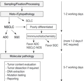
Before mutation testing, the presence of tumor cells in the sample must be assessed by a pathologist. The ratio of tumor cells to normal cells is crucial for adequate mutation testing. For direct sequencing, the percentage of tumor cells in the sam-ple should ideally be at least 50%, although reliable results can be influenced by a variety of factors. Thus, determination of the Sampling/Fixation/Processing Histo-/Cytopathology Molecular pathology Poorly differentiated 1-2 working days (more 1-2 days if IHC required) 5-7 working days - Tumor content evaluation
- Tumor dissection if required - DNA extraction - Mutation testing - Reporting Immmunohistochemistry NSCLC ADC SQC SCLC Favor ADC NSCLC-NOS Favor SQC
Fig. 2. Overall process for pathologic diagnosis and molecular analysis with recommended turnaround times. SCLC, small cell lung carcinoma; NSCLC, non-small cell lung carcinoma; ADC, ad-enocarcinoma; SQC, squamous cell carcinoma; NSCLC-NOS, non-small cell carcinoma-not otherwise specified; IHC, immuno-histochemistry.
percentage of tumor cells in a given tissue or cell specimen is recommended. Macro- or microdissection may be used to in-crease the ratio of tumor to normal tissues if required. A study performed by Sun et al. showed that the following parameters correlate with the most reliable EGFR mutation results when using cytology samples: DNA concentration >25 µg/µL, con-tent of >30 tumor cells, and tumor percentage >30%.23 The
minimum number of tumor cells required for adequate testing and the minimum ratio of tumor to normal cells is influenced by the testing method (see below).
METHODS FOR EGFR MUTATION TESTING
Various methods can be used for detecting EGFR muta-tions.33-35 Pathologists should consider the available facilities
and the pros and cons of each method, including the sensitivity and turnaround time. In addition, new techniques must be ap-proved by the Korean government.
Direct sequencing is considered to be the gold standard for
EGFR mutation analysis. In Korea, most pathology laboratories
perform direct sequencing for the detection of EGFR mutations using FFPE tissue samples. However, for directing DNA se-quencing, a high ratio of tumor tissue to normal tissue content is required (more than 50% tumor content). In contrast, real time polymerase chain reaction (PCR)-based methods exhibit
high sensitivity, requiring a mutant DNA content of only 1%.33
However, these methods can only detect previously known mu-tations or targeted sites. The peptide nucleic acid (PNA)-medi-ated PCR clamping method was recently developed and ap-proved in Korea. The PNA clamping method exhibits high sensitivity compared with direct sequencing, and clinical out-comes are not significantly different between groups harboring
EGFR mutations detected by direct sequencing or
PNA-medi-ated PCR clamping.36-38 Highly sensitive methods can also be
useful for detection of EGFR mutations associated with acquir-ed resistance, such as T790M.39
TURNAROUND TIME
Gefitinib is approved as a first-line treatment for advanced NSCLC-harboring EGFR mutations. Thus, the results of muta-tion analysis should be made available to physicians as soon as possible. Pathologic diagnosis and molecular testing are a com-bined and continuous process and should be supervised by a pa-thologist (Fig. 2). It is recommended that testing be completed within five to seven working days after ordering EGFR
muta-tion testing.
Several factors may influence turnaround time. In general, pathologic diagnoses and EGFR mutation testing performed within the same department rather than at separate laboratories would have a shorter turnaround time. When mutation testing is performed by an outside laboratory, communication and co-ordination between the pathology department and the external laboratory are encouraged.
REPEATED EXAMINATION
Several criteria for repeated examination of EGFR mutation status have been recommended. The test should be repeated in cases of poor sequencing data, a cycle threshold too close to the defined cut-off limit (with pyrosequencing or PNA clamp kit), and/or mutation results that are not matched with previously well-defined clinical-pathologic characteristics. Specifically, pa-thologists should carefully interpret the results of EGFR muta-tions found in heavy smokers, solid or mucinous cancer types, or when EGFR mutations are concurrent with other exclusive driver mutations.
REPORTING FORMAT
Molecular testing reports should contain the following infor-mation: pathologic number, age, sex, hospital unit number, bi-opsy site, sample source, requesting physician, requesting de-partment, adequacy for testing (estimated tumor cell content), receipt day, report day, methodology used, exons tested and as-sociated range of detectable mutations, mutation status, com-ments, testing technician, and corresponding pathologist.
PATHOLOGIST’S ROLES
The pathologist plays an essential role in EGFR mutation testing.40 The pathologist can either perform the test at the
home institution or transfer the tissue to a reference laboratory for external examination. In both situations, the pathologist is responsible through the procedures. First, the pathologist should choose the most appropriate tissue to be tested.7,8,23 Second, the
pathologist should verify that the selected tissue block for EGFR mutation testing contains sufficient tumor cells required for analysis. The proportion of the tumor cells in the tissue or cy-tology samples is very important to prevent contamination with non-tumor cells.7,23 Lastly, the pathologist is responsible
from routing diagnostic information (such as histologic diagno-sis), as well as from EGFR mutation testing. If the test is per-formed by an external reference laboratory, the pathologist inte-grates the test results into the pathology report of his/her
insti-tute.40 We recommend all patients with NSCLC having an
ade-nocarcinoma component or NSCLC-not-otherwise-specified af-ter immunohistochemistry should be tested for EGFR muta-tion. In the cases of squamous cell carcinomas or small cell car-cinomas arising from never-smokers, mutation testing can be performed. As for the small biopsy or cytology specimens, mac-ro- or microdissection may enhance mutation testing sensitivity.
PERSPECTIVES AND ADDITIONAL RECOMMENDATIONS
EGFR mutations and ALK rearrangements are currently
used as predictive biomarkers for targeted lung cancer therapy. In addition, other driver mutations are now receiving attention,
including ROS1 rearrangement,41 BRAF mutation,42,43 HER2
mutation,44 and RET rearrangement.45 In terms of molecular
diagnostics, these other targetable mutations have developed the need for multiplex mutational testing. However, because most patients with lung cancer present with advanced-stage disease at the time of diagnosis, the diagnosis of lung cancer is often based on small specimens from a biopsy or cytology alone. Thus, each pathology department must develop a strategy to
manage clinical samples and collaborate with clinicians.9 As
mentioned above, these strategies include minimization of di-agnostic stains in order to maximize the available tissue for mo-lecular studies9,12 and reduction of the number of trimmings for
slide sections.
CONCLUSION
As targetable mutations are discovered and corresponding targeted agents are developed, molecular diagnostics using clin-ical samples has become increasingly important. EGFR muta-tions are the most robust predictive factors for response to EGFR TKIs. Thus, each pathology department should maintain an optimal organization for the entire workflow of EGFR muta-tion testing, from sample collecmuta-tion to the final report. Lastly, pathologists should keep in mind that personalized medicine is driven by pathology and molecular diagnostics.
Conflicts of Interest
No potential conflict of interest relevant to this article was
reported.
Acknowledgments
The authors appreciate all members of the Korean Cardio-Pulmonary Pathology Study Group, their support, and their excellent opinions. This research was conducted with support from an Investigator Sponsored Study Programme of AstraZen-eca; Partly supported by a grant from the Korea Healthcare Technology R&D Project, Ministry of Health and Welfare, Re-public of Korea (A111405, to JH Chung).
REFERENCES 1. Cagle PT, Chirieac LR. Advances in treatment of lung cancer with targeted therapy. Arch Pathol Lab Med 2012; 136: 504-9. 2. Janku F, Stewart DJ, Kurzrock R. Targeted therapy in non-small-cell lung cancer: is it becoming a reality? Nat Rev Clin Oncol 2010; 7: 401-14. 3. Linardou H, Dahabreh IJ, Bafaloukos D, Kosmidis P, Murray S. So-matic EGFR mutations and efficacy of tyrosine kinase inhibitors in NSCLC. Nat Rev Clin Oncol 2009; 6: 352-66. 4. Mok TS, Wu YL, Thongprasert S, et al. Gefitinib or carboplatin-pa-clitaxel in pulmonary adenocarcinoma. N Engl J Med 2009; 361: 947-57. 5. Fukuoka M, Wu YL, Thongprasert S, et al. Biomarker analyses and final overall survival results from a phase III, randomized, open-la- bel, first-line study of gefitinib versus carboplatin/paclitaxel in clini-cally selected patients with advanced non-small-cell lung cancer in Asia (IPASS). J Clin Oncol 2011; 29: 2866-74. 6. D’Angelo SP, Pietanza MC, Johnson ML, et al. Incidence of EGFR exon 19 deletions and L858R in tumor specimens from men and cigarette smokers with lung adenocarcinomas. J Clin Oncol 2011; 29: 2066-70.
7. Sun PL, Seol H, Lee HJ, et al. High incidence of EGFR mutations in Korean men smokers with no intratumoral heterogeneity of lung adenocarcinomas: correlation with histologic subtypes, EGFR/TTF-1 expressions, and clinical features. J Thorac Oncol 2012; 7: 323-30. 8. Pirker R, Herth FJ, Kerr KM, et al. Consensus for EGFR mutation testing in non-small cell lung cancer: results from a European work-shop. J Thorac Oncol 2010; 5: 1706-13. 9. Travis WD, Brambilla E, Noguchi M, et al. International Association for the Study of Lung Cancer/American Thoracic Society/European Respiratory Society international multidisciplinary classification of lung adenocarcinoma. J Thorac Oncol 2011; 6: 244-85. 10. Salto-Tellez M, Tsao MS, Shih JY, et al. Clinical and testing protocols for the analysis of epidermal growth factor receptor mutations in
East Asian patients with non-small cell lung cancer: a combined clinical-molecular pathological approach. J Thorac Oncol 2011; 6: 1663-9. 11. Rekhtman N, Ang DC, Sima CS, Travis WD, Moreira AL. Immuno-histochemical algorithm for differentiation of lung adenocarcinoma and squamous cell carcinoma based on large series of whole-tissue sections with validation in small specimens. Mod Pathol 2011; 24: 1348-59. 12. Noh S, Shim H. Optimal combination of immunohistochemical markers for subclassification of non-small cell lung carcinomas: a tissue microarray study of poorly differentiated areas. Lung Cancer 2012; 76: 51-5. 13. Yatabe Y. EGFR mutations and the terminal respiratory unit. Can-cer Metastasis Rev 2010; 29: 23-36. 14. Zakowski MF, Hussain S, Pao W, et al. Morphologic features of ad-enocarcinoma of the lung predictive of response to the epidermal growth factor receptor kinase inhibitors erlotinib and gefitinib. Arch Pathol Lab Med 2009; 133: 470-7. 15. Shim HS, Lee da H, Park EJ, Kim SH. Histopathologic characteris- tics of lung adenocarcinomas with epidermal growth factor recep-tor mutations in the International Association for the Study of Lung Cancer/American Thoracic Society/European Respiratory Society lung adenocarcinoma classification. Arch Pathol Lab Med 2011; 135: 1329-34. 16. Park SH, Ha SY, Lee JI, et al. Epidermal growth factor receptor mu-tations and the clinical outcome in male smokers with squamous cell carcinoma of lung. J Korean Med Sci 2009; 24: 448-52. 17. Miyamae Y, Shimizu K, Hirato J, et al. Significance of epidermal growth factor receptor gene mutations in squamous cell lung carci-noma. Oncol Rep 2011; 25: 921-8. 18. Tatematsu A, Shimizu J, Murakami Y, et al. Epidermal growth fac-tor receptor mutations in small cell lung cancer. Clin Cancer Res 2008; 14: 6092-6. 19. Shiao TH, Chang YL, Yu CJ, et al. Epidermal growth factor receptor mutations in small cell lung cancer: a brief report. J Thorac Oncol 2011; 6: 195-8. 20. Rekhtman N, Brandt SM, Sigel CS, et al. Suitability of thoracic cytol- ogy for new therapeutic paradigms in non-small cell lung carcino-ma: high accuracy of tumor subtyping and feasibility of EGFR and KRAS molecular testing. J Thorac Oncol 2011; 6: 451-8. 21. Navani N, Brown JM, Nankivell M, et al. Suitability of endobron- chial ultrasound-guided transbronchial needle aspiration speci-mens for subtyping and genotyping of non-small cell lung cancer: a multicenter study of 774 patients. Am J Respir Crit Care Med 2012; 185: 1316-22. 22. Allegrini S, Antona J, Mezzapelle R, et al. Epidermal growth factor receptor gene analysis with a highly sensitive molecular assay in routine cytologic specimens of lung adenocarcinoma. Am J Clin Pathol 2012; 138: 377-81. 23. Sun PL, Jin Y, Kim H, Lee CT, Jheon S, Chung JH. High concordance of EGFR mutation status between histologic and corresponding cy-tologic specimens of lung adenocarcinomas. Cancer Cytopathol 2012 Dec 5 [Epub].http://dx.doi.org/10.1002/cncy.21260. 24. da Cunha Santos G, Saieg MA, Geddie W, Leighl N. EGFR gene status in cytological samples of nonsmall cell lung carcinoma: con-troversies and opportunities. Cancer Cytopathol 2011; 119: 80-91. 25. Taniguchi K, Okami J, Kodama K, Higashiyama M, Kato K. Intra- tumor heterogeneity of epidermal growth factor receptor muta-tions in lung cancer and its correlation to the response to gefitinib. Cancer Sci 2008; 99: 929-35. 26. Schmid K, Oehl N, Wrba F, Pirker R, Pirker C, Filipits M. EGFR/ KRAS/BRAF mutations in primary lung adenocarcinomas and cor-responding locoregional lymph node metastases. Clin Cancer Res 2009; 15: 4554-60. 27. Gow CH, Chang YL, Hsu YC, et al. Comparison of epidermal growth factor receptor mutations between primary and corresponding me-tastatic tumors in tyrosine kinase inhibitor-naive non-small-cell lung cancer. Ann Oncol 2009; 20: 696-702. 28. Yatabe Y, Matsuo K, Mitsudomi T. Heterogeneous distribution of EGFR mutations is extremely rare in lung adenocarcinoma. J Clin Oncol 2011; 29: 2972-7. 29. Chen ZY, Zhong WZ, Zhang XC, et al. EGFR mutation heterogene-ity and the mixed response to EGFR tyrosine kinase inhibitors of lung adenocarcinomas. Oncologist 2012; 17: 978-85. 30. Bai H, Wang Z, Chen K, et al. Influence of chemotherapy on EGFR mutation status among patients with non-small-cell lung cancer. J Clin Oncol 2012; 30: 3077-83. 31. Eberhard DA, Giaccone G, Johnson BE; Non-Small-Cell Lung Can-cer Working Group. Biomarkers of response to epidermal growth factor receptor inhibitors in Non-Small-Cell Lung Cancer Working Group: standardization for use in the clinical trial setting. J Clin Oncol 2008; 26: 983-94. 32. Nicholson AG, Gonzalez D, Shah P, et al. Refining the diagnosis and EGFR status of non-small cell lung carcinoma in biopsy and cytologic material, using a panel of mucin staining, TTF-1, cytoker-atin 5/6, and p63, and EGFR mutation analysis. J Thorac Oncol 2010; 5: 436-41. 33. Pao W, Ladanyi M. Epidermal growth factor receptor mutation test-ing in lung cancer: searching for the ideal method. Clin Cancer Res 2007; 13: 4954-5. 34. Ellison G, Zhu G, Moulis A, Dearden S, Speake G, McCormack R.
EGFR mutation testing in lung cancer: a review of available meth- ods and their use for analysis of tumour tissue and cytology sam-ples. J Clin Pathol 2013; 66: 79-89. 35. Lee HJ, Xu X, Kim H, et al. Comparison of direct sequencing, PNA clamping-real time polymerase chain reaction, and pyrosequencing methods for the detection of EGFR mutations in non-small cell lung carcinoma and the correlation with clinical responses to EGFR tyro-sine kinase inhibitor treatment. Korean J Pathol 2013; 47: 52-60. 36. Kim HJ, Kim WS, Shin KC, et al. Comparative analysis of peptide nucleic acid (PNA)-mediated real-time PCR clamping and DNA direct sequencing for EGFR mutation detection. Tuberc Respir Dis 2011; 70: 21-7. 37. Kim HJ, Lee KY, Kim YC, et al. Detection and comparison of pep- tide nucleic acid-mediated real-time polymerase chain reaction clam- ping and direct gene sequencing for epidermal growth factor re-ceptor mutations in patients with non-small cell lung cancer. Lung Cancer 2012; 75: 321-5. 38. Han HS, Lim SN, An JY, et al. Detection of EGFR mutation status in lung adenocarcinoma specimens with different proportions of tu- mor cells using two methods of differential sensitivity. J Thorac On-col 2012; 7: 355-64. 39. Arcila ME, Oxnard GR, Nafa K, et al. Rebiopsy of lung cancer pa-tients with acquired resistance to EGFR inhibitors and enhanced detection of the T790M mutation using a locked nucleic acid-based assay. Clin Cancer Res 2011; 17: 1169-80. 40. van Krieken JH, Jung A, Kirchner T, et al. KRAS mutation testing for predicting response to anti-EGFR therapy for colorectal carcino-ma: proposal for an European quality assurance program. Virchows Arch 2008; 453: 417-31. 41. Bergethon K, Shaw AT, Ou SH, et al. ROS1 rearrangements define a unique molecular class of lung cancers. J Clin Oncol 2012; 30: 863-70. 42. Marchetti A, Felicioni L, Malatesta S, et al. Clinical features and out-come of patients with non-small-cell lung cancer harboring BRAF mutations. J Clin Oncol 2011; 29: 3574-9. 43. Paik PK, Arcila ME, Fara M, et al. Clinical characteristics of patients with lung adenocarcinomas harboring BRAF mutations. J Clin On-col 2011; 29: 2046-51. 44. Arcila ME, Chaft JE, Nafa K, et al. Prevalence, clinicopathologic as- sociations, and molecular spectrum of ERBB2 (HER2) tyrosine ki-nase mutations in lung adenocarcinomas. Clin Cancer Res 2012; 18: 4910-8. 45. Wang R, Hu H, Pan Y, et al. RET fusions define a unique molecular and clinicopathologic subtype of non-small-cell lung cancer. J Clin Oncol 2012; 30: 4352-9.