R E S E A R C H
Open Access
Change of the upper airway after
mandibular setback surgery in patients
with mandibular prognathism and anterior
open bite
Kyungjin Lee
1and Soon Jung Hwang
2*Abstract
Purpose: It has been reported before that the amount of pharyngeal airway space (PAS) significantly decreases following mandibular setback (MS) surgery in patients with mandibular prognathism (MP). Further, MP patients with an anterior open-bite (AOB) presentation may show a larger decrease in PAS compared with those without AOB. However, studies on postoperative PAS changes in MP patients with AOB remain rare. This study sought to evaluate changes in PAS and hyoid bone positioning following MS surgery in MP patients with and without AOB. Patients and methods: Twenty patients who underwent two jaw surgery involving MS movement were included. Patients were divided into a non-AOB group (n = 10; overbite > 2 mm) and an AOB group (n = 10; overbite < − 4 mm). Three-dimensional changes in PAS and hyoid bone positioning were compared and statistically evaluated pre- and postoperatively using computed tomography (CT).
Results: The mean magnitude of MS was 6.0 ± 2.8 mm and 5.6 ± 3.2 mm in the non-AOB group and AOB group, respectively. The oropharyngeal volume and upper hypopharyngeal volume were significantly reduced after surgery in both the groups (p = 0.006 and p = 0.003), while the retroglossal cross-sectional area was significantly reduced only in the AOB group (p = 0.028). Although the AOB group showed a larger decrease in PAS, the difference was not statistically significant between the groups. The position of the hyoid bone showed significant posterior and inferior displacement only in the AOB group, while the vertical displacement of the hyoid bone showed a statistically significant difference between the two groups.
Conclusion: PAS was significantly decreased after MS in both the groups, while only the AOB group presented a statistically significant reduction in the retroglossal cross-sectional area. Vertical displacement of the hyoid bone showed a statistically significant difference between the groups, while the PAS change was not. Surgeons should be aware of potential postoperative airway problems that may arise when performing MS surgeries.
Keywords: Mandibular setback, Pharyngeal airway space, Mandibular prognathism, Open-bite, Hyoid bone
© The Author(s). 2019 Open Access This article is distributed under the terms of the Creative Commons Attribution 4.0 International License (http://creativecommons.org/licenses/by/4.0/), which permits unrestricted use, distribution, and reproduction in any medium, provided you give appropriate credit to the original author(s) and the source, provide a link to the Creative Commons license, and indicate if changes were made.
* Correspondence:[email protected]
2HSJ Dental Clinic for Oral and Maxillofacial Surgery, Wannam Building 2,3F,
349 Gangnam-daero, Seocho-gu, Seoul 06626, Republic of Korea Full list of author information is available at the end of the article
Introduction
Mandibular setback (MS) surgery is usually performed for aesthetic and functional correction in patients with mandibular prognathism (MP). Because MS involves backward movement of the mandible along with muscle and other soft tissue attachments, the pharyngeal airway space (PAS) and positioning of the tongue are inevitably affected [1]. One of the main concerns of these conse-quences is their potential to cause obstructive sleep apnea (OSA) [2]. OSA is known to increase the risk of hypertension, ischemic myocardial diseases, and cerebral vascular diseases and thus is considered potentially life-threatening [3].
Earlier studies on MS and PAS changes were per-formed using lateral cephalometric analysis. These stud-ies repeatedly showed that the PAS decreased after MS surgery [1, 4–6], and the degree of PAS reduction was smaller with bimaxillary surgery than with mandible-only surgery [5]. In contrast, in certain cases, particularly in patients with AOB, the joint movement of the man-dible to maxilla could scale down the reduction of the PAS. However, cephalometric study for the analysis of change in the PAS is limited to two-dimensional mea-surements on the sagittal plane, while the PAS is a three-dimensional (3D) structure that requires 3D vol-ume and cross-sectional analysis for proper assessment [7, 8]. Some studies using computed tomography (CT) reported no decrease in PAS [9] or even increase in the total PAS after bimaxillary surgery involving posterior movement of the mandible [10,11], but the majority of them confirmed reduced oropharyngeal space, cross-sectional area posterior to the tongue and soft palate, or decreased lateral and frontal dimensions of the PAS [7,
8, 10–14]. It has also been speculated that MS may in-duce OSA [5, 7]. A recent study by Yang et al. [14] found that four of twelve patients who underwent orthognathic surgery involving substantial MS presented with new-onset OSA in the postoperative period.
Patients with MP and AOB commonly present with a higher mandibular plane angle, higher gonial angle, forward tongue position, and macroglossia [15, 16]. Anterior tongue placement in patients with AOB is regarded as a physiological adaptation [15, 17]. Moreover, patients with AOB have more constricted upper airways [18, 19] and inferiorly positioned hyoid bones [20, 21]. Partial glossectomy is often warranted when posterior movement of the mandible is planned [16, 22]. Given the fact that patients with AOB have a higher ratio of tongue volume to oral space volume [15], it is rational to speculate that the PAS can be-come constricted following backward movement of the anteriorly positioned and enlarged tongue after surgical closure of AOB when compared with those without AOB. Nevertheless, studies on postoperative
PAS changes in MP patients with AOB are only rarely reported.
The purpose of this study was to evaluate the changes in PAS and hyoid bone positioning after MS using 3D facial multislice CT scans and to compare the results be-tween patients with and without AOB.
Patients and methods
Patients
This study was conducted under the Institutional Re-view Board approval from Seoul National University Dental Hospital (CRI12036). The study procedure was performed in accordance with Helsinki Declaration revised in 2008. This study included 20 patients with skeletal class III malocclusion who underwent bilat-eral sagittal split ramus osteotomy (BSSRO) and Le Fort I osteotomy with or without genioplasty at the Department of Oral and Maxillofacial Surgery, Seoul National University Dental Hospital in Seoul, Korea. All patients showed < 10 mm of MS at the B point and < 4 mm of maxillary movement at the A point in the immediate postoperative lateral cephalogram. Pa-tients were divided into two groups: an AOB group (n = 10) with < − 4 mm of preoperative overbite and a non-AOB group (n = 10) with an overbite of > 2 mm. Those who underwent BSSRO alone were ex-cluded, and all surgeries were performed by a single surgeon. In all patients, multislice CT scans were taken 1 month before (T1) and 3–6 months after the surgery (T2). The mean age of the patients at the time of surgery was 20.1 ± 6.1 years (range 16–26 years) and the sex ratio was 11:9 (male:female). All patients received pre- and postoperative orthodontic treatment, and semirigid fixation was completed using four-hole titanium miniplates and screws.
Cephalometric analysis
To measure surgical changes and the degree of over-bite, the lateral cephalograms of each patient were taken in the maximum intercuspal position at a mag-nification ratio of 1.1:1 prior to surgery (T0) and im-mediately after surgery (T1). Each cephalogram was traced on an acetate paper. Cephalometric analysis was conducted using the superimposition technique. Reference points included the sella (S), nasion (N), A point (A), B point (B), upper incision (U1), and lower incision (L1). All reference points were transferred to the lateral cephalograms taken at T1. An x- and y-co-ordinate system was established, wherein the x-axis was constructed by rotating S–N clockwise by 7° (SN7), while the y-axis was constructed by drawing a line through the sella, perpendicular to SN7 (SN7v). The linear parameters consisted of the x and y coor-dinates of A and B.
Evaluation of pharyngeal airway in CT
3D facial multislice CT scans were obtained from all pa-tients using a CT scanner (SOMATOM sensation 10; Siemens, Munich, Germany) with the following pa-rameters: 120 kVp, 80 mAs, and a slice thickness of 0.75 mm. Digital image files were saved in the Digital Imaging and Communications in Medicine format and imported into the Invivo Dental software pro-gram (Anatomage, San Jose, CA, USA). CT images were rendered into volumetric images. The recon-structed sagittal, axial, and coronal slices and 3D im-ages were then obtained. To standardize the measurements and minimize errors, the Frankfort horizontal (FH) plane was constructed to reorient the 3D images. The FH plane was constructed from the right and left porions and the right orbitale.
The airway study in CT consisted of three compo-nents: (1) changes in distance and area in axial CT, (2) changes in airway volume in 3D images, and (3) changes in hyoid bone positioning.
To assess the linear distance and cross-sectional area of the posterior airway, pre- and postoperative measure-ments were collected at two different levels, specifically at the retropalatal level (the level of the most posterior point of the soft palate parallel to the FH plane) and the retroglossal level (the level of the most posterior point of the tongue base parallel to the FH plane). For both levels, the largest mesiodistal width (LMD),
anteroposter-ior length (LAP), and cross-sectional area (S) were
measured.
To evaluate the airway volume, a range of − 1024 to − 600 Hounsfield units was set as the threshold value of the CT image, where the pharyngeal airway could be effectively differentiated from the neighbor-ing soft tissue. The pharyngeal airway was divided into the following two regions using cervical vertebra (CV) reference points, with the CVn plane defined as the plane parallel to the FH plane passing through the most inferior point of the CVn: (1) the oropha-ryngeal volume between CV1 and CV2 planes (Vo)
and (2) the upper hypopharyngeal volume between the CV2 and CV3 planes (Vh). The pharyngeal airway
volumes of all patients were measured by the same examiner using the Invivo Dental software program (Anatomage, San Jose, CA, USA), and postoperative volume changes were calculated (Fig. 1).
To evaluate the hyoid bone positioning before and after surgery, two reference lines were defined. The x-axis consisted of the line tangent to the inferior portion of the sella turcica and parallel to the FH plane, while the y-axis consisted of the line perpendicular to the x-axis and passing through nasion. The horizontal and ver-tical positions of the hyoid bone were measured using the distances between the most anterosuperior point of
hyoid bone to the x-axis (Hx) and y-axis (Hy),
respect-ively (Fig.2).
Statistical analysis
The descriptive statistics of the preoperative and postopera-tive measurements were processed using the SPSS for Win-dows version 21 software program (IBM Corp., Armonk, NY, USA). Kolmogorov–Smirnov analysis was performed for all measured parameters attested to the normal distribution of values. The preoperative and postoperative cephalometric measurements and pharyngeal airway volume, length, and area were analyzed using the Wilcoxon signed-rank test. A pairedt test was performed to compare the differences be-tween the groups. Differences were considered to be signifi-cant atp < 0.05
Results
Surgical changes of maxilla and mandible
On average, the mandible was moved backward by 6.0 ± 2.76 mm and upward by 1.3 ± 1.6 mm in the non-AOB group and backward by 5.6 ± 3.2 mm and upward by 5.4 ± 2.2 mm in the AOB group at the B point. Further-more, the maxilla was moved forward by 1.4 ± 2.2 mm and upward by 1.9 ± 2.3 mm in the non-AOB group and forward by 2.0 ± 2.0 mm and upward by 3.9 ± 1.7 mm in the AOB group at the A point (Table1.)
Pharyngeal airway
The postoperative airway volume was significantly de-creased in both the oropharynx and upper hypopharynx in both the groups (p < 0.05). The average change in the airway volume was − 12.16% ± 28.18% in the non-AOB group and − 14.62% ± 10.60% in the AOB group in the oropharynx and − 14.71% ± 20.02% in the non-AOB group and − 17.43% ± 20.18% in the AOB group in the upper hypopharynx, respectively. Although there was a greater reduction of PAS in the AOB group in both the oropharynx and upper hypopharynx, the difference was not statistically significant (Table2; Fig.3).
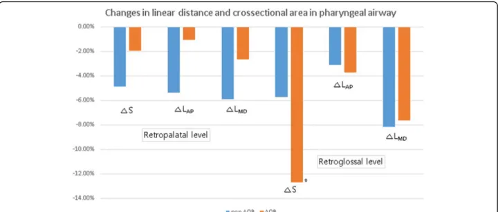
The linear distance and area were also decreased after surgery in both the groups. At both the retropalatal and retroglossal levels, LMD, LAP, and S decreased, but there
was no statistically significant difference in these changes except for S at the retroglossal level in the AOB group (p = 0.03). Intergroup analysis showed no significant dif-ference present in all parameters at both levels (Table2; Fig.4).
Hyoid bone positioning
The hyoid bone was displaced backward by 2.02 ± 5.13 mm and downward by 0.32 ± 2.93 mm in the non-AOB group and backward by 4.63 ± 4.80 mm and upward by 4.02 ± 4.40 mm in the AOB group (p > 0.05). Vertical
and horizontal changes in the hyoid bone in the AOB group demonstrated statistical significance (p < 0.05) (Fig. 5). There was a statistically significant difference between the two groups in terms of vertical displace-ment of the hyoid bone (p = 0.02).
Discussion
Changes in the airway space after MS surgery have gained attention in recent decades and have been widely studied, yet the outcomes vary among studies. Some of the earlier studies did not report any signifi-cant changes being present in the airway space after MS [9, 23, 24]. Several other studies performed using CT, however, suggested that PAS was significantly de-creased after MS [2, 3, 10–13, 25–27]. For example, Lee et al. achieved a decrease of 14.07% in the total volume following a mean MS of 9.20 mm [11], while Kim et al. reported a decrease of 15.80% in total PAS and decreases of 22.08%, 8.10%, and 12.43% in the oropharynx, nasopharynx, and hypopharynx, respect-ively, after a mean MS of 8.25 mm [28]. Elsewhere, Yang et al. showed a decrease of 22.30% in the oro-pharynx and of 25.24% in the hypooro-pharynx after a mean MS of 10.53 mm [14]. In the present study with mean MS values of 6.0 mm in the non-AOB group
Fig.2 Vertical and horizontal positioning of the hyoid bone was measured in CT (mm). Thex-axis was defined as the line tangent to the inferior point of the sella turcica and parallel to the FH plane, while they-axis was defined as the line passing through the nasion and perpendicular to thex-axis
Fig. 1 Measurements in volume, linear distance, and area in the pharyngeal airway. The largest transverse width, anteroposterior length, and cross-sectional area were measured at the level of the most constricted point of the soft palate and tongue base. The airway was divided as follows, and each volume was measured for the oropharyngeal area (between CV1 line and CV2 line) and the upper hypopharyngeal area (between CV2 line and CV3 line), with the CVn lines being parallel to the FH plane
and 5.6 mm in the AOB group, the PAS was signifi-cantly decreased in both the groups at the oropharynx (12.10% and 14.62% in the non-AOB and AOB groups, respectively) and upper hypopharynx (14.71% and 17.42% in the non-AOB and AOB groups, respectively).
Cases of AOB present common skeletal characteris-tics including a negative overbite, augmented anterior facial height, steep mandibular plane, and increased gonial angle [29–31]. Patients with AOB also have different characteristics of the airway, hyoid bone, and tongue. Chang et al. and Cho et al. reported that the tongue was positioned more anteriorly in MP patients with AOB [32, 33], and Lee et al. showed that the hyoid bone was positioned more inferiorly in MP pa-tients with AOB [21]. Tarkar et al. determined in their study that the PAS was significantly narrower and the position of the dorsum of the tongue was sig-nificantly higher in AOB patients compared with non-AOB patients [19], while Abu Allhaija et al. showed that the anteroposterior dimension of PAS was nar-rower in AOB patients than in non-AOB patients [34]. These features are unfavorable for comfortable respiration and may influence the changes in PAS after MS surgery. However, studies on postoperative PAS changes in MP patients with AOB are hard to find in the literature. According to the results of the present study, there was a greater reduction in the oropharyngeal and upper hypopharyngeal spaces and the sectional space area at the tongue base level in the AOB group, but the difference was not statisti-cally significant. Further studies with larger sample sizes are warranted to elucidate differences in PAS changes between the two groups.
The hyoid bone is a movable hard tissue suspended via muscles and ligaments connecting it to the skull base, mandible, pharynx, and tongue. Thus, its pos-ition is easily influenced by the movement of sur-rounding tissues [35, 36]. A positional assessment of the hyoid bone is used to evaluate the physiological equilibrium of the suprahyoid muscles, infrahyoid muscles, and tissues surrounding the hyoid bone [6,
37]. Both posterior and inferior displacements of the hyoid bone have been generally noted after MS sur-gery [26, 38]. Such movements are believed to be the result of soft tissue (e.g., muscle) adaptation [26, 39], and collectively represent one of the most important factors in the phenomenon of PAS narrowing. PAS narrowing is a very important clinical consequence because it can cause upper airway crowding and col-lapse [3]. In the present study, the hyoid bone showed posterior and inferior displacement in both the groups, which is in accordance with the majority of findings of previous studies. However, the
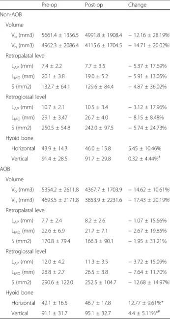
Table 2 Results of pharyngeal airway and hyoid bone position measurements
Pre-op Post-op Change Non-AOB Volume Vo(mm3) 5661.4 ± 1356.5 4991.8 ± 1908.4 − 12.16 ± 28.19%* Vh(mm3) 4962.3 ± 2086.4 4115.6 ± 1704.5 − 14.71 ± 20.02%* Retropalatal level LAP(mm) 7.4 ± 2.2 7.7 ± 3.5 − 5.37 ± 17.69% LMD(mm) 20.1 ± 3.8 19.0 ± 5.2 − 5.91 ± 13.05% S (mm2) 132.7 ± 64.1 129.6 ± 84.4 − 4.87 ± 36.02% Retroglossal level LAP(mm) 10.7 ± 2.1 10.5 ± 3.4 − 3.12 ± 17.96% LMD(mm) 29.1 ± 3.47 26.7 ± 4.0 − 8.15 ± 8.48% S (mm2) 250.5 ± 54.8 242.0 ± 97.5 − 5.74 ± 24.73% Hyoid bone Horizontal 43.9 ± 14.3 46.0 ± 15.8 5.45 ± 10.46% Vertical 91.4 ± 28.5 91.7 ± 29.8 0.32 ± 4.44%# AOB Volume Vo(mm3) 5354.2 ± 2611.8 4367.7 ± 1703.9 − 14.62 ± 10.61%* Vh(mm3) 4693.5 ± 2171.8 3853.9 ± 2231.6 − 17.43 ± 20.19%* Retropalatal level LAP(mm) 7.7 ± 2.4 8.2 ± 2.6 − 1.07 ± 15.66% LMD(mm) 22.6 ± 6.9 21.7 ± 7.1 − 2.67 ± 19.85% S (mm2) 170.8 ± 79.4 166.3 ± 90.1 − 1.95 ± 31.21% Retroglossal level LAP(mm) 12.0 ± 4.2 11.3 ± 3.5 − 3.72 ± 15.09% LMD(mm) 28.8 ± 2.7 26.5 ± 3.8 − 7.64 ± 11.70% S (mm2) 290.6 ± 122.0 252.5 ± 104.7 − 12.68 ± 14.97%* Hyoid bone Horizontal 42.1 ± 16.5 46.7 ± 17.8 12.77 ± 9.61%* Vertical 91.1 ± 31.7 95.1 ± 32.7 4.4 ± 5.11%*#
*p < 0.05 in Wilcoxon singed-rank tests
#
p < 0.05 in paired t test
Table 1 Surgical movements following two jaw surgery for mandibular setback movement
Surgical movement non-AOB AOB Mandible Horizontal − 5.95 ± 2.67 − 5.55 ± 3.24 Vertical 1.25 ± 1.57 5.35 ± 2.17 Maxilla Horizontal 1.35 ± 2.19 1.95 ± 1.96 Vertical 1.90 ± 2.32 3.90 ± 1.66
magnitude of vertical and horizontal displacement showed statistically significant differences only in the AOB group. In the intergroup comparison, only the vertical displacement of the hyoid bone showed a sta-tistically significant difference.
Conclusion
Following MS surgery in MP patients, oropharyngeal and upper hypopharyngeal airway volume were signifi-cantly decreased in both the AOB and non-AOB groups. Meanwhile, the hyoid bone showed significant inferior and posterior displacement in the AOB group
only. In a comparison between the two groups, the hyoid bone showed a significantly larger inferior dis-placement in the AOB group. Although the difference was not statistically significant, the magnitude of PAS reduction was larger in the AOB group in both the oropharynx and upper hypopharynx. The results of this study suggest that the possibility of the onset of a respiratory disturbance such as OSA following MS may be greater in MP patients with AOB than in those without AOB. Therefore, surgeons should be aware of potential postoperative airway problems when per-forming MS surgeries.
Fig. 3 The average change in the pharyngeal airway volume. The postoperative airway volume was significantly decreased in both the groups (*p = 0.006 and *p = 0.003), even though the difference between the two groups was not statistically significant
Fig. 4 The average changes in linear distance and cross-sectional area in the pharyngeal airway. All parameters were decreased postoperatively in the retropalatal level and retroglossal level, while S at the retroglossal level was significantly decreased in the AOB group (*p = 0.028)
Abbreviations
2D:Two-dimensional; 3D: Three-dimensional; AOB: Anterior open-bite; BSSRO: Bilateral sagittal split ramus osteotomy; CSA: Cross-sectional area; CT: Computed tomography; CV: Cervical vertebra; MP: Mandibular prognathism; MS: Mandibular setback; OSA: Obstructive sleep apnea; PAS: Pharyngeal airway space
Acknowledgements Not Applicable Authors’ contributions Not Applicable Authors’ information Not Applicable Funding Not Applicable
Availability of data and materials
All data generated or analyzed during this study are included in this article and its supplementary information files.
Ethics approval and consent to participate
This study was approved by the Institutional Review Board of Seoul National University Dental
Hospital (CRI12036).
Consent for publication Not Applicable
Competing interests
The authors declare that they have no competing interests. Author details
1
Department of Oral and Maxillofacial Surgery, School of Dentistry, Seoul National University, Seoul, Republic of Korea.2HSJ Dental Clinic for Oral and
Maxillofacial Surgery, Wannam Building 2,3F, 349 Gangnam-daero, Seocho-gu, Seoul 06626, Republic of Korea.
Received: 2 September 2019 Accepted: 23 September 2019
References
1. Muto T, Yamazaki A, Takeda S et al (2008) Effect of bilateral sagittal split ramus osteotomy setback on the soft palate and pharyngeal airway space. Int J Oral Maxillofac Surg 37:419–423
2. Mattos CT, Vilani GN, Sant'Anna EF et al (2011) Effects of orthognathic surgery on oropharyngeal airway: a meta-analysis. Int J Oral Maxillofac Surg 40:1347–1356
3. Hasebe D, Kobayashi T, Hasegawa M et al (2011) Changes in oropharyngeal airway and respiratory function during sleep after orthognathic surgery in patients with mandibular prognathism. Int J Oral Maxillofac Surg 40:584–592 4. Chen F, Terada K, Hanada K et al (2005) Predicting the pharyngeal airway
space after mandibular setback surgery. J Oral Maxillofac Surg 63:1509–1514 5. Chen F, Terada K, Hua Y et al (2007) Effects of bimaxillary surgery and
mandibular setback surgery on pharyngeal airway measurements in patients with Class III skeletal deformities. Am J Orthod Dentofacial Orthop 131:372–377
6. Kawakami M, Yamamoto K, Fujimoto M et al (2005) Changes in tongue and hyoid positions, and posterior airway space following mandibular setback surgery. J Craniomaxillofac Surg 33:107–110
7. Kim HS, Kim GT, Kim S et al (2016) Three-dimensional evaluation of the pharyngeal airway using cone-beam computed tomography following bimaxillary orthognathic surgery in skeletal class III patients. Clin Oral Investig 20:915–922
8. Park SB, Kim YI, Son WS et al (2012) Cone-beam computed tomography evaluation of short- and long-term airway change and stability after orthognathic surgery in patients with Class III skeletal deformities: bimaxillary surgery and mandibular setback surgery. Int J Oral Maxillofac Surg 41:87–93
9. Jakobsone G, Neimane L, Krumina G (2010) Two- and three-dimensional evaluation of the upper airway after bimaxillary correction of Class III malocclusion. Oral Surg Oral Med Oral Pathol Oral Radiol Endod 110:234–242 10. Gokce SM, Gorgulu S, Gokce HS et al (2014) Evaluation of pharyngeal airway
space changes after bimaxillary orthognathic surgery with a 3-dimensional simulation and modeling program. Am J Orthod Dentofacial Orthop 146: 477–492
11. Lee Y, Chun YS, Kang N et al (2012) Volumetric changes in the upper airway after bimaxillary surgery for skeletal class III malocclusions: a case series study using 3-dimensional cone-beam computed tomography. J Oral Maxillofac Surg 70:2867–2875
12. Kawamata A, Fujishita M, Ariji Y et al (2000) Three-dimensional computed tomographic evaluation of morphologic airway changes after mandibular Fig. 5 Vertical and horizontal changes of the hyoid bone location. The hyoid bone was displaced backward and downward in both the groups. Vertical and horizontal changes in the hyoid bone positioning in the AOB group showed statistical significance (*p = 0.022 and *p = 0.028), and there was a statistically significant difference between the two groups in terms of vertical displacement of the hyoid bone (#p = 0.02)
setback osteotomy for prognathism. Oral Surg Oral Med Oral Pathol Oral Radiol Endod 89:278–287
13. Foltan R, Hoffmannova J, Donev F et al (2009) The impact of Le Fort I advancement and bilateral sagittal split osteotomy setback on ventilation during sleep. Int J Oral Maxillofac Surg 38:1036–1040
14. Yang HJ, Jung YE, Kwon IJ et al (2019) Airway changes and prevalence of obstructive sleep apnoea after bimaxillary orthognathic surgery with large mandibular setback. Int J Oral Maxillofac Surg.
15. Shenoy U, Hazarey P, Akhare PJ et al (2015) Cephalometric appraisal of tongue and related soft tissues in normal and open bite subjects at rest. J Clin Diagn Res 9:ZC16–ZC20
16. Mehtap Karamese OA, Selimoglu MN, Abacı M, Akatekin A, Tosun Z (2014) Combined Therapy for Mandibular Prognathism: Sagittal Split Osteotomy with Excision of Tongue. Modern Plastic Surgery 4:5
17. Proffit WR (1972) Lingual pressure patterns in the transition from tongue thrust to adult swallowing. Arch Oral Biol 17:555–563
18. de Freitas MR, Alcazar NM, Janson G et al (2006) Upper and lower pharyngeal airways in subjects with Class I and Class II malocclusions and different growth patterns. Am J Orthod Dentofacial Orthop 130:742–745 19. Tarkar JS, Parashar S, Gupta G et al (2016) An evaluation of upper and lower
pharyngeal airway width, tongue posture and hyoid bone position in subjects with different growth patterns. J Clin Diagn Res 10:ZC79–ZC83 20. Tourne LP (1991) Growth of the pharynx and its physiologic implications.
Am J Orthod Dentofacial Orthop 99:129–139
21. Lee YSBH, Lee KJ, Yu HS (2009) The structural change in the hyoid bone and upper airway after orthognathic surgery for skeletal class III anterior open bite patients using 3-dimensional computed tomography. Korean J Orthodontics 39:11
22. Yucel G, Marsan G, Cura N et al (2009) Treatment of a patient with a severe Class III and open bite: a case report. World J Orthod 10:57–66
23. Lee DKKS (1989) Study on the changes in the upper airway following osteotomy for the mandibular prognathism. J Korean Dental Assoc 27:1143 24. Chin KSSW (1993) The relationships between the postoperative stability and
the changes in the tongue position, the hyoid bone position and the upper airway size after orthognathic surgery in patients with mandibular prognathism. Korean J Orthodontics 23:13
25. Hong JS, Park YH, Kim YJ et al (2011) Three-dimensional changes in pharyngeal airway in skeletal class III patients undergoing orthognathic surgery. J Oral Maxillofac Surg 69:e401–e408
26. Gokce SM, Gorgulu S, Gokce HS et al (2012) Changes in posterior airway space, pulmonary function and sleep quality, following bimaxillary orthognathic surgery. Int J Oral Maxillofac Surg 41:820–829
27. Lee UL, Oh H, Min SK et al (2017) The structural changes of upper airway and newly developed sleep breathing disorders after surgical treatment in class III malocclusion subjects. Medicine (Baltimore) 96:e6873
28. Kim MA, Kim BR, Choi JY et al (2013) Three-dimensional changes of the hyoid bone and airway volumes related to its relationship with horizontal anatomic planes after bimaxillary surgery in skeletal Class III patients. Angle Orthod 83:623–629
29. Sandler PJ, Madahar AK and Murray A (2011) Anterior open bite: aetiology and management. Dent Update 38:522-524, 527-528, 531-522.
30. Arriola-Guillen LE, Flores-Mir C (2014) Molar heights and incisor inclinations in adults with Class II and Class III skeletal open-bite malocclusions. Am J Orthod Dentofacial Orthop 145:325–332
31. Kim K, Choy K, Park YC et al (2018) Prediction of mandibular movement and its center of rotation for nonsurgical correction of anterior open bite via maxillary molar intrusion. Angle Orthod 88:538–544
32. YI C (1987) A radiographic study of the hyoid bone position in malocclusion. Korean J Orthodontics 17:8.
33. Cho IJRB (1990) A cephalometric study on the position of the hyoid bone in cleft lip and palate individuals. Korean J Orthodontics 20:11
34. Abu Allhaija ES, Al-Khateeb SN (2005) Uvulo-glosso-pharyngeal dimensions in different anteroposterior skeletal patterns. Angle Orthod 75:1012–1018 35. Athanasiou AE, Toutountzakis N, Mavreas D et al (1991) Alterations of hyoid
bone position and pharyngeal depth and their relationship after surgical correction of mandibular prognathism. Am J Orthod Dentofacial Orthop 100:259–265
36. Enacar A, Aksoy AU, Sencift Y et al (1994) Changes in hypopharyngeal airway space and in tongue and hyoid bone positions following the surgical correction of mandibular prognathism. Int J Adult Orthodon Orthognath Surg 9:285–290
37. Lew KK (1993) Changes in tongue and hyoid bone positions following anterior mandibular subapical osteotomy in patients with Class III malocclusion. Int J Adult Orthodon Orthognath Surg 8:123–128
38. Sears CR, Miller AJ, Chang MK et al (2011) Comparison of pharyngeal airway changes on plain radiography and cone-beam computed tomography after orthognathic surgery. J Oral Maxillofac Surg 69:e385–e394
39. Tselnik M, Pogrel MA (2000) Assessment of the pharyngeal airway space after mandibular setback surgery. J Oral Maxillofac Surg 58:282–285 discussion 285-287
Publisher’s Note
Springer Nature remains neutral with regard to jurisdictional claims in published maps and institutional affiliations.