A Thesis
For The Degree of Master of Veterinary Medicine
Melatonin ameliorates autoimmune
encephalomyelitis through suppression of
intercellular adhesion molecule-1
Graduate School, Cheju National University
Department of Veterinary Medicine
Jongchul Kang
Melatonin ameliorates autoimmune
encephalomyelitis through suppression of
intercellular adhesion molecule-1
Jongchul Kang
(Supervised by Professor Taekyun Shin)
A thesis submitted in partial fulfillment of the requirements for the degree of Master of Veterinary Medicine.
2001. 10.
This thesis has been examined and approved.
Thesis director, Du-Sik Lee, Prof. of Veterinary Medicine
Jong-Hee Bae, Prof. of Veterinary Medicine
Tae-Kyun Shin, Prof. of Veterinary Medicine
2001. 12 .
Department of Veterinary Medicine GRADUATE SCHOOL CHEJU NATIONAL UNIVERSITY
Abstract
Melatonin ameliorates autoimmune encephalomyelitis
through suppression of intercellular adhesion
molecule-1
Advised by professor Taekyun Shin
Jongchul Kang
Department of Veterinary Medicine
Graduate School, Cheju National University, Jeju, Korea
Melatonin (N-acetyl-5-methoxytryptamine), a pineal neurohormone, is a hydroxyl radical scavenger and antioxidant, and plays an important role in the immune system. We studied the effect of exogenous melatonin on the pathogenesis of experimental autoimmune encephalomyelitis (EAE). EAE was induced in Lewis rats by immunization with rat spinal cord homogenates. Subsequent oral administration of melatonin at 5 mg/kg significantly reduced the clinical severity of EAE paralysis compared with administration of the vehicle alone (p < 0.01). Infiltration of ED1+ macrophages and CD4+ T cells into spinal cords occurred both in the absence and presence of melatonin treatment, but melatonin-treated rats had less spinal cord infiltration of inflammatory cells than did the control
group. Both intercellular adhesion molecule(ICAM)-1 and lymphocyte function-associated antigen(LFA)-1α immunoreactivity in the blood vessels of EAE lesions was decreased in melatonin-treated rats compared to vehicle-treated rats.
These findings suggest that exogenous melatonin ameliorates EAE via a mechanism involving reduced expression of ICAM-1 and LFA-1α in autoimmune target organs.
... Keywords: Melatonin, Experimental autoimmune encephalomyelitis, Intercellular adhesion molecule-1.
CONTENTS
1.
Introduction ---
1
2.
Materials and Methods ---
3
3.
Results ---
6
4.
Discussion --- 14
Ⅰ. Introduction
Experimental autoimmune encephalomyelitis (EAE) is an autoimmune disease of the central nervous system (CNS) that is a model for human demyelinating diseases, such as multiple sclerosis (Shin et al., 1995). The clinical course of EAE is characterized by weight loss, ascending paralysis, and spontaneous recovery. EAE is characterized by T cell and macrophage infiltration and, at the peak stage of paralysis, increased expression of intracellular cell adhesion molecules (Kohji et al., 1998). EAE-induced paralysis is associated with inflammatory cytokines, including IL-1, and the effect of radicals such as nitric oxide on neurons, and thus it is ameliorated or prevented by the action of anti-inflammatory factors, including IL-4 (Piccirillo and Prudhomme, 1999), suppressor cells (Badger et al., 1989), and NK cells (Matsumoto et al., 1998).
The neuroendocrine-immune system axis is one that has gained attention in recent years. Melatonin (N-acetyl-5-methoxytryptamine) is a neuro-modulator that is synthesized in the pineal gland (Guerrero and Reiter, 1992). It is known to play roles in many physiological processes. And it is important in the transmission of photoperiod information, control of reproduction (Arendt, 1986), and by direct binding to melatonin receptors on T helper cells, the modulation of immune response (Currier et al., 2000; Gonzalez-Haba et al., 1995). Melatonin is also involved in the inhibition of aging (Pierpaoli and Regelson, 1994) and the scavenging of free hydroxyl radicals (Reiter, 1995). Recent studies have shown that melatonin activates non-specific immunity by activating natural killer cells (Constantinescu et
al., 1997), stimulating IL-4 production (Maestroni, 1995), inhibiting nuclear
factor κB (NF-κB) (Chuang et al., 1996; Gilad et al., 1998), and
suppressing intercellular adhesion molecule (ICAM)-1 (Matsumoto et al., 1988).
Since many of effects of melatonin described above are associated with the modulation of the immune system, melatonin may be associated with the suppression of autoimmune diseases, including EAE (Kawai et al., 1996; Matsumoto et al., 1988; Racke et al., 1994). Despite this possible link, previous studies upon the functional role of exogenous melatonin with respect to the induction of EAE are limited. In this study, we sought to determine whether exogenous melatonin affects the pathogenesis of autoimmune encephalomyelitis, an animal model for human multiple sclerosis.
Ⅱ. Materials and Methods
1. Induction of EAE and the assessment of clinical signs
Lewis rats of both sexes (8-12 weeks old) were obtained from Harlan (Sprague-Dawley, Indianapolis, IN) and bred in our animal facility. The animals weighting 160-200g were used throughout the experiments. EAE was induced in 8-12 week-old male rats. Briefly, each rat was inoculated subcutaneously in both hind footpads with 100 µl of an emulsion containing 1 mg of fresh Lewis rat spinal cord homogenate in phosphate-buffered saline per ml of complete Freunds adjuvant (CFA; Mycobacterium tuberculosis H37Ra, Difco, Detroit, Michigan). Control animals received CFA
only. Immunized rats were observed daily for signs of paralysis, which is the clinical manifestation of EAE. Paralysis was graded in five stages of severity (grade 0, no signs; grade 1, floppy tail; grade 2, mild paraparesis; grade 3, severe paraparesis; grade 4, tetraparesis or moribund condition). Duration was also noted for paralysis grades > 2, as described in a previous paper (Ruuls et al., 1995).
2. Administration of melatonin to rats
Melatonin (Sigma, St. Louis, MO) was dissolved in ethanol (5% w/v) and administered orally to each rat at a dose of 5 mg/kg. The melatonin-treated group consisted of 9 rats. As controls, the rats in the vehicle-treated group (n=8) received 5% ethanol orally. Melatonin was administered from the day of immunization (day 0) to day 14 post-immunization (PI).
3. Antisera
The following antisera were used in this study: Monoclonal antibodies against intercellular adhesion molecule(ICAM)-1 and lymphocyte function-associated antigen(LFA)-1α were obtained from Seikagaku Corp (Tokyo). Monoclonal antibodies OX22 (anti-leukocyte common antigen), ED1 for macrophages, and 3.2.3(anti-NKR-P1) for rat NK cells(Currier et al., 2000) were obtained from Serotec (London, UK). Rabbit antibody against glial fibrillary acidic protein (GFAP), which was used for staining astrocytes, was from Dakopatte (Copenhagen, Denmark).
4. Tissue sampling
Three rats from each group were sacrificed with ether on days 14 and 21 PI, i.e., at the peak and recovery stages of EAE, respectively. Tissue samples were taken from each rat. The spinal cords were removed and several segments of the spinal cords were flash-frozen in OCT compound (Sakura Fine Technical Co., Ltd., Tokyo, Japan). Ten µm thick sections were cut and stored at -80oC until use.
5. Immunohistochemistry
Frozen sections of the spinal cord were air-dried and fixed in ether for 10 min. After three washes with PBS, the sections were exposed to normal goat serum for 30 min and then incubated in optimally-diluted mouse primary antisera (anti-ICAM-1 at 1:100, anti-LFA-1α at 1:100, OX-22 at 1:800, ED1 at 1:3200, or anti-NK cell at 1:400) for 1 h at room temperature. To identify cell types, rabbit anti-GFAP (1:800) (for
astrocytes) and ED1 (1:800) (for macrophages) were applied to adjacent sections. After three washes in PBS, the sections were incubated with biotinylated anti-rabbit or anti-mouse antibody and then with an avidin-biotin reagent (Vector, Burlingame, CA) and the chromogen diaminobenzidine. Slides were counter-stained with hematoxylin, dehydrated, and mounted in balsam (Sigma).
6. Statistical analysis
Statistical comparisons among groups were made using the Student-Newman-Keuls Multiple Comparisons test. Differences with
Ⅲ. Results
1. Effect of melatonin on the course of EAE paralysis
All animals, with or without melatonin treatment, developed some degree of paralysis, signifying EAE. Oral administration of melatonin significantly reduced the severity and duration of the paralysis compared with the control, vehicle-treated group (Table 1).
Table 1. Effect of melatonin administered orally from day 0 to day 14 after immunization on the expression of clinical signs during EAE
Treatment Incidence Clinical score≥2 Mean peak clinical score
a (scale from 0 to 5) Duration of paralysis a(days) Vehicle 7 / 8 3 / 8 1.63±0.42 3.63±0.89 Melatonin (5mg/kg) 1 / 9 1 / 9 0.33±0.33* 0.44±0.44** a
Data are expressed as mean±s.e.m, *p< 0.05 and **p< 0.01, melatonin-treated compared with vehicle-treated, Students unpaired, two-tailed t-test.
2. Immunohistochemistry
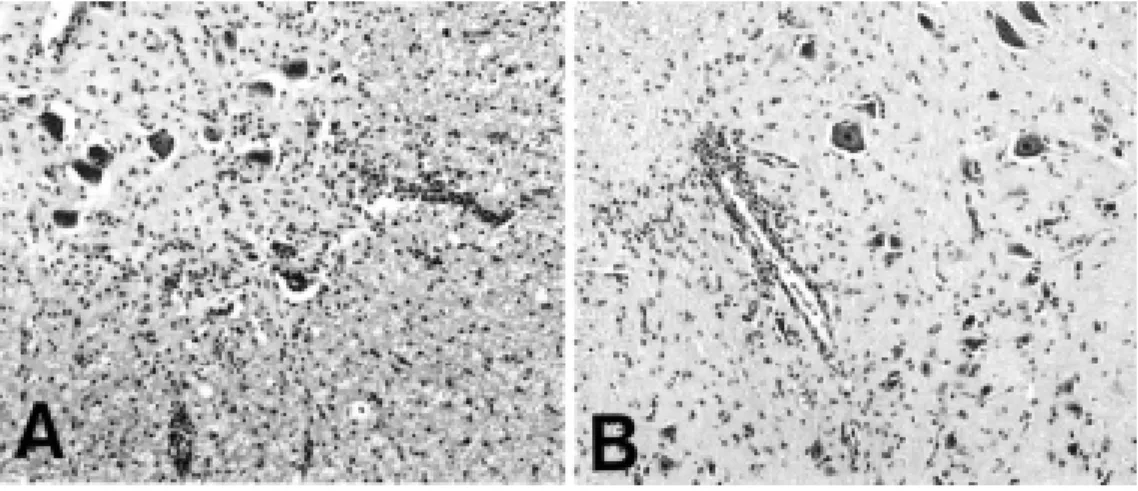
Histological lesions in melatonin-treated animals were nearly identical to those of the control animals at the peak stage of EAE (day 14 PI) (Fig. 1). The inflammatory cells consisted primarily of ED1+ macrophages, CD4+ T cells, and CD8+ T cells (data not shown). NK+ cells and OX22+ cells were also present. The inflammatory cells were less abundant in melatonin-treated rats than in vehicle-treated rats. The proportion of NK+ cells to total inflammatory cells was higher (about 2-fold) in the melatonin-treated animals than in the vehicle-treated control group (Fig. 2).
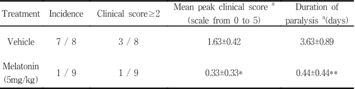
Fig. 1. Histological findings in spinal cords from vehicle-treated control (A) and melatonin-treated Lewis rats (B) with EAE (day 14 PI). A and B show inflammatory lesions in the spinal cord that are typical of EAE, but a comparison of A with B shows that many more inflammatory cells infiltrate the parenchyma in A. H & E stain, Magnification: ×200.
Fig. 2. Immunohistochemical detection of OX22+ and NK+ cells in vehicle-treated (A and B) and melatonin-treated (C and D) groups at the peak stage (day 14 PI) of EAE. OX22+ (A and C) and NK+ (B and D) cells were mainly located in the perivascular region but some were in the parenchyma. Counterstained with hematoxylin. Magnification: ×132.
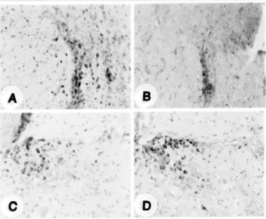
Adhesion molecules, including LFA-1α and ICAM-1, were examined in both groups. The intensity of ICAM-1 immunoreactivity in the blood vessels of EAE lesions was weaker in melatonin-treated rats than in vehicle-treated rats, whereas the intensity of LFA-1α immunoreactivity (indicating inflammatory cells) was similar in both groups (Fig. 3)(Table 2).
Fig.3. Immunohistochemical detection of LFA-1α and ICAM-1 in melatonin-treated (B and D) and vehicle-treated (A and C) rats at the peak stage (day 14 PI) of EAE. LFA-1α positive cells were mainly located in the perivascular region, but some were in the parenchyma (A and B). ICAM-1 immunoreactivity was mainly detected in the blood vessels of the vehicle-treated (C) rats, but it was rarely detected in spinal cords of the melatonin-treated rats (D). Counterstained with hematoxylin. Magnification: 400.
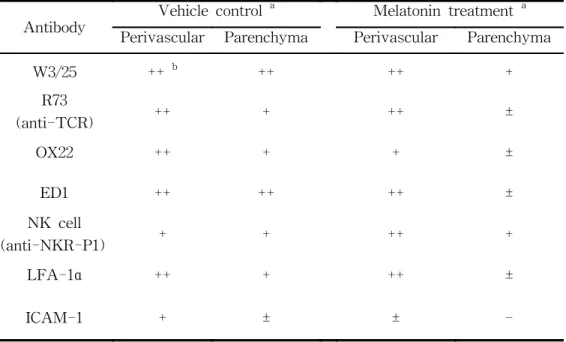
Table 2. Immunohistochemical localization of W3/25, R73, OX22, ED1, LFA-1α and ICAM-1 positive cells, and NK cells in the spinal cords of vehicle controls and melatonin treated EAE rats at the peak stage (D14PI).
Antibody
Vehicle control a Melatonin treatment a Perivascular Parenchyma Perivascular Parenchyma
W3/25 ++ b ++ ++ + R73 (anti-TCR) ++ + ++ ± OX22 ++ + + ± ED1 ++ ++ ++ ± NK cell (anti-NKR-P1) + + ++ + LFA-1α ++ + ++ ± ICAM-1 + ± ± -a
Three to five animals were examined in each group. b
Stained sections were scored on the number of cells per field that were positive. The number of positive cells was defined in the average of 5 randomly chosen 100 fields: -, no positive cells; ±, <10 cells per field; +, <30 cells; ++, ≥30 cells.
Ⅳ. Discussion
This study constitutes the first clinical demonstration that exogenous melatonin affects the progression of autoimmune disease in model EAE. The underlying mode of melatonin action in preventing EAE paralysis remains controversial. A recent study has suggested that melatonin causes non-specific immunity by inducing the production of NK cells (Currier et
al., 2000) and IL-4, which are important mediators in the amelioration or
prevention of EAE-induced paralysis (Constantinescu et al., 1997). Melatonin is also implicated in reducing the expression of NF-κB (Gilad et
al., 1998) and ICAM-1 (Kawai et al., 1996), and thus may ameliorate the
progression of EAE by blocking these factors.
By demonstrating that melatonin induces the repression of ICAM-1 expression, this study suggests another possible mechanism for EAE amelioration by melatonin. We suggest that the suppression of cell adhesion molecules is an important factor in reducing cell infiltration into CNS tissues. Adhesion molecules are important in the peripheral leukocytes into the CNS, which is a major event in the pathogenesis of the inflammatory demyelinating disease, multiple sclerosis (Duran et al., 1999; Elovaara et al., 2000; Rose et al., 1999). In our previous study (Shin et al., 1995), we found that ICAM-1 significantly increased in the early stage of EAE.
Although this study does not quantify the increase in NK cells, the trend in our immunohistochemical data shows that NK cells are present in higher amounts in melatonin-treated rats than in vehicle-treated rats. This aspect of NK cell biology needs further quantitative study to confirm this
phenomenon.
We did not expect that melatonin treatment would completely block the onset of EAE, since this disease is caused by the homing of auto-reactive T cells to the target tissue (spinal cord), and melatonin does not prevent the generation of autoreactive T cells, even though it reduces their homing ability. However, the findings of this study suggest that melatonin has an anti-inflammatory effect on EAE, caused by suppression of ICAM-1 and possibly by the induction of NK cells production.
Although we found amelioration of EAE in melatonin-treated rats, Constantinescu et al. (1997) reported that melatonin receptor antagonists ameliorated EAE paralysis in rats, suggesting that melatonin is in fact detrimental in EAE, and that it may play a role in increasing inflammation. We speculate that these seemingly contradictory results may arise from experimental differences in the severity state of EAE. Whereas Constantinescu et al. used myelin basic protein as an immunogen, we used rat spinal cord homogenates as an immunogen. Myelin basic protein may cause more severe inflammation than rat spinal cord homogenate, which caused mild EAE in our system. This idea gains support from the observation that melatonin stimulates the production of inflammatory cytokines in human CD4+ cells (Gonzalez-Haba et al., 1995). The exact function of melatonin in modulating immune system response needs further study.
Taking all of these observations into consideration, we postulate that melatonin suppresses autoimmune diseases, including mild EAE, by suppressing the production of ICAM-1 in the target organ, the spinal cord.
Ⅴ. References
Arendt, J., 1986. Role of the pineal gland and melatonin in seasonal reproductive function in mammals. Oxf. Rev. Reprod. Biol. 8, 266-320.
Badger, A.M., Dimartino, M.J., Talmadge, J.E., Picker, D.H., Schwartz, D.A., Dorman, J.W., Mirabelli, C.K., Hanna, N., 1989. Inhibition of animal models of autoimmune disease and the induction of non-specific suppressor cells by SK&F 105685 and related azaspiranes. Int. J. Immunopharmacol. 7, 839-846.
Chuang, J.I., Mohan, N., Meltz, M.L., Reiter, R.J., 1996. Effect of melatonin on NF-kappa B DNA-binding activity in the Rat spleen. Cell Biology International. 20, 687-692.
Constantinescu, C.S., Hilliard, B., Ventura, E., Rostami, A. Luzindole,, 1997. a melatonin receptor antagonist, suppresses experimental autoimmune encephalomyelitis. Pathobiology. 65,190-194.
Currier, N.L., Sun, L.Z.Y., Miller, S.C., 2000. Exogenous melatonin: Quantitative enhancement in vivo of cells mediating non-specific immunity. J. Neuroimmunol. 104, 101-108.
Cuzzocrea, S., Costantion, G., Mazzon, E., Micali, A., De Sarro, A., Caputi, AP., 2000. Beneficial effects of melatonin in a rat model of splanchnic
artery occlusion and reperfusion. J. Pineal. Res.28, 52-63.
Duran, I., Martinez-Caceres, E.M., Rio, J., Barbera, N., Marzo, M.E., Montalban, X., 1999. Immunological profile of patients with primary progressive multiple sclerosis: Expression of adhesion molecules. Brain 122, 2297-2307.
Elovaara, I., Ukkonen, M., Leppakynnas, M., Lehtimaki, T., Luomala, M., Peltola, J., Dastidar, P., 2000. Adhesion molecules in multiple sclerosis: relation to subtypes of disease and methylprednisolone therapy. Arch. Neurol. 57, 546-551.
Gilad, E., Wong, H.R., Zingarelli, B., Virag, L., OConnor, M., Salzman, A.L., Szabo, C., 1998. Melatonin inhibits expression of the inducible isoform of nitric oxide synthase in murine macrophages: role of inhibition of NF-kappa B activation. J. FASEB. 12, 685-693.
Gonzalez-Haba, M.G., Garcia-Maurino, S., Calvo, J.R., Goberna, R., Guerrero, J.M., 1995. High-affinity binding of melatonin by human circulating T lymphocytes (CD4+). J. FASEB. 9, 1331-1335.
Guerrero, J.M., Reiter, R.J., 1992. A brief survey of pineal gland-immune system interrelationships. Endocr. Res. 18, 91-113.
Guerrero, J.M., Reiter, R,J., 1992. Iodothyronine 5'-deiodinating activity in the pineal gland. Int. J. Biochem. 24, 1513-1523.
Kawai, K., Kobayashi, Y., Shiratori, M., Sobue, G., Tamatani, T., Miyasaka, M., Yoshikai, Y., 1996. Intrathecal administration of antibodies against LFA-1 and against ICAM-1 suppresses experimental allergic encephalomyelitis in rats. Cell. Immunol. 171, 262-268.
Kohji, T., Tanuma, N., Aikawa, Y., Kawazoe, Y., Suzuki, Y., Kohyama, K., Matsumoto, Y., 1998. Interaction between apoptotic cells and reactive brain cells in the central nervous system of rats with autoimmune encephalomyelitis. J. Neuroimmunol. 82,168-174.
Maestroni, G.J., 1995. T-helper-2 lymphocytes as a peripheral target of melatonin. J. Pineal Res. 18, 84-89.
Matsumoto, N., Ribaudo, R.K., Abastado, J.P., Margulles, D.H., Yokoyama, W.M., 1988. The lectin-like NK cell receptor Ly-49A recognizes a carbohydrate-independent epitope on its MHC class I ligand. Immunity 8, 245-254.
Matsumoto, Y., Kohyama, K., Aikawa, Y., Shin, T., Kawazoe, Y., Suzuki, Y., Tanuma N., 1998. Role of natural killer cells and TCR T cells in acute autoimmune encephalomyelitis. Eur. J. Immunol. 28, 1681-1688.
Piccirillo, C.A., Prudhomme, G.J., 1999. Prevention of experimental allergic encephalomyelitis by intramuscular gene transfers with cytokine-encoding plasmid vectors. Hum. Gene Ther. 10, 1915-1922.
Pierpaoli, W., Regelson, W., 1994. Pineal control of aging: effect of melatonin and pineal grafting on aging mice. Proc. Natl. Acad. Sci., U.S.A. 91, 787-791.
Racke, M.K., Bonomo, A., Scott, D.E., Cannella, B., Levine, A., Raine, CS., Shevach, E.M., Rocken, M., 1994. Cytokine-induced immune deviation as a therapy for inflammatory autoimmune disease. J. Exp. Med. 180, 1961-1966.
Reiter, R.J., 1995. Functional pleiotropy of the neurohormone melatonin: antioxidant protection and neuroendocrine regulation. Front Neuroendocrinol. 16, 383-415.
Rose, J.W., Welsh, C.T., Hill, K.E., Houtchens, M.K., Fujinami, R.S., Townsend, J.J., 1999. Contrasting effects of anti-adhesion molecule therapy in experimental allergic encephalomyelitis and Theilers murine encephalomyelitis. J. Neuroimmunol. 97, 110-118.
Ruuls, S.R., Bauer, J., Sontrop, K., Huitinga, I., 't Hart, B.A., Dijkstra, C.D., 1995. Reactive oxygen species are involved in the pathogenesis of experimental allergic encephalomyelitis in Lewis rats. J. Neuroimmunol. 56, 207-217.
Shin, T., Kojima, T., Tanuma, N., Ishihara, Y., Matsumoto, Y., 1995. The subarachnoid space as a site for precursor T cell proliferation and effector T cell selection in experimental autoimmune encephalomyelitis. J.
초 록
Melatonin이 adhesion molecules 억제를 통한
자기면역성 뇌척수염의 완화
(지도교수: 신태균)
강 종 철
제주대학교 대학원 수의학과
melatonin(N-acethyl-5-methoxytryptamine)은 송과체의 신경 호르몬으로 서 면역계에서 수산화 잔기 제거와 항산화에 중요하게 작용한다. 본 실험에서 는 외인성 멜라토닌이 자기면역성 뇌척수염(Experimental autoimmune encephalomyelitis, EAE)에 어떤 영향을 미치는지 연구하였다. 랫트 뇌척수 조직을 균질화 한 후 면역원으로 하여 EAE를 유발시킨 후 멜라토닌(5mg/kg)을 경구 투여 하였다. 투여 결과 EAE임상 증상이 대조군에 비해 유의성 있게 완화되는 경향을 나타내었다(p< 0.01). 면역염색 결과 큰포식세포와 CD4+ T세포는 멜라토닌 투여군과 Vehicle 투여 군 모두에서 혈관 및 실질 조직 내에 침윤 되었지만, 멜라토닌 투여군에 서는 Vehicle 투여군 보다 적게 침윤되는 경향을 볼 수 있었다.멜라토닌 투여군과 Vehicle 투여군의 EAE 병변에서 adhesion molecules인 intercellular adhesion molecule-1(ICAM-1)과 lymphocyte function-associated antigen-1α (LFA-1α)를 면역염색 한 결과 멜라토닌 투여군의 EAE 병변에
서 이들 인자가 억제됨을 알 수 있었다.
결과적으로 외인성 멜라토닌이 자기면역성 뇌척수염의 진행 과정 중 ICAM-1과 LFA-1α를 억제시켜 EAE 병변을 완화 시켰으며, 외인성 멜라토 닌은 자기면역성 질병의 예방과 치료에 효과가 있을 것으로 생각된다.