Computational Study on the Binding of Aux/IAA17 and ARF5
Involved in Auxin’s Transcriptional Regulation using Molecular
Docking
Sohee Kwon†, Gyu Rie Lee, and Chaok Seok*
Department of Chemistry, Seoul National University, 1 Gwanak-ro, Gwanak-gu, Seoul, 151-742 E-mail: [email protected], [email protected]
Abstract Auxin response factor (ARF) and Aux/IAA transcriptional repressor family proteins play a major role in auxin’s signalling process. Using the GALAXY protein modelling programs, monomer, dimer and oligomer structures of Aux/IAA17 and ARF5 protein were predicted based on the known experimental structures. By analysing the proposed complex structures, key interacting residues on binding site could be determined, and further suggestions for experimental studies were made.
Introduction
Auxin is a class of plant hormone, essential for regulation of plant growth and organ development. Auxin’s signalling process is mediated by several protein families including TIR1 (transport inhibitor response 1), ARF (auxin response factor), Aux/IAA transcriptional repressors and ubiquitin ligase complex. When auxin is absent, Aux/IAA and ARF together repress transcriptional activity. However, in the presence of auxin, Aux/IAA, TIR1 and auxin binds to cause proteosomal degradation of Aux/IAA protein. With low concentration, or ithout Aux/IAA present, ARF is free to activate gene expression.
From previous experimental studies on interaction between Aux/IAA and ARF, it has been shown that Aux/IAA and ARF can form homo-oligomers as well as hetero-oligomers. Also from multiple sequence alignment information, it has been revealed that Aux/IAA and ARF both share a unique domain Ⅲ-Ⅳ,
also known as Phox and Bem 1 (PB1) domain. The PB1 domain is common in many biological processes. It helps proteins form dimers in a front to back manner via charged residues in the binding domain. Since the charged residues in PB1 domain are highly conserved in both Aux/IAA and ARF proteins, it has been suggested that PB1 domain plays a major role in Aux/IAA and ARF protein interactions.
Such theory was supported by several experimental data. The crystal structure of monomer ARF7 (PDB ID: 4NJ6)[1] was found,
which has a similar fold as PB1 domain, and later crystal structure of ARF5 oligomerized domain (PDB ID: 4CHK)[2] which bound on the expected charged binding site was revealed. Also mutation on several key charged residues consistent with PB1 domain and ARF proteins, as well as Aux/IAA proteins showed that such residues played a key role in dimerization of ARF and Aux/IAA protein families.[3]
Also there were experimental data supporting the theory on how transcriptional regulation works. It has been revealed that the binding affinity of Aux/IAA17, ARF5 hetero-dimer is much larger than that of Aux/IAA17 and ARF5 homo-dimer. This data implies that in case of none or low concentration of auxin, both ARF and Aux/IAA proteins exist and they will bind as hetero-oligomer rather than as homo-oligomer.[3]
Among various Aux/IAA and ARF proteins, the structures of monomer Aux/IAA17 (PDB ID: 2MUK) and oligomerized domain of ARF5 (PDB ID: 4CHK) is known. Based on the known experimental structures, this study attempts to explain the result of the experimental data using computational programs of GALAXY[4, 5]. Moreover, based on the computational results, suggestions for yet unknown protein dimer and oligomer structures will be made.
Theoretical Background
Protein Homology modeling
Protein homology modelling, also known as template-based modelling (TBM), uses information of known protein 3D structures to model the unknown structure of a protein based on the assumption that the sequence of a protein determines its 3D structure. Though it is possible to get protein structure data from X-ray crystallography or NMR, such experimental methods require large amounts of time and effort to come up with the structure of a single protein. However using computational tools like homology modelling, it is possible to get considerably well predicted structures of proteins with less time and effort.
When given a protein sequence, a homology modelling method searches the protein structure database to find homologous proteins with related sequences. The template and target protein sequences are then aligned. Based on the alignment, target protein structure is modelled and refined. During the refinement process the unaligned parts are modelled using ab initio or knowledge-based energy functions. For this study, homology modelling program called GalaxyTBM[6] is used. Protein-protein docking
Protein-protein docking models possible complex structures. The main goal for protein-protein docking is to predict the best possible binding pose of the complex. Docking is mostly done by sampling various possible complex structures and then scoring them using energy functions to find the structure with lowest energy. The energy function can be thought as the potential of mean force for the protein complex system, with an assumption that the structure with lowest free energy is the most stable structure.
When docking, it is important to consider the flexibility of protein structures, because it is possible for the monomer protein structure to change during docking to better interact with the binding protein. But considering full protein flexibility is difficult due to limits in computational time and power. Instead many protein-protein docking techniques consider partial protein structure flexibility to improve docking results. For this study, in-house protein-protein docking program called GalaxyTongDock[7], with similar approach to ZDOCK[8] is used.
Methods
Outline of overall procedure
Figure 1. Outline of overall procedure
Figure 1 shows the outline of overall procedure. Computation was performed using known structures of a single protein structure from oligomerized domain of ARF5 and monomer structure of Aux/IAA17. First, using GalaxyTBM[6], known protein structures were
mutated on the key charged residues. Then, homo-dimer and hetero-dimer structures of both wild and mutated ARF5 and Aux/IAA17 were made using protein-protein docking program GalaxyTongDock. Finally, the results of GalaxyTongDock were evaluated using several evaluation measures.
Protein homology modeling of Aux/IAA17 and ARF5 mutants using GalaxyTBM
To check the experimental data that Aux/IAA and ARF family proteins both bind on the charged sites of PB1 domain, Aux/IAA17 and ARF5’s conserved key charged residues were mutated. Based on the alignment data of various ARF and Aux/IAA family proteins[3],
three residues (key residues) were selected for mutation. For the positive charged site lysine was mutated and for the negative site two aspartic acids were mutated. Such mutation for Aux/IAA17 accounts for K114M mutation
(Aux/IAA17 M1) and D183N/D187N mutation (Aux/IAA17 M2). Also for ARF5 it accounts for K797M (ARF5 M1) and D847N/D851N (ARF5 M2).
Using experimentally known structures wild type ARF5 (ARF5 W, PDB ID: 4CHK) and Aux/IAA17 M2 (PDB ID: 2MUK) as templates, unknown structures ARF5 M1 and M2, Aux/IAA17 W and M1 were modelled.
Protein-protein docking of Aux/IAA17 and ARF5 proteins using GalaxyTongDock
After getting wild and mutated structures for both ARF5 and Aux/IAA17, docking of various combinations were tried. The protein-protein docking was mainly carried out using GalaxyTongDock. However like most protein-protein docking programs, GalaxyTongDock cannot effectively consider protein backbone fluctuation. Thus, in the cases when initial structure used for docking seemed less reliable or further alternation on initial structure seemed required for docking, ensemble docking was used. Ensemble docking first models various probable structures of the receptor and dock ligand to them to find out the best likely complex structure. For ensemble docking, loop modelling program GalaxyLongLoop[9] was
used to model loop ensemble, and then GalaxyTongDock was used for docking.
For ARF5 and Aux/IAA17 homodimers of both wild type structures (WW), M1 mutated structures (M1M1) and M2 mutated structures (M2M2) were modelled to find out the structure of each complex as well as to reveal the effect of mutated residues. For ARF5, Aux/IAA17 heterodimer, besides the • mutation of protein using
homology modeling
GalaxyTBM
• protein-protein docking of dimer complex structures
GalaxyTongDock
• Structure, RMSD, TM-score, Fnat, oligomer possibility Evaluation
combinations made for homodimers, additional complexes with both M1 and M2 mutated structures (M1M2, M2M1) were made. This additional dimer structures were constructed to check the binding of ARF5 and Aux/IAA17 on each predicted positive and negative binding sites.
Analysis Method
After docking, 50 models obtained from GalaxyTongDock were evaluated. In the case of ARF5 homodimer modelling, since we have experimental data on the dimer structure, it is easier to analyse the result. Using crystallographic structure as a reference, we can calculate the RMSD (root-mean-square deviation) on all-atom bases to check how close the modelled structure is to the known structure. In addition, TM-score[0] can be used
to measure the structural similarity of the modelled and experimental structures. It is known that RMSD is more sensitive to local structure variations, and TM-score measures global fold similarity better. TM-score ranges from 0 to 1, where 1 means a perfect match. For protein complex structures, when the orientation of the complex is different from the reference structure, the calculated TM-score is close to 0.5.
Using RMSD and TM-score measures is difficult for Aux/IAA17 homodimer and heterodimer of ARF5 and Aux/IAA17, because there are no structural data given to use as a reference state. Instead a different measure Fnat[11] can be used to access whether the
binding site is conserved. Fnat is originally
defined as the number of native residue– residue contacts in the predicted complex
divided by the number of native contacts in the target structure.[11] F
nat for this study had
to be slightly modified, since only native structure for ARF5 homodimer is available, and mutations was introduced. Accordingly using known ARF5 homodimer structure as the native structure, we counted conserved residue-residue contacts based on the alignment data. Residue-residue contact was defined as contact within 5 Å of any heavy atom in the residue of interest. Contacting residue pairs for native structure were 39 in total, involving 29 residues. One of 29 residues couldn’t be aligned with Aux/IAA17 which engaged in one residue pair.
To identify if a modelled structure can be thought as plausible structure, its oligomerized structure was checked. Though only the dimer structure was modelled, in the real plant system, ARF5 and Aux/IAA17 are both expected to exist in a long oligomerized form in one of homo- or hetero- oligomer state. Therefore using the modelled dimer structure, possible oligomer structure was checked to see if the structure can also work on a longer oligomer state.
Result and Discussion
ARF5 homodimer RMSD WW M1M1 M2M2 OVER 40 38% 50% 10% 25-40 20% 14% 80% 10-25 20% 22% 10% 4-10 16% 14% - 0-4 6% - - TM-score WW M1M1 M2M2
0-0.6 66% 72% 100% 0.6-0.75 14% 2% - 0.75-0.85 8% 12% - 0.85-1.0 14% 18% - Fnat WW M1M1 M2M2 0.0-0.15 22% 50% 100% 0.15-0.3 14% 2% - 0.3-0.45 8% 12% - 0.45-0.6 14% 18% - 0.6-0.75 8% 18% - 0.75-0.9 16% - - 0.9-1.0 18% - -
Table 1. Docking evaluation results of ARF5 homodimer (RMSD, TM-score, Fnat)
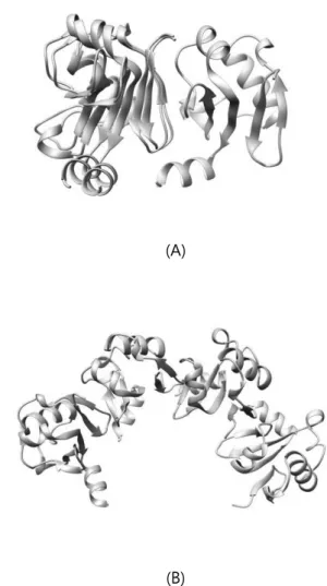
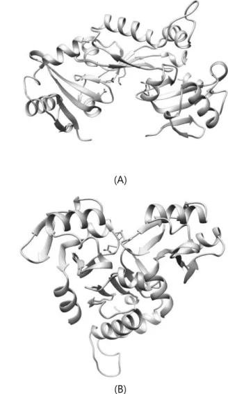
Table 1 shows RMSD, TM-score, Fnat result for docking result of ARF5 homodimer structures. WW homodimer structures showed the best score for all three evaluation measures. Especially several structures of WW homodimer result had RMSD less than 10Å, showing high resemblance in local structure level to known crystal structure. Modelled structure shown in Figure 2 has RMSD of 3.646, 1.0 Fnat score and TM-score of 0.94. Also, when matching several modelled ARF5 WW homodimer structures, ARF5’s oligomer structure could be found. On the contrary, none of M2M2 homodimer structures docked as the native structure. This tells us that the two aspartic acid residues on M2 mutated site are crucial for the binding of ARF5 homodimer.
(A)
(B)
Figure 2. ARF5 WW homodimer (A: beige = native / blue = model) and homooligomer structure (B)
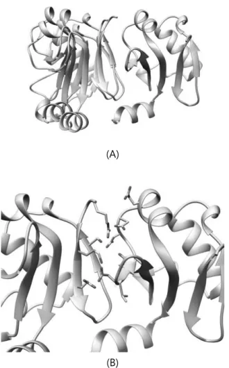
Unlike M2M2 homodimer, few structures of M1M1 homodimer turned out to be similar to the native structure. This is because M1 mutation site only has one mutated residue compared to the two mutated residues on M2 site. Besides the few key residues which we mutated there are additional polar and hydrogen interactions nearby[2] which helps binding of ARF5 proteins as shown in Figure 3. But since less modelled structure resembled native structure, it can be concluded that the mutated lysine residue is also important for
0-0.6 66% 72% 100% 0.6-0.75 14% 2% - 0.75-0.85 8% 12% - 0.85-1.0 14% 18% - Fnat WW M1M1 M2M2 0.0-0.15 22% 50% 100% 0.15-0.3 14% 2% - 0.3-0.45 8% 12% - 0.45-0.6 14% 18% - 0.6-0.75 8% 18% - 0.75-0.9 16% - - 0.9-1.0 18% - -
Table 1. Docking evaluation results of ARF5 homodimer (RMSD, TM-score, Fnat)
Table 1 shows RMSD, TM-score, Fnat result for docking result of ARF5 homodimer structures. WW homodimer structures showed the best score for all three evaluation measures. Especially several structures of WW homodimer result had RMSD less than 10Å, showing high resemblance in local structure level to known crystal structure. Modelled structure shown in Figure 2 has RMSD of 3.646, 1.0 Fnat score and TM-score of 0.94. Also, when matching several modelled ARF5 WW homodimer structures, ARF5’s oligomer structure could be found. On the contrary, none of M2M2 homodimer structures docked as the native structure. This tells us that the two aspartic acid residues on M2 mutated site are crucial for the binding of ARF5 homodimer.
(A)
(B)
Figure 2. ARF5 WW homodimer (A: beige = native / blue = model) and homooligomer structure (B)
Unlike M2M2 homodimer, few structures of M1M1 homodimer turned out to be similar to the native structure. This is because M1 mutation site only has one mutated residue compared to the two mutated residues on M2 site. Besides the few key residues which we mutated there are additional polar and hydrogen interactions nearby[2] which helps binding of ARF5 proteins as shown in Figure 3. But since less modelled structure resembled native structure, it can be concluded that the mutated lysine residue is also important for
dimerization of ARF5.
(A)
(B)
Figure 3. ARF5 M1M1 homodimer structure (beige = native / blue = model / orange = key residues)
Aux/IAA17 homodimer
Unlike modelled ARF5 homodimers, crystal structure for Aux/IAA homodimer is unknown, so only the Fnat evaluation measure was used. Fnat score for all modelled structure of Aux/IAA17 homodimer was below 0.15. Though all modelled structures checked one by one, none of Aux/IAA proteins docked near the expected binding site.
Such failure of docking was due to initial structure. ARF5 dimer docking was easier because the initial structure used for
protein-protein docking was extracted from known dimerized structure. However the initial structure for Aux/IAA17 docking was an NMR monomer structure, so the structure for dimerized Aux/IAA17 can differ from the initial structure used. Since we had 20 NMR structure of Aux/IAA17 monomer, it was possible to find out which site of Aux/IAA17 fluctuate the most. The most fluctuating site detected using NMR structure data was a loop (residue 148-163). Based on an assumption that the loop will fluctuate while docking to better dimerize, three additional loop structures shown in Figure 4 were modelled using GalaxyLongLoop and each used as initial structure for docking.
Figure 4. Aux/IAA17 loop modelling result (beige = native / blue = loop1 / pink = loop2 / green = loop3)
Fnat Loop1 Loop2 Loop3
0~0.15 92% 70% 100%
0.15~0.3 8% 20% -
0.3~0.45 - 10% -
Table 2. Ensemble docking Fnat result of Aux/IAA17 WW homodimer
structures, protein with loop2 showed the best result. Lower Fnat score compared to ARF5 homodimer can be explained by the orientation difference of Aux/IAA homodimer. Among the structures with high Fnat score, structures with slightly farther key residue distance oligomerized better. Figure5 shows an example of such WW homodimer of Aux/IAA17 and its possible oligomer structure. Though the orange coloured residues of two proteins seem far, additional lysine helps charged interaction between two proteins. Also the binding site is relatively well conserved with Fnat score near 0.3.
(A)
(B)
Figure 5. Aux/IAA17_loop2 WW homodimer (A: orange = key residues) and homo-oligomer (B) structure
Furthermore the Fnat results of M1M1 and M2M2 homodimer from Table 3 imply that the mutated residues also play an important role on binding of Aux/IAA17 dimer.
Fnat WW M1M1 M2M2
0~0.15 70% 100% 100%
0.15~0.3 20% - -
0.3~0.45 10% - -
Table 3. Ensemble docking Fnat result of Aux/IAA17_loop2 homodimer
Aux/IAA17, ARF5 heterodimer
Considering the docking result of Aux/IAA17 homodimer, possible dimerized structure of Aux/IAA17 and ARF5 were checked beforehand as shown in Figure6. Accordingly docking of heterodimer of Aux/IAA17 and ARF5 was done using loop modelled structure used for Aux/IAA17 homodimer docking.
Figure 6. Expected structure of heterodimer of ARF5 W (beige) and Aux/IAA17 W (blue), compared to that of Aux/IAA17_loop2 (pink)
Fnat score for various heterodimer structures were all below 0.2, more than 90% of the structures with Fnat score below 0.1. Based on Fnat score result, it seemed that
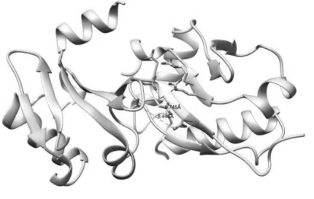
heterodimerization didn’t occur near the expected binding site. However looking at each WW heterodimer structure, it was found that for most structures, two proteins bound near the expected sight, just not close enough. Like shown in Figure 7, the distance between key residues along with other native contacted residues were more than 5Å, resulting in near 0 Fnat scores. However, also from Figure 7, we can see that the key residues still played an important role in binding of two proteins, just paired with other nearby residues. Accordingly, it could still be said that the binding site was fairly conserved for heterodimer as well.
Figure 7. Heterodimer structure of ARF5 M1, Aux/IAA17_loop2 M2(orange = key residues)
To determine the possible structure, modelled structures were checked to see their oligomer structure. Unlike homo-oligomer structures, hetero-oligomer structures have different binding site and orientation on each side. In order to check each side selectively, modelled structures for M1M2 heterodimer and M2M1 heterodimer were used to figure out the possible oligomer structure.
Most of the structures couldn’t be oligomerized due to its binding orientation. As
shown in Figure 8, when matching certain M1M2 and M2M1 heterodimer structure, they clash. Such dimer structures, since it cannot oligomerize, can be eliminated.
Figure 8. Clashed hetero-oligomer structure of ARF5 and Aux/IAA17 (clash between blue and pink)
Eliminating most of the structure through oligomerization possibility, the final selected dimer structure was like Figure 9. In a zig-zag like form, Aux/IAA17 and ARF5 can be oligomerized. Also looking more closely at the binding interface, while the mutated residues were more than 5 Å apart, marked as uncontacted residue pair in Fnat calculation, other charged residues like arginine, glutamic acid and aspartic acid helped form a tighter interaction. Also at the binding interface, several hydrophobic chains were closely packed, further enhancing the intensity of the charged interaction nearby. Such interaction between charged residues and enhancement of charge interaction through interaction between hydrophobic residues was only found on heterodimers, therefore can be viewed as explanation for larger binding affinity of heterodimers compared to homodimers.
(A)
(B)
Figure 9. Heterodimer(A: orange = key residues / hydrophobic residues = green) and hetero-oligomer (B) structure of ARF5 and Aux/IAA17_loop2
As a further suggestion, R205M/R207M mutation of Aux/IAA17 were made, which on the suggested structure, seemed to highly affect the interaction between ARF5 M1 and Aux/IAA17 M2. After mutating by GalaxyTBM and docking by GalaxyTongDock, the end result of heterodimer structure changed completely. Most structure didn’t bind near the expected binding site. Even the few that bound near the site bound in a totally
different position, unable to form oligomer structure due to steric hindrance as shown in Figure 10.
(A)
(B)
Figure 10. Heterodimer (A: beige = without additional mutation / blue = with additional mutation) and hetero-oligomer(B) structure of ARF5 and Aux/IAA17_loop with additional R205M/ R207M mutation of Aux/IAA17
Apart from the docking result, Arg205 and Arg207 are also known to be conserved only in Aux/IAA17 family proteins and not in ARF family proteins[3]. Also Arg205 and Arg207 don’t involve in binding between two Aux/IAA17 proteins (Figure 11). So such
(A)
(B)
Figure 9. Heterodimer(A: orange = key residues / hydrophobic residues = green) and hetero-oligomer (B) structure of ARF5 and Aux/IAA17_loop2
As a further suggestion, R205M/R207M mutation of Aux/IAA17 were made, which on the suggested structure, seemed to highly affect the interaction between ARF5 M1 and Aux/IAA17 M2. After mutating by GalaxyTBM and docking by GalaxyTongDock, the end result of heterodimer structure changed completely. Most structure didn’t bind near the expected binding site. Even the few that bound near the site bound in a totally
different position, unable to form oligomer structure due to steric hindrance as shown in Figure 10.
(A)
(B)
Figure 10. Heterodimer (A: beige = without additional mutation / blue = with additional mutation) and hetero-oligomer(B) structure of ARF5 and Aux/IAA17_loop with additional R205M/ R207M mutation of Aux/IAA17
Apart from the docking result, Arg205 and Arg207 are also known to be conserved only in Aux/IAA17 family proteins and not in ARF family proteins[3]. Also Arg205 and Arg207 don’t involve in binding between two Aux/IAA17 proteins (Figure 11). So such
residues can be thought to be only involved in key interaction between Aux/IAA and ARF family proteins, making heterodimer interaction stronger than homodimer interaction.
Figure 11. Aux/IAA17_loop2 WWhomodimer (A: orange = key residues / green = Arg205 and Arg207)
Conclusion
Using computational tools of GALAXY, binding between Aux/IAA17 and ARF5 were studied. By docking proteins with several mutated residues, we could confirm that such residues play an important role in the interaction between the two proteins. Docking of ARF5 homodimer illustrated the accuracy of computational docking, by reproducing the known complex structure within RMSD less than 4 Å. In the cases of Aux/IAA17 homodimer and Aux/IAA17-ARF5 heterodimer, suggestions for both homodimer and homo-oligomer structures were made. Moreover, we could see how protein can fluctuate while making stronger interactions with the other protein.
The proposal for structures of yet unknown protein complexes can mean more than just a
possible structure. Like in the case of heterodimer structure for Aux/IAA17 and ARF5, further suggestions on key residues of interaction can be made. In this study only the importance of Arg205 and Arg207 on Aux/IAA17 was checked. However other residues like hydrophobic residues near binding site could be further checked to see how they affect the binding of Aux/IAA17 and ARF5 more than the binding of homodimers.
Acknowledgement
This research was supported by the EDISON Program through the National Research Foundation of Korea (NRF) funded by the Ministry of Science, ICT & Future Planning (NRF-2011-0020576)
Reference
[1] Korasick, D.A. and Westfall, C.S., Proc. Natl. Acad. Sci. USA 111: 5427-5432 (2014).
[2] Nanao, M.H. and Vinos-Poyo T., Nat.Commun. 5:3617 (2014).
[3] Han M. et al., Proc. Natl. Acad. Sci. USA, 111: 18613-18618.
[4] http://galaxy.seoklab.org. Galaxy website. [5] http://www.edison.re.kr. EDISON website.
[6] Ko J., Park H. and Seok C, BMC Bioinformatics, 13:198 (2012).
[7] GalaxyTongDock, unpublished.
[8] Pierce BG. et al., Bioinformatics 30(12): 1771-3. (2014).
[9] D. Lee, C. Seok and J. Lee, J. Korean Phys. Soc. 52, 1137 (2008).
[10] Y. Zhang, J. Skolnick, Proteins, 57: 702-710 (2004).
[11] Mendez et. al., PROTEINS: Structure, Function, and Genetics 52:51–67 (2003).