The Rockefeller University Press J. Cell Biol. Vol. 193 No. 5 805–807
www.jcb.org/cgi/doi/10.1083/jcb.201104140 JCB 805
JCB: Comment
Blood flow generates shear stress on vascular endothelial cells, which potently regulates endothelial morphology and function, including cell death and growth as well as inflammatory and thrombotic responses. The importance of shear stress in vascular biology and pathophysiology is highlighted by the protective role of stable flow (unidirectional and high shear stress) against atherosclerosis (atheroprotective) and the contrasting role of unstable flow (low and oscillatory shear stress) in promoting atherosclerosis (proatherogenic; Nam et al., 2009).
Endothelial cells contain numerous mechanosensors that detect local shear stress forces and transduce them into a variety of cell signaling pathways (Fig. 1 A). Via these mechanosen-sors, stable flow induces cell cycle arrest and inhibits apoptosis and inflammation in endothelial cells through acute and chronic mechanisms. Acute pathways include production of several fac-tors, including nitric oxide (NO) from endothelial nitric oxide synthase (eNOS), which acts as a key mediator of the protective effect of stable flow. Long-term stable flow up-regulates athero-protective genes, such as Klf2, eNOS, and antioxidant genes, coupled with the down-regulation of proinflammatory and pro-atherogenic genes (Ni et al., 2010).
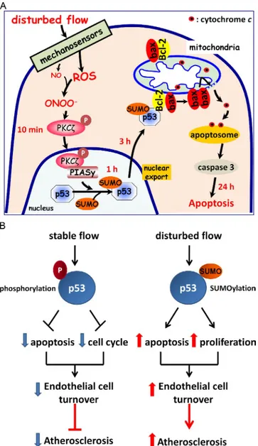
In contrast, disturbed flow increases endothelial apoptosis and proliferation, resulting in a high turnover rate that corresponds to hot spots of increased endothelial permeability, inflammation, and atherosclerosis (Chiu and Chien, 2011). The mechanism by which disturbed flow regulates these endothelial responses involves the production of reactive oxygen species (ROS), which react with NO to form peroxynitrite and regulation of Disturbed blood flow induces apoptosis of vascular endo-thelial cells, which causes atherosclerosis. In this issue, Heo et al. (2011. J. Cell Biol. doi:10.1083/jcb.201010051) sheds light on p53’s role in this phenomenon. Disturbed flow induces peroxynitrite production, which activates protein kinase C and it’s binding to the E3 SUMO (small ubiquitin-like modifier) ligase PIASy (protein inhibitor of activated STATy). This leads to p53 SUMOylation and its export to the cytosol, where it binds to the antiapoptotic protein Bcl-2 to induce apoptosis.
W. Takabe and N. Alberts-Grill contributed equally to this paper. Correspondence to Hanjoong Jo: hjo@emory.edu
proatherogenic genes (Sorescu et al., 2004; Ni et al., 2010). Despite intense study, however, the mechanisms by which flow regulates endothelial turnover are unclear.
The tumor suppressor p53 plays a crucial role in deter-mining the fate of apoptosis or cell cycle arrest in response to various stresses. In response to DNA damage or stress, cells increase p53 levels in the nucleus to up-regulate the expression of proapoptotic genes or cell cycle–regulating genes (Lee and Bernstein, 1995; Mihara et al., 2003; Teodoro et al., 2006). The p53 protein is also known to induce apoptosis by a nonnuclear, mitochondrial-dependent mechanism. p53 binds to and inhib-its antiapoptotic members of the Bcl-2 family that reside in the mitochondrial surface, such as Bcl-2 or Bcl-xL, resulting in increased mitochondrial membrane permeability, the release of mitochondrial cytochrome c into the cytosol, and the initiation of the apoptotic caspase cascade (Fig. 1 A; Mihara et al., 2003).
The roles of p53 in atherosclerosis and flow-sensitive en-dothelial biology are confusing. In human atherosclerotic plaques, p53 expression is increased in endothelial cells, implying a role for p53 as an atherosclerosis-promoting factor (Ihling et al., 1998). Also, overexpression of p53 in a transgenic mouse line has been shown to induce endothelial dysfunction and inflam-mation by down-regulating transcription of klf2, an important antiatherogenic gene (Kumar et al., 2011). Surprisingly, however, p53 overexpression did not exacerbate atherosclerosis in trans-genic mice (Sanz-González et al., 2007). To further confuse matters, p53 knockout enhanced atherosclerosis in mice, despite reducing vascular cell turnover (Guevara et al., 1999). These conflicting results may be caused by differential roles of p53 in different cell types involved in atherosclerosis, such as endothe-lial cells, smooth muscle cells, and macrophages, or even in the same cell under different stress conditions (Mercer et al., 2007).
In endothelial cells, the role of p53 in flow-dependent reg-ulation of endothelial apoptosis and cell cycle arrest has only been partially described. Stable flow causes cell cycle arrest in a p53-dependent manner by stimulating JNK-mediated phos-phorylation of p53, which in turn up-regulates expression of the cell cycle–inhibitory proteins GADD45 and p21cip1, resulting in cell cycle arrest (Fig. 1 B; Lin et al., 2000). However, the
Disturbed flow: p53 SUMOylation in the turnover
of endothelial cells
Wakako Takabe,1 Noah Alberts-Grill,2 and Hanjoong Jo1,2,3
1Wallace H. Coulter Department of Biomedical Engineering, Georgia Institute of Technology, and 2Division of Cardiology, Emory University, Atlanta, GA 30322 3Department of Bioinspired Science, Ewha Womans University, Seodaemun-gu, Seoul 120-750, South Korea
© 2011 Takabe et al. This article is distributed under the terms of an Attribution– Noncommercial–Share Alike–No Mirror Sites license for the first six months after the publication date (see http://www.rupress.org/terms). After six months it is available under a Creative Commons License (Attribution–Noncommercial–Share Alike 3.0 Unported license, as de-scribed at http://creativecommons.org/licenses/by-nc-sa/3.0/).
THE
JOURNAL
OF
CELL
BIOLOGY
on October 19, 2016 Downloaded from Published May 30, 2011JCB • VOLUME 193 • NUMBER 5 • 2011
806
(b) PKC is activated in flow-disturbed endothelium in the porcine aorta (Magid and Davies, 2005); (c) human atherosclerotic endo-thelium shows increased levels of p53 expression (Ihling et al., 1998); and (d) both atherosclerotic lesions and flow-disturbed re-gions show evidence of increased levels of peroxynitrite (Patel et al., 2000; Hsiai et al., 2007).
To test these hypotheses, the authors developed a cone and plate shear device that enables the exposure of endothelial cells to laminar or disturbed shear stress. Using this system, they showed that disturbed flow phosphorylates PKC on T410 and T560 residues in a time-dependent manner in human umbilical vein endothelial cells (HUVECs). Then, they showed that disturbed flow induces HUVEC apoptosis by a PKC-dependent mechanism by using PKC siRNA or dominant-negative PKC. Next, they tested whether peroxynitrite produced in endothelial cells by disturbed flow was responsible for PKC activation and apop-tosis. Using both chemical peroxynitrite and reagents affecting peroxynitrite levels (ebselen, N-nitro-l-arginine methyl ester, or Mn (III)tetrakis(4-benzoic acid)porphyrin chloride), they showed that this reactive nitrogen species mediated the activa-tion of PKC and apoptosis. Unexpectedly, however, they found that neither disturbed flow nor peroxynitrite up-regulated p53 level and that peroxynitrite actually inhibited p53 transcription. By using a series of elegant molecular biological approaches and immunoprecipitation experiments, they determined that disturbed flow induced p53 SUMOylation, which led to the translocation of nuclear p53 into the cytoplasm where it bound Bcl-2. p53 SUMOylation was mediated by PKC binding to the E3 SUMO ligase PIASy. The binding between PKC and PIASy was then mapped to the C-terminal segment of the PKC kinase domain and the RING domain of PIASy. Surprisingly, although PIASy-mediated SUMOylation of p53 required PKC binding, PIASy was not phosphorylated by PKC, indicating a phosphorylation-independent activation of PIASy by PKC. Importantly, point mutations of p53 SUMOylation sites or a truncation mutant lacking the p53 nuclear export sequence abolished translocation of p53 to the cytosol and apoptosis induced by disturbed flow, indicating the critical importance of SUMOylation and nuclear export into the cytosol for p53’s apoptotic action.
These findings were validated in vivo by staining experi-ments with flow-disturbed lesser curvature and stable greater cur-vature regions of the mouse aortic arch. Endothelial cells in the lesser curvature regions show higher levels of apoptosis, total PKC, phosphorylated PKC, and nitrotyrosine staining, a marker of peroxynitrite. They also found increased perinuclear localization of p53 in lesser curvature regions compared with greater curvature regions, which is consistent with their in vitro findings.
These novel insights raise several questions: First, it is not clear how and where SUMOylated p53 binds to Bcl-2. The cur-rent immunostaining study in HUVECs suggests that disturbed flow and peroxynitrite induce translocation of p53 into the cytosol, but its specific subcellular location is not clear. However, p53 is known to bind Bcl-2 or Bcl-xL on the mitochondrial sur-face, leading to mitochondrial pore formation and cytochrome c release (Mihara et al., 2003). Whether disturbed flow works by this same mechanism needs to be clarified. Second, the authors show that p53 is only transiently SUMOylated in response to role of p53 in shear-induced apoptosis has not been determined.
Now, Heo et al. (in this issue) reports a novel mechanism by which disturbed flow induces apoptosis of endothelial cells via SUMOylation of p53 in a PKC-dependent manner.
In this work, the authors tested whether disturbed flow induces apoptosis by p53- and PKC-dependent mechanisms in endothelial cells. They further hypothesized that peroxynitrite produced in response to disturbed flow mediates the apoptotic pathway in these cells. These hypotheses were based on the following previous observations: (a) flow-disturbed regions show enhanced endothelial cell apoptosis (Zeng et al., 2009);
Figure 1. p53 coordinates the opposing effects of stable and disturbed blood flow on endothelial cell turnover. (A) A proposed pathway including a timeline by which disturbed flow is sensed by mechanosensors, which induces peroxynitrite (ONOO) production, PKC phosphorylation, activation
of E3 SUMO ligase PIASy, SUMOylation of p53, and its translocation to the cytosol, where it binds Bcl-2. Upon binding SUMOylated p53, Bcl-2 likely releases bax, which stimulates cytochrome c release from mitochon-dria, leading to apoptosome formation, caspase activation, and subsequent apoptosis. (B) Posttranslational modification of p53 (phosphorylation and SUMOylation under stable and disturbed flow, respectively) determines cell turnover and atherosclerosis. P, phosphorylation of p53.
on October 19, 2016
Downloaded from
807 Death by flow: p53 SUMOylation in endothelium • Takabe et al.
Sanz-González, S.M., L. Barquín, I. García-Cao, M. Roque, J.M. González, J.J. Fuster, M.T. Castells, J.M. Flores, M. Serrano, and V. Andrés. 2007. Increased p53 gene dosage reduces neointimal thickening induced by me-chanical injury but has no effect on native atherosclerosis. Cardiovasc.
Res. 75:803–812. doi:10.1016/j.cardiores.2007.05.002
Sorescu, G.P., H. Song, S.L. Tressel, J. Hwang, S. Dikalov, D.A. Smith, N.L. Boyd, M.O. Platt, B. Lassègue, K.K. Griendling, and H. Jo. 2004. Bone morphogenic protein 4 produced in endothelial cells by oscillatory shear stress induces monocyte adhesion by stimulating reactive oxygen species production from a nox1-based NADPH oxidase. Circ. Res. 95:773–779. doi:10.1161/01.RES.0000145728.22878.45
Teodoro, J.G., A.E. Parker, X. Zhu, and M.R. Green. 2006. p53-mediated inhibi-tion of angiogenesis through up-regulainhibi-tion of a collagen prolyl hydroxy-lase. Science. 313:968–971. doi:10.1126/science.1126391
Zeng, L., A. Zampetaki, A. Margariti, A.E. Pepe, S. Alam, D. Martin, Q. Xiao, W. Wang, Z.G. Jin, G. Cockerill, et al. 2009. Sustained activation of XBP1 splicing leads to endothelial apoptosis and atherosclerosis development in response to disturbed flow. Proc. Natl. Acad. Sci. USA. 106:8326–8331. doi:10.1073/pnas.0903197106
disturbed flow (at 3 h). By 6 h, p53 is no longer SUMOylated, suggesting an active de-SUMOylation event. What is the under-lying mechanism? Third, the question still remains how p53 determines the balance between apoptosis, cell survival, and cell proliferation and how this ultimately controls endothelial cell turnover under various flow environments. Lastly, although the current study provides correlative in vivo evidence, cell type–specific knockout, knockin, or overexpression models of p53 in mice will be required to fully understand its role in flow-dependent regulation of cell turnover and vascular disease.
H. Jo’s work was supported by funding from National Institutes of Health grants HL87012, HL75209, and HHSN268201000043C and a World Class University Project from the Ministry of Education, Science and Technology of South Korea.
Submitted: 28 April 2011 Accepted: 10 May 2011
References
Chiu, J.J., and S. Chien. 2011. Effects of disturbed flow on vascular endothelium: pathophysiological basis and clinical perspectives. Physiol. Rev. 91:327– 387. doi:10.1152/physrev.00047.2009
Guevara, N.V., H.S. Kim, E.I. Antonova, and L. Chan. 1999. The absence of p53 accelerates atherosclerosis by increasing cell proliferation in vivo. Nat.
Med. 5:335–339. doi:10.1038/6585
Heo, K.-S., H. Lee, P. Nigro, T. Thomas, N.-T. Le, E. Chang, C. McClain, C.A. Reinhart-King, M.R. King, B.C. Berk, K. Fujiwara, C.-H. Woo, and J.-i. Abe. 2011. PKC mediates disturbed flow-induced endothelial apoptosis via p53 SUMOylation. J. Cell Biol. 193: 867–884.
Hsiai, T.K., J. Hwang, M.L. Barr, A. Correa, R. Hamilton, M. Alavi, M. Rouhanizadeh, E. Cadenas, and S.L. Hazen. 2007. Hemodynamics influences vascular peroxynitrite formation: Implication for low-density lipoprotein apo-B-100 nitration. Free Radic. Biol. Med. 42:519–529. doi:10.1016/j.freeradbiomed.2006.11.017
Ihling, C., J. Haendeler, G. Menzel, R.D. Hess, G. Fraedrich, H.E. Schaefer, and A.M. Zeiher. 1998. Co-expression of p53 and MDM2 in human atherosclerosis: implications for the regulation of cellularity of ath-erosclerotic lesions. J. Pathol. 185:303–312. doi:10.1002/(SICI)1096-9896(199807)185:3<303::AID-PATH106>3.0.CO;2-P
Kumar, A., C.S. Kim, T.A. Hoffman, A. Naqvi, J. Dericco, S.B. Jung, Z. Lin, M.K. Jain, and K. Irani. 2011. p53 impairs endothelial function by tran-scriptionally repressing Kruppel-Like Factor 2. Arterioscler. Thromb.
Vasc. Biol. 31:133–141. doi:10.1161/ATVBAHA.110.215061
Lee, J.M., and A. Bernstein. 1995. Apoptosis, cancer and the p53 tumour suppres-sor gene. Cancer Metastasis Rev. 14:149–161. doi:10.1007/BF00665797 Lin, K., P.P. Hsu, B.P. Chen, S. Yuan, S. Usami, J.Y. Shyy, Y.S. Li, and S. Chien.
2000. Molecular mechanism of endothelial growth arrest by laminar shear stress. Proc. Natl. Acad. Sci. USA. 97:9385–9389. doi:10.1073/ pnas.170282597
Magid, R., and P.F. Davies. 2005. Endothelial protein kinase C isoform identity and differential activity of PKCzeta in an athero-susceptible region of porcine aorta. Circ. Res. 97:443–449. doi:10.1161/01.RES .0000179767.37838.60
Mercer, J., M. Mahmoudi, and M. Bennett. 2007. DNA damage, p53, apoptosis and vascular disease. Mutat. Res. 621:75–86.
Mihara, M., S. Erster, A. Zaika, O. Petrenko, T. Chittenden, P. Pancoska, and U.M. Moll. 2003. p53 has a direct apoptogenic role at the mitochondria.
Mol. Cell. 11:577–590. doi:10.1016/S1097-2765(03)00050-9
Nam, D., C.W. Ni, A. Rezvan, J. Suo, K. Budzyn, A. Llanos, D. Harrison, D. Giddens, and H. Jo. 2009. Partial carotid ligation is a model of acutely in-duced disturbed flow, leading to rapid endothelial dysfunction and athero-sclerosis. Am. J. Physiol. Heart Circ. Physiol. 297:H1535–H1543. doi:10.1152/ajpheart.00510.2009
Ni, C.W., H. Qiu, A. Rezvan, K. Kwon, D. Nam, D.J. Son, J.E. Visvader, and H. Jo. 2010. Discovery of novel mechanosensitive genes in vivo using mouse carotid artery endothelium exposed to disturbed flow. Blood. 116:e66– e73. doi:10.1182/blood-2010-04-278192
Patel, R.P., D. Moellering, J. Murphy-Ullrich, H. Jo, J.S. Beckman, and V.M. Darley-Usmar. 2000. Cell signaling by reactive nitrogen and oxygen spe-cies in atherosclerosis. Free Radic. Biol. Med. 28:1780–1794. doi:10.1016/ S0891-5849(00)00235-5
on October 19, 2016
Downloaded from