Inhibition of Epstein–Barr virus-induced growth
proliferation by a nuclear antigen EBNA2-TAT peptide
Christopher J. Farrell*, Jae Myun Lee†, Eui-Cheol Shin‡, Marek Cebrat*§, Philip A. Cole*†, and S. Diane Hayward*†¶
*Department of Pharmacology and Molecular Sciences and†The Sidney Kimmel Comprehensive Cancer Center, Johns Hopkins University School of
Medicine, 1650 Orleans Street, Baltimore, MD 21231;‡Department of Microbiology, Yonsei University College of Medicine, Seoul 120-752, Republic of
Korea; and§Faculty of Chemistry, University of Wroclaw, 50-373, Wroclaw, Poland
Edited by Elliott D. Kieff, Harvard University, Boston, MA, and approved January 21, 2004 (received for review October 14, 2003) Epstein–Barr virus (EBV) causes infectious mononucleosis and is
associated with cancers in immunocompromised populations. An-tiviral drugs targeted against lytic viral replication have limited efficacy in these disease settings. EBV infection of peripheral blood mononuclear cells induces growth proliferation and the EBV la-tency Epstein–Barr virus-encoded nuclear antigen (EBNA)2 tran-scriptional transactivator (TAT) is essential for this response. EBNA2 targets the cellular DNA-binding protein CBF1 to mimic activated Notch signaling. A 10-aa peptide from the CBF1 interac-tion domain of EBNA2 was synthesized as a fusion with the protein transduction domain of HIV-1 TAT. The EBNA2-TAT peptide blocked EBNA2-CBF1 interaction in an in vitro GST affinity assay and labeling with fluorescein confirmed that the EBNA2-TAT peptide efficiently entered cultured B cells. Neither EBNA2-TAT, nor a mutant peptide with a 2-aa substitution that was unable to block the EBNA2–CBF1 interaction, significantly affected the growth of non-EBNA2-expressing EBV(ⴚ) B cells or Burkitt’s lymphoma Akata cells. However, treatment of an EBV-immortalized lymphoblastoid cell line with the EBNA2-TAT peptide stopped cell growth and reduced cell viability. RT-PCR analyses of gene expression in the peptide-treated lymphoblastoid cell line cultures revealed that EBNA2-TAT treatment down-regulated the EBNA2-responsive viral LMP1 and LMP2 genes and cellular CD23, intercellular adhesion molecule 1, BATF, and Cdk1 genes while up-regulating expression of the cyclin-dependent kinase inhibitor p21. EBV-induced out-growth of B cells from cultured peripheral blood mononuclear cells was also blocked in a dose-responsive manner by the EBNA2-TAT peptide. This study suggests that cell-permeable EBNA2 peptides may have potential as novel anti-EBV therapeutics.
I
nfection with Epstein–Barr virus (EBV) can have a wide range of health consequences, from inapparent, when the individual is infected in childhood, to temporarily disabling, in the case of infectious mononucleosis in young adults, to the rarer but graver outcome of EBV-associated malignancy. Pathogenesis is pre-dominantly associated with the proliferation of latently infected cells and EBV encodes multiple genes that together orchestrate this response. The full growth proliferative program, also called latency III, comprises the nuclear proteins EBV-encoded nu-clear antigens (EBNA)1, EBNA2, EBNA3A, -3B, and -3C, and EBNA-LP, plus the latent membrane protein (LMP)1 and LMP2A, the products of the BamHII-A rightward transcripts, and the noncoding polymerase III EBER RNAs (1). This program is expressed in in vitro EBV-infected lymphoblastoid cell lines (LCLs), in naive B cells in the tonsils of healthy carriers (2, 3), in the peripheral blood mononuclear cells (PBMCs) of patients with mononucleosis (4), and in malignancies arising in situations of immunocompromise, such as posttransplant (5–7)-and AIDS-related malignancies (8, 9), as well as in lymphoma in the aged (10). A more restricted pattern of EBV gene expression, latency I or latency II, occurs in tumors arising in immunocom-petent individuals. A feature of latencies I and II is the lack of expression of EBNA2, one consequence of which is a loss of expression of immunodominant viral epitopes (11).EBNA2 is essential for EBV-immortalization of B cells and contributes to the process as a transcriptional activator and a mediator of cell survival. In both of these roles, EBNA2 mimics aspects of activated Notch signaling (12–16). EBNA2 is one of the first viral genes expressed after infection and regulates the expres-sion of the other EBNA and LMP genes as well as reprogramming cell gene expression. Like activated Notch, EBNA2 targets respon-sive promoters predominantly through interaction with the CSL family [CBF1, Su(H), Lag-1] DNA-binding protein CBF1 (17–20) with additional interactions with proteins such as Spi兾Pu.1 (21, 22), AUF1 (23), SKIP (24), DP103 (25), the SWI兾SNF complex (26), and histone acetyl transferases (27) also influencing promoter responsiveness. The critical importance of the EBNA2–CBF1 interaction is highlighted by the observation that EBV carrying a mutated EBNA2 unable to bind CBF1 is incapable of immortal-izing B cells (28). The antiapoptotic function shared by EBNA2 and activated Notch is mediated through binding to the immediate-early response factor Nur77 and prevention of Nur77-induced cell death (29, 30).
The dependence on CBF1 as a partner for EBNA2 promoter targeting raises the possibility that pharmacological disruption of their interaction might represent a way to impact on the B cell growth proliferative response induced by latent EBV infection. Comparative protein sequence analysis revealed several short amino acid sequences within the EBNA2–CBF1 interaction domain that are highly conserved across the different EBV strains and EBV-related lymphocryptoviruses (31–33). In earlier work, peptides representing these conserved sequences were tested as competitors for EBNA2 binding by using an in vitro EMSA assay. A 10-aa peptide was identified that was an effective competitor for EBNA2 binding to CBF1. A version of this peptide in which the two tryptophan residues were changed to serine and arginine did not affect the interaction (34). However, assessing the potential of this reagent to disrupt EBNA2 function in EBV-infected cells requires a means of delivering the peptide across the cell plasma membrane and into the nucleus.
Recently, short basic regions termed protein transduction domains (PTDs) have been identified and shown to be able to transport linked cargo such as peptides, proteins, or nucleic acid into living cells (35–37). Three proteins that contain PTDs are HIV transcriptional transactivator (TAT), Drosophila antenna-pedia, and herpes simplex virus VP22 (38–41). A synthetic oligoarginine peptide also acts as an effective PTD (42–44). We generated an EBNA2-TAT PTD peptide and tested this reagent
This paper was submitted directly (Track II) to the PNAS office.
Abbreviations: EBV, Epstein–Barr virus; EBNA, EBV-encoded nuclear antigen; TAT, tran-scriptional transactivator; EBNA(mt)-TAT, mutant peptide with a 2-aa substitution; F-EBNA2-TAT; an N-terminal fluorescein-conjugated version of the EBNA2-TAT peptide; LCL, lymphoblastoid cell line; PBMC, peripheral blood mononuclear cell; PTD, protein transduction domain; NotchIC, intracellular domain of Notch; ICAM-1, intercellular adhe-sion molecule 1; LMP, latent membrane protein; cdk, cyclin-dependent kinase.
¶To whom correspondence should be addressed at: The Sidney Kimmel Comprehensive
Cancer Center, CRB 308, Johns Hopkins University School of Medicine, 1650 Orleans Street, Baltimore, MD 21231. E-mail: [email protected].
© 2004 by The National Academy of Sciences of the USA
for its ability to enter B cells and to affect B cell outgrowth induced by EBV infection. The peptide had minimal toxicity even at high concentration and antagonized EBNA2 activity in both established EBV⫹ LCL cell lines and in primary B cell
outgrowth assays. The results suggest that, with further devel-opment, PTD-based peptides may have therapeutic potential as anti-EBV reagents in situations of immunocompromise or se-vere acute disease where the EBV growth proliferative program places the individual at risk for subsequent development of EBV-associated malignancies.
Materials and Methods
Peptide Synthesis and Detection. Peptides were synthesized by standard fluorenylmethoxycarbonyl methodology, were purified by RP-HPLC, and were analyzed by MS. Concentrations were based on peptide mass. An N-terminal aminohexanoic acid linker to fluorescein was incorporated to reduce intramolecular interactions.
Intracellular localization of an N-terminal fluorescein-conjugated version of the EBNA2-TAT peptide (F-EBNA2-TAT) was performed by pulsing the cells with 100M peptide in PBS, pH 7.4, plus 2% FCS for 20 min at room temperature. The cells were then washed with PBS, were resuspended in 50l of PBS, were spotted onto slides, were fixed in 4% paraformal-dehyde, were mounted with 4⬘,6-diamidino-2-phenylindole (Vector Laboratories), and were imaged by using a fluorescence microscope.
For cellular uptake and stability, the cells were pulsed with 50 M peptide in RPMI medium 1640 with 10% FCS, were washed with PBS, were lysed in Tris-tricine loading buffer [1 M Tris䡠HCl (pH 6.8)兾40% (vol兾vol) glycerol兾14% (wt兾wt) SDS兾0.3 M DTT兾0.06% (wt兾vol) Coomassie blue stain], were normalized by a BCA protein assay (Pierce), and were run on a 10% Tris-tricine gel. Peptide levels were imaged and quantitated with FluorChem (Alpha Innotech, San Leandro, CA).
GST Affinity Assay.In vitro transcription兾translation of CBF1 was carried out by using a TNT T7 quick-coupled transcription兾 translation system (Promega) and the plasmid pSG5-CBF1 (JH261). GST-tagged EBNA2 (252–425) (PDL115) was prepared by standard procedures. Purified GST-EBNA2 (252–425) was incubated with 2l of35S-labeled CBF1 in the absence or presence
of competitor EBNA2-TAT peptides at 0.1, 1, 10 or 100g for 1 h at room temperature. The beads were washed three times in NETN buffer [100 mM NaCl兾1 mM EDTA兾0.2% Nonidet P-40兾20 mM Tris䡠HCl (pH 8.0)兾0.2 mM PMSF], and was added to 30l of sample buffer. Samples were boiled and electrophoresed through SDS兾12% PAGE gels, which were dried and exposed to x-ray film. Images were quantitated with FluorChem (Alpha Innotech). Growth and Survival Assays.DG75, Akata, and an Akata virus-immortalized LCL were fed daily with fresh medium or medium containing peptide. Cell viability was determined by using Trypan blue exclusion, and metabolic activity was measured by using the CellTiter-Glo (Promega) assay.
Proliferation Assays. The EBV virus was obtained by treating Akata or Akata Bx1 cells [gift of L. Hutt-Fletcher (Feist–Weiller Cancer Center, Louisiana State University Health Sciences Center, Shreveport, LA) (45) with 50g兾ml anti-IgG (Cappel)]. Virus supernatant was concentrated by using Centricon Plus-80 filters (Millipore). PBMCs (Johns Hopkins Oncology Center Cell Procurement Bank) were infected with concentrated virus for 3 h and were cultured in RPMI medium 1640 plus 10% FCS for 7 days. Medium, plus or minus peptide, was replaced daily. Assays were performed in 5- or 10-well replicates. Colony outgrowth and EBV-GFP(⫹) cells were monitored by using fluorescence microscopy. For fluorescence-activated cell sorter
analysis, PBMCs were incubated with anti-CD19 phycoerythrin-conjugated antibody (Becton Dickinson Pharmingen).
RNA Extraction and RT-PCR. RNA was extracted from untreated and peptide-treated LCLs by using RNeasy (Qiagen, Valencia, CA). RNA was treated with Dnase (Invitrogen) according to manufacturer’s instructions. cDNA synthesis was carried out with AMV RT (Promega). PCR conditions were 2 min at 95°C, 30 sec at 55°C, 30 sec at 72°C, 30 sec at 95°C for 35 cycles, then for 10 min at 72°C. The following PCR primers were used: LMP1 5⬘-GTGATTCTGACGAAGCCAGAG-3⬘ and 5⬘-CGT-GGGGCGCCCCAGGCACCA-3⬘; LMP2A 5⬘-GACTAT-CAACCACTAGGAAC-3⬘ and 5⬘-CTGCCAAGAGTA-GAAGTGAG-3⬘; CD23 5⬘-GTTGTCAGGGAGTGAGTGC-3⬘ and 5⬘-GCTCGAAGTTCCTCCAGTTC-3⬘; cyclin-dependent kinase (Cdk)1 5⬘-GGCTCTTGGAAATTGAGCGGA-3⬘ and 5⬘-AGGAACCCCTTCCTCTTCACT-3⬘; BATF 5⬘-GACAA-GAGAGCCCAGAGGTG-3⬘ and 5⬘-GTAGAGCCGCGTTCT-GTTTC-3⬘; p21 5⬘-GTCCGTCAGAACCCATGCGGC-3⬘ and 5⬘-TGACAGGTCCACATGGTCTTC-3⬘; intercellular adhe-sion molecule 1 (ICAM-1)
5⬘-GTAATACTGGGGAACCA-GAG-3⬘ and 5⬘-GTCTCCTCGGTCCCTTCT5⬘-GTAATACTGGGGAACCA-GAG-3⬘; -actin
5⬘-TTGGCCTTGGGGTTCAGGGGGG-3⬘ and 5⬘-ATG-GAACGCGACCTTGAGAG-3⬘; and TATA box-binding pro-tein 5⬘-CACGAACCACGGCACTGATT-3⬘ and 5⬘-TTTTCT-TGCTGCCAGTCTGGAC-3⬘.
Southern blots were performed to confirm the specificity of the RT-PCR products by using the following oligonucleotide probes: LMP1 5⬘-GTCTCCTCGGTCCCTTCTGAG-3⬘; LMP2A GGTCACAACGGTACTAACTG-3⬘; CD23 5⬘-CATCGGGAGAATCCAAGCAG-3⬘; and Cdk1 5⬘-CTAC-CATACCCATTGACTAAC-3⬘.
Results
Synthesis of EBNA2-TAT Peptides.Previously (34), we had identified a peptide sequence from conserved region 6 of EBNA2, which blocked interaction in vitro between viral EBNA2 and the cellular protein CBF1 that targets EBNA2 to responsive pro-moters. We wished to determine whether this peptide would be capable of interfering with EBNA2 function in EBV-infected cells. However, such an analysis requires that the peptide be delivered across the cell membrane and into living B cells. We synthesized the EBNA2 peptide as a fusion with TAT amino acid 47–57 (35) (PSGPPWWPPV-YGRKKRRQRRR). Two control peptides were also synthesized; F-EBNA2-TAT, in which a fluorescein moiety was conjugated through an aminohexanoate linker and an EBNA2-TAT fusion in which the two tryptophans in the EBNA2 peptide were changed to serine and arginine to generate a mutant peptide [EBNA2(mt)-TAT] (PSGPPSRPPV-YGRKKRRQRRR). HPLC analysis revealed a single peptide species in each case and MS analysis confirmed the identity of the peptides (data not shown).
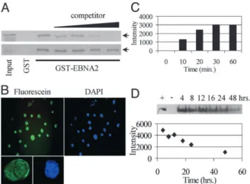
To verify that the addition of a TAT fusion partner did not alter the ability of the EBNA2 peptide to interfere with EBNA2 binding to CBF1, a GST affinity assay was performed and the EBNA2-TAT peptide and EBNA2(mt)-TAT peptides were tested as competitors. The CBF1-binding domain of EBNA2, was expressed as a GST fusion protein, GST-EBNA2 (252–425), and interaction between GST-EBNA2 (252–425) and in vitro-translated35S-labeled CBF1 was demonstrated (Fig. 1A). Added
EBNA2-TAT peptide was able to compete with 35S-labeled
CBF1 for binding to GST-EBNA2 (252–425) with an IC50 of
⬍10M, whereas the control EBNA2(mt)-TAT peptide was an
ineffective competitor.
EBNA2-TAT Peptide Uptake and Stability.To monitor uptake, the fluorescein modified peptide F-EBNA2-TAT was incubated with EBV(⫺) DG75 B cells for 20 min after which the cells were
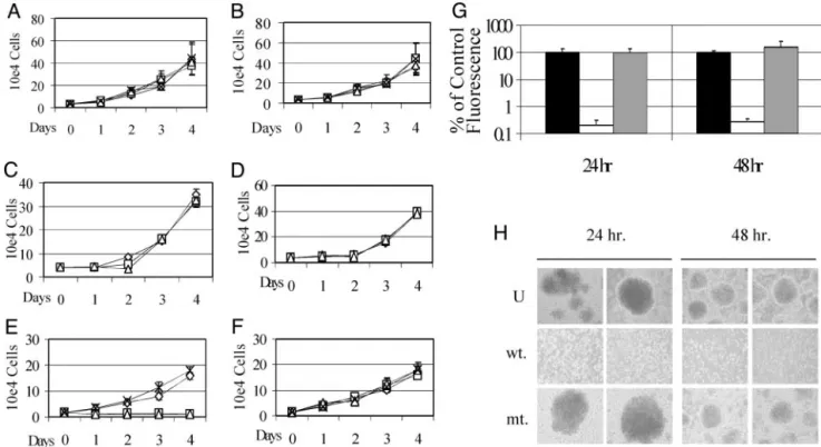
washed, spotted onto slides, fixed, and examined by fluorescence microscopy (Fig. 1B). Uptake was very efficient, with essentially all cells showing fluorescence throughout both the cytoplasm and the nucleus. The time course of uptake of the F-EBNA2-TAT peptide was examined in EBV(⫺) DG75 B cells. The intracellular concentration of F-EBNA2-TAT peptide was mea-sured by harvesting cells at the indicated times after peptide exposure and subjecting the cell extracts to gel electrophoretic separation. The relative amount of intracellular peptide was quantified by using FluorChem to measure the fluorescence intensity in the peptide band. Half-maximal peptide uptake occurred at⬇15 min after exposure to the cells at 37°C (Fig. 1C). In the same way, we examined the stability of the peptide in DG75 cells continuously exposed to peptide. An effective half-life of 24 h was observed for the intracellular peptide (Fig. 1D). TAT Peptide Affects the Growth and Survival of EBNA2-Expressing LCLs. The effect of different concentrations of EBNA2-TAT and EBNA2(mt)-TAT peptides on the growth of EBV(⫺) and EBV(⫹) B cell lines was next examined. Over a 4-day period, treatment of EBV(⫺) DG75 cells with either peptide at 1-, 25-, or 50-M concentrations had little effect on cell proliferation (Fig. 2 A and B). Akata cells, which are EBV(⫹) but are EBNA2(⫺), were also not significantly affected by treatment with either peptide (Fig. 2 C and D). A newly established EBV⫹LCL cell line was also insensitive to treatment
with mutant peptide at the same three concentrations (Fig. 2F). However, treatment with wild-type EBNA2-TAT peptide at 25-or 50-M concentrations dramatically reduced proliferation of the LCL cells (Fig. 2E). The metabolic activity of LCLs treated
with 25M EBNA2-TAT or EBNA2(mt)-TAT peptides was
examined in a CellTiter-Glo assay in which intracellular ATP
levels are measured by using a luciferase readout. Cells treated with EBNA2-TAT peptide had significantly reduced metabolic activity at both 24 and 48 h after treatment (Fig. 2G). In contrast, cells treated with mutant peptide showed metabolic activity comparable to controls in this assay (Fig. 2G). EBV⫹LCLs have
up-regulated surface adhesion molecules and form macroscopic clumps in culture. Examination of the peptide treated versus the untreated LCL cultures revealed that the EBNA2(mt)-TAT peptide did not affect this growth phenotype, whereas in the EBNA2-TAT peptide-treated culture, the clumps were com-pletely dispersed (Fig. 2H).
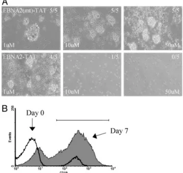
In culture, EBV(⫹) LCLs express the type III latency genes, which include the membrane proteins LMP1 and LMP2A (1). LMP1 is particularly important for cellular proliferative re-sponses and is also responsible for the up-regulation of cell adhesion antigens (46). Because LMP1 and LMP2A expression in B cells is driven by EBNA2, we examined the effect of the EBNA2-TAT peptides on their expression by using RT-PCR, followed by Southern blotting of the PCR products. LCLs were treated with 50-M concentrations of either wild-type or mutant peptide for 24 and 48 h. Treatment with EBNA2-TAT peptide significantly reduced the level of both transcripts at 48 h, whereas treatment with mutant peptide did not alter transcript abun-dance (Fig. 3A). EBNA2 also modulates cellular gene expression and cell genes known to be EBNA2-responsive include CD23 and Cdk1. These genes were also expressed at reduced levels in EBNA2-TAT peptide-treated LCLs (Fig. 3A). RNA expression in peptide-treated cells was also examined by real-time RT-PCR (Fig. 3B). LMP1, CD23, and Cdk1 were down-regulated in this analysis, as was BATF and ICAM-1. In contrast, expression of p21 was increased in peptide-treated cells at both 24 and 48 h. EBNA2-TAT Peptide Blocks Outgrowth of Colonies from EBV-Infected PBMCs.The EBNA2-TAT peptide interfered with the prolifer-ation of already immortalized B cells that depended on the type III latency program for continued growth. To evaluate the ability of the EBNA2-TAT peptide to prevent EBV-induced initiation of B cell proliferation, a PBMC proliferation assay was estab-lished. Human PBMCs were incubated in the presence or absence of EBV virus obtained from induced Akata BX-1 cells. This virus contains a GFP marker inserted in the BXLF1 ORF (45). After 7 days of incubation, the wells were examined for outgrowth of clumps of self-adherent proliferating cells. In wells that were left uninfected, most of the cells were dying and no macroscopic colonies were visible. In contrast, wells that were infected with EBV virus contained large numbers of macro-scopic colonies that were GFP(⫹), demonstrating EBV infection (data not shown). The effect of the EBNA2-TAT and EBNA2(mt)-TAT peptides on B cell outgrowth was assessed by using 1-, 10-, and 50-M concentrations of peptide. The peptide was added immediately after virus infection and replenished daily. The EBNA2(mt)-TAT peptide had no discernable effect on B cell colony outgrowth at any of the three doses, and in each case, five of five wells contained macroscopic B cell colonies (Fig. 4A). On the other hand, the EBNA2-TAT peptide had a dose-responsive effect on colony formation. At the lowest con-centration (1M), four of five wells contained visible colonies; at 10M, only one of five wells was positive and no colonies were detected in the wells receiving 50M EBNA2-TAT peptide (Fig.
4A). The IC50of the EBNA2-TAT peptide was⬍10M, which
is consistent with the concentration required to block the interaction of EBNA2 and CBF1 in a GST affinity assay (Fig. 1 A).
To verify that the assay was measuring B cell outgrowth, untreated PBMCs were taken at day 0 and at 7 days after EBV infection, and were analyzed by fluorescence-activated cell sorter for expression of the pan B cell marker CD19 (Fig. 4B).
Fig. 1. Peptide in vitro activity and intracellular availability. (A) GST affinity assay in which in vitro-translated,35S-labeled CBF1 (arrows) was incubated
with GST-EBNA2 (252– 425) or control GST protein and binding was examined in the presence of increasing amounts (0.125, 1.25, 12.5, and 125M) of competitor EBNA2-TAT (Upper) or EBNA2(mt)-TAT peptide (Lower). (B) Pho-tomicrographs demonstrating incorporation of F-EBNA2-TAT into DG75 B cells after 20 min of peptide exposure at room temperature. (Left Upper) Fluorescein. (Right Upper) Cell nuclei stained with 4 ⬘,6-diamidino-2-phenylindole. (Lower) Confocal image of a single cell showing nuclear plus cytoplasmic distribution of F-EBNA2-TAT (Left) and 4 ⬘,6-diamidino-2-phenylindole nuclear staining (Right). (C) Time course of F-EBNA2-TAT pep-tide uptake into DG75 cells incubated with F-EBNA2-TAT peppep-tide for the indicated times. Intracellular F-EBNA2-TAT peptide levels were measured as described in D. (D) F-EBNA2-TAT peptide bioavailability in DG75 cell cultures incubated in medium containing 50M peptide added at time 0. (Upper) Visualization, by UV excitation, of electrophoretically separated intracellular peptide. (Lower) Time course of loss of intracellular F-EBNA2-TAT peptide.
At day 0, the PBMCs contained 5% CD19(⫹) B cells. By day 7 after EBV infection, 70% of the live cells were CD19(⫹). Continuous Peptide Exposure Is Necessary to Prevent Colony Out-growth. The EBNA2-TAT peptide is proposed to function by competing with EBNA2 for CBF1 binding, and hence, down-regulating EBNA2-responsive viral and cellular genes whose expression is necessary for the growth proliferative response. Such a mechanism of physical interference should require the continuous presence of the peptide. The effect on B cell out-growth of a regimen of short-term exposure followed by culture in the absence of EBNA2-TAT peptide was therefore examined. In this experiment, 10-well replicates were scored for macro-scopic colony outgrowth. The peptide-treated wells were incu-bated in medium containing EBNA2-TAT peptide at 10- or 100-M concentrations for 1 week after which the cultures were switched to peptide free medium for another 3 weeks. No colony outgrowth was detected in the uninfected PBMC cultures at any of the time points examined (1, 3, and 4 weeks), whereas the EBV-infected wells all contained macroscopic colonies (Table 1). In the wells treated with the EBNA2-TAT peptide, no colony outgrowth was visible after 1 week of continuous treatment with either 10- or 100-M peptide concentrations. However, when the cultures were maintained for another 3 weeks in the absence of peptide, colony outgrowth rebounded. Ten of 10 wells treated
initially with 10M EBNA2-TAT peptide and 8 of 10 wells
treated initially with 100M EBNA2-TAT peptide contained macroscopic colonies at the 4-week postinfection time point. This result indicates that continuous exposure to peptide is needed to interfere with EBNA2 function and provides
addi-tional support for the proposed mechanism of action of the EBNA2-TAT peptide.
Discussion
PTDs have the ability to mediate cell entry in a concentration-dependent manner that is inconcentration-dependent of receptors or trans-porters and consequently operates in a wide variety of cell types. The mechanism of cell entry is not fully understood but may involve avid binding of the positively charged residues to cell-surface polyanions such as heparin sulfate and internalization through an endocytosis related process (35, 47). PTDs have been used to deliver fusion proteins and peptides that have shown biological activity (48–58). The EBNA2-TAT peptide severely impaired the growth of cultured EBV(⫹) LCLs. The LCL cultures used in our experiments were newly immortalized and the W latency promoter was still active (data not shown). The W latency promoter is not CBF1- or EBNA2-responsive and hence W latency promoter-driven EBNA2 expression would not be affected by the EBNA2-TAT peptide. The use of newly immor-talized LCLs in these experiments allowed us to evaluate the impact of interfering with EBNA2 activation of CBF1-responsive promoters in a setting in which normal EBNA2 expression was retained. This result differs from previous anal-yses of EBNA2 function that have studied the effects of loss of EBNA2 through genetic deletion or through conditional nuclear localization.
LCLs are dependent on the transcriptional activation function of EBNA2 for continued growth. LCLs immortalized with a virus expressing an estrogen-regulated EBNA2 stop growing when estrogen is removed from the culture medium (59). These cells can be rescued by transduction with a vector expressing
Fig. 2. EBNA2-TAT affects EBNA2(⫹) LCL growth and metabolic activity. Proliferation assays showing the effect of EBNA2-TAT (A, C, and E) and EBNA2(mt)-TAT (B, D, and F) peptides on cell growth. X, Untreated;〫, 1M peptide;䊐, 25M peptide;‚, 50M peptide. Neither EBNA2-TAT nor EBNA2(mt)-TAT peptide significantly affected growth of EBV(⫺) DG75 B cells (A and B) or EBV(⫹), but EBNA2(⫺) Akata cells (C and D). Growth of EBV and EBNA2(⫹) LCLs was inhibited by EBNA2-TAT peptide at 25- and 50-M concentrations (E) but not by EBNA2(mt)-TAT peptide (F). (G) Comparison of the metabolic activity of EBV⫹LCLs
untreated or treated for 24 or 48 h with 25M EBNA2-TAT or EBNA2(mt)-TAT peptide. Metabolic activity, as measured in a CellTiter-Glo assay, was drastically reduced by EBNA2-TAT treatment. Filled bars, untreated; open bars, 25 mM EBNA2-TAT; shaded bars, 25M EBNA2(mt)-TAT. (H) Phase contrast photomicro-graphs of EBV⫹LCLs showing that EBNA2-TAT, but not EBNA2(mt)-TAT, at a concentration of 50M, prevents LCLs from growing as macroscopic colonies. U, untreated; wt, EBNA2-TAT 50 mM; mt, EBNA2(mt)-TAT 50 mM.
wild-type EBNA2, but cannot be rescued by an EBNA2 that is mutated in the conserved region 6 motif and is unable to interact with CBF1 (13). The estrogen-regulated LCLs can also be
rescued by transduction of activated intracellular domain of Notch (NotchIC) in circumstances in which LMP1 is either also provided or is selectively up-regulated (13, 14, 60). This obser-vation highlights both the high degree of overlap in EBNA2- and NotchIC-regulated cell genes and the importance for B cell growth of EBNA2 activation of the viral LMP1 gene (which is poorly responsive to NotchIC). RT-PCR analyses revealed that LMP1 was down regulated by the EBNA2-TAT peptide and this is likely to be a significant component of the EBNA2-TAT peptide’s negative effect on LCL growth. Although EBNA2 and NotchIC both alter cellular gene expression through interactions with CBF1, the EBNA2-TAT peptide is designed to be specific for the EBNA2–CBF1 interaction. EBNA2 and NotchIC bind to adjacent but distinct regions of CBF1, and mutagenesis studies have identified amino acids that affect only NotchIC or EBNA2 interaction (61, 62).
The EBNA2-regulated viral LMP2A gene also showed re-duced expression in the presence of the EBNA2-TAT peptide. LMP2A inhibits lytic viral reactivation and provides cell survival signals through activation of Akt (63, 64). Cell genes tested that were known to be either EBNA2-regulated or responsive to the combination of EBNA2 and LMP1 were also down-regulated in the presence of the EBNA2-TAT peptide. ICAM-1 (65) medi-ates cell–cell contacts and contributes to B cell growth as clumps in culture, soluble CD23 (46) acts as an autocrine growth factor, and BATF (66), an AP-1 family member, may have a role in repression of the EBV lytic cycle. Cdk1兾Cdc2 (59) interacts with A and B cyclins to regulate the mitotic phase of the cell cycle (67) and was the most significantly affected of the cellular genes evaluated here. The Cdk1 promoter is regulated by nuclear factor Y, and nuclear factor Y has recently been found to be up-regulated by EBNA2 in conditionally EBNA2-expressing cells (68). With the exception of Cdk1, the real-time RT-PCR analyses showed a relatively small down-regulation of the tested genes by the EBNA2-TAT peptide. This observation suggests that cessation of EBV-driven LCL growth can be achieved through the accumulated effects of down-regulating multiple genes without necessarily completely ablating expression of individual EBNA2-regulated genes. The increased expression of p21 observed in the peptide treated cells is consistent with induction of growth arrest.
The EBNA2-TAT peptide also prevented the proliferation of primary B cells infected in vitro with EBV. The effect was specific in that the EBNA2(mt)-TAT peptide did not have this property and continuous exposure to peptide was required to block outgrowth of proliferating B cell colonies. Improved bioavail-ability of the peptide could be addressed in the future by using peptidomimetic approaches or modifications such as cyclization. Peptidomimetics contain nonnatural building blocks such as D-amino acids or -amino acids (69–71). The ability of the EBNA2-TAT peptide to affect the growth of EBV-infected LCLs as well as to prevent expansion of newly infected B cells suggests that the peptide may have therapeutic potential. Early-onset posttransplant lymphoproliferative disease is strongly EBV-associated and has a high mortality. The disease is a
Fig. 3. EBNA2-TAT down-regulates expression of known EBNA2-responsive viral and cellular genes. (A) Southern blots of RT-PCR products amplified using primers for the EBNA2-regulated EBV LMP1 and LMP2A genes and cell CD23 and Cdk1 genes. The amplified products were hybridized with32P-labeled
oligonucleotide probes specific for the individual genes. Ethidium bromide-stained-actin cDNA served as a loading control. U, untreated; wt, 50 mM EBNA2-TAT; mt, 50 mM EBNA2(mt)-TAT. (B) Changes in gene expression in peptide-treated LCLs as measured by real-time RT-PCR. Results are shown as the relative fold difference between LCLs treated with 50M EBNA2-TAT or EBNA2 (mt)-TAT, with TATA box-binding protein as the internal standard. Cells were treated for 24 h (open bar) or 48 h (filled bar). The data are representative of three experiments.
Fig. 4. EBNA2-TAT prevents EBV-induced B cell proliferation. (A) Phase contrast photomicrographs showing PBMC 7 days after EBV infection in the presence of 1-, 10-, or 50-M concentrations of EBNA2-TAT or EBNA2(mt)-TAT peptide. The infections were performed in five-well replicates. The fraction of the wells showing B cell outgrowth is indicated. B cell outgrowth was not affected by EBNA2(mt)-TAT but was reduced at 1- and 10-M EBNA2-TAT concentrations and was abolished by 50M EBNA2-TAT peptide. (B) Fluores-cence-activated cell sorter profile showing that proliferating colonies are B cells. Expression of the CD19 B cell marker at days 0 and 7 after EBV infection is shown. Bar, CD19⫹cells.
Table 1. Continuous EBNA2-TAT is required to block EBV-induced B cell proliferation
Experimental conditions
Colony outgrowth at
1 week 3 weeks 4 weeks
Infected 10 of 10 10 of 10 9 of 9
Uninfected 0 of 10 0 of 10 0 of 10
Infected, 10M EBNA2-TAT* 0 of 10 0 of 10 10 of 10 Infected, 100M EBNA2-TAT* 0 of 10 0 of 10 8 of 10 *Treated for 1 week.
particular problem in children who are more likely to be EBV-seronegative at the time of transplant (6). There is some heterogeneity in viral gene expression in the tumor cells but EBNA2-driven expansion is a significant component of the disease. Current treatment centers on reduction in immunosup-pression, which carries an associated risk of graft rejection. Other treatments such as adoptive immunotherapy and clear-ance of B cells by using anti-B cell antibodies show promise but there remains a need for additional treatment options (72). Infectious mononucleosis normally resolves with only
symptom-atic treatment. However, infectious mononucleosis can have an extended recovery time in severe cases and new intervention strategies targeting the expansion of latently infected cells may also be relevant to primary EBV-associated disease.
We thank Lindsey Hutt-Fletcher for Akata Bx1 cells, Leslie Metzler for assistance with microscopy, and Deborah Nguyen for assistance with the CellTiter-Glo assay. This work was supported by U.S. Public Health Service Grants R37 CA42245 (to S.D.H.) and GM 62437 (to P.A.C.) and by Johns Hopkins Lymphoma SPORE Grant P50 CA96888.
1. Kieff, E. (1996) in Fields Virology, eds. Field, B. N., Knipe, D. M. & Howley, P. M. (Raven, New York), Vol. 2, pp. 2343–2396.
2. Joseph, A. M., Babcock, G. J. & Thorley-Lawson, D. A. (2000) J. Virol. 74, 9964–9971.
3. Babcock, G. J., Hochberg, D. & Thorley-Lawson, A. D. (2000) Immunity 13, 497–506.
4. Tierney, R. J., Steven, N., Young, L. S. & Rickinson, A. B. (1994) J. Virol. 68, 7374–7385.
5. Rogatsch, H., Bonatti, H., Menet, A., Larcher, C., Feichtinger, H. & Dirnhofer, S. (2000) Am. J. Surg. Pathol. 24, 614–621.
6. Holmes, R. D. & Sokol, R. J. (2002) Pediatr. Transplant. 6, 456–464. 7. Timms, J. M., Bell, A., Flavell, J. R., Murray, P. G., Rickinson, A. B.,
Traverse-Glehen, A., Berger, F. & Delecluse, H. J. (2003) Lancet 361, 217–223. 8. Boshoff, C. & Weiss, R. (2002) Nat. Rev. Cancer 2, 373–382.
9. Ambinder, R. F. (2001) Eur. J. Cancer 37, 1209–1216.
10. Oyama, T., Ichimura, K., Suzuki, R., Suzumiya, J., Ohshima, K., Yatabe, Y., Yokoi, T., Kojima, M., Kamiya, Y., Taji, H., et al. (2003) Am. J. Surg. Pathol.
27,16–26.
11. Kelly, G., Bell, A. & Rickinson, A. (2002) Nat. Med. 8, 1098–1104. 12. Hsieh, J. J.-D., Henkel, T., Salmon, P., Robey, E., Peterson, M. G. & Hayward,
S. D. (1996) Mol. Cell. Biol. 16, 952–959.
13. Gordadze, A. V., Peng, R., Tan, J., Liu, G., Sutton, R., Kempkes, B., Bornkamm, G. W. & Ling, P. D. (2001) J. Virol. 75, 5899–5912.
14. Hofelmayr, H., Strobl, L. J., Marschall, G., Bornkamm, G. W. & Zimber-Strobl, U. (2001) J. Virol. 75, 2033–2040.
15. Hofelmayr, H., Strobl, L. J., Stein, C., Laux, G., Marschall, G., Bornkamm, G. W. & Zimber-Strobl, U. (1999) J. Virol. 73, 2770–2780.
16. Hayward, S. D. (1999) EBV Report 6, 151–157.
17. Henkel, T., Ling, P. D., Hayward, S. D. & Peterson, M. G. (1994) Science 265, 92–95.
18. Grossman, S. R., Johannsen, E., Tong, X., Yalamanchili, R. & Kieff, E. (1994)
Proc. Natl. Acad. Sci. USA 91, 7568–7572.
19. Zimber-Strobl, U., Strobl, L. J., Meitinger, C., Hinrichs, R., Sakai, T., Fu-rukawa, T., Honjo, T. & Bornkamm, G. W. (1994) EMBO J. 13, 4973–4982. 20. Waltzer, L., Logeat, F., Brou, C., Israel, A., Sergeant, A. & Manet, E. (1994)
EMBO J. 13, 5633–5638.
21. Johannsen, E., Koh, E., Mosialos, G., Tong, X., Kieff, E. & Grossman, S. R. (1995) J. Virol. 69, 253–262.
22. Laux, G., Adam, B., Strobl, L. J. & Moreau-Gachelin, F. (1994) EMBO J. 13, 5624–5632.
23. Fuentes-Panana, E. M., Peng, R., Brewer, G., Tan, J. & Ling, P. D. (2000)
J. Virol. 74, 8166–8175.
24. Zhou, S., Fujimuro, M., Hsieh, J. J., Chen, L. & Hayward, S. D. (2000) J. Virol.
74,1939–1947.
25. Voss, M. D., Hille, A., Barth, S., Spurk, A., Hennrich, F., Holzer, D., Mueller-Lantzsch, N., Kremmer, E. & Grasser, F. A. (2001) J. Virol. 75, 11781–11790.
26. Wu, D. Y., Krumm, A. & Schubach, W. H. (2000) J. Virol. 74, 8893–8903. 27. Wang, L., Grossman, S. R. & Kieff, E. (2000) Proc. Natl. Acad. Sci. USA 97,
430–435.
28. Yalamanchili, R., Tong, X., Grossman, S., Johannsen, E., Mosialos, G. & Kieff, E. (1994) Virology 204, 634–641.
29. Osborne, B. & Miele, L. (1999) Immunity 11, 653–663.
30. Lee, J. M., Lee, K. H., Weidner, M., Osborne, B. A. & Hayward, S. D. (2002)
Proc. Natl. Acad. Sci. USA 99, 11878–11883.
31. Ling, P. D., Rawlins, D. R. & Hayward, S. D. (1993) Proc. Natl. Acad. Sci. USA
90,9237–9241.
32. Peng, R., Gordadze, A. V., Fuentes Panana, E. M., Wang, F., Zong, J., Hayward, G. S., Tan, J. & Ling, P. D. (2000) J. Virol. 74, 379–389. 33. Cho, Y. G., Gordadze, A. V., Ling, P. D. & Wang, F. (1999) J. Virol. 73,
9206–9212.
34. Ling, P. D. & Hayward, S. D. (1995) J. Virol. 69, 1944–1950.
35. Leifert, J. A. & Lindsay Whitton, J. (2003) Mol. Ther. 8, 13–20. 36. Futaki, S. (2002) Int. J. Pharm. 245, 1–7.
37. Denicourt, C. & Dowdy, S. F. (2003) Trends Pharmacol. Sci. 24, 216–218. 38. Elliott, G. & O’Hare, P. (1997) Cell 88, 223–233.
39. Fawell, S., Seery, J., Daikh, Y., Moore, C., Chen, L. L., Pepinsky, B. & Barsoum, J. (1994) Proc. Natl. Acad. Sci. USA 91, 664–668.
40. Green, M. & Loewenstein, P. M. (1988) Cell 55, 1179–1188. 41. Frankel, A. D. & Pabo, C. O. (1988) Cell 55, 1189–1193.
42. Mitchell, D. J., Kim, D. T., Steinman, L., Fathman, C. G. & Rothbard, J. B. (2000) J. Pept. Res. 56, 318–325.
43. Futaki, S., Suzuki, T., Ohashi, W., Yagami, T., Tanaka, S., Ueda, K. & Sugiura, Y. (2001) J. Biol. Chem. 276, 5836–5840.
44. Wender, P. A., Mitchell, D. J., Pattabiraman, K., Pelkey, E. T., Steinman, L. & Rothbard, J. B. (2000) Proc. Natl. Acad. Sci. USA 97, 13003–13008. 45. Molesworth, S. J., Lake, C. M., Borza, C. M., Turk, S. M. & Hutt-Fletcher, L. M.
(2000) J. Virol. 74, 6324–6332.
46. Wang, F., Gregory, C., Sample, C., Rowe, M., Liebowitz, D., Murray, R., Rickinson, A. B. & Kieff, E. (1990) J. Virol. 64, 2309–2318.
47. Sandgren, S., Cheng, F. & Belting, M. (2002) J. Biol. Chem. 277, 38877–38883. 48. Schwarze, S. R., Ho, A., Vocero-Akbani, A. & Dowdy, S. F. (1999) Science 285,
1569–1572.
49. Asoh, S., Ohsawa, I., Mori, T., Katsura, K., Hiraide, T., Katayama, Y., Kimura, M., Ozaki, D., Yamagata, K. & Ohta, S. (2002) Proc. Natl. Acad. Sci. USA 99, 17107–17112.
50. Kilic, E., Dietz, G. P., Hermann, D. M. & Bahr, M. (2002) Ann. Neurol. 52, 617–622.
51. Cao, G., Pei, W., Ge, H., Liang, Q., Luo, Y., Sharp, F. R., Lu, A., Ran, R., Graham, S. H. & Chen, J. (2002) J. Neurosci. 22, 5423–5431.
52. Dietz, G. P., Kilic, E. & Bahr, M. (2002) Mol. Cell. Neurosci. 21, 29–37. 53. Vocero-Akbani, A. M., Heyden, N. V., Lissy, N. A., Ratner, L. & Dowdy, S. F.
(1999) Nat. Med. 5, 29–33.
54. Jo, D., Nashabi, A., Doxsee, C., Lin, Q., Unutmaz, D., Chen, J. & Ruley, H. E. (2001) Nat. Biotechnol. 19, 929–933.
55. Yan Liu, X., Robinson, D., Veach, R. A., Liu, D., Timmons, S., Collins, R. D. & Hawiger, J. (2000) J. Biol. Chem. 275, 16774–16778.
56. Kiosses, W. B., Hood, J., Yang, S., Gerritsen, M. E., Cheresh, D. A., Alderson, N. & Schwartz, M. A. (2002) Circ. Res. 90, 697–702.
57. Shibagaki, N. & Udey, M. C. (2003) Eur. J. Immunol. 33, 850–860. 58. Wang, H. Y., Fu, T., Wang, G., Zeng, G., Perry-Lalley, D. M., Yang, J. C.,
Restifo, N. P., Hwu, P. & Wang, R. F. (2002) J. Clin. Invest. 109, 1463–1470. 59. Kempkes, B., Spitkovsky, D., Jansen-Durr, P., Ellwart, J. W., Kremmer, E., Delecluse, H.-J., Rottenberger, C., Bornkamm, G. W. & Hammerschmidt, W. (1995) EMBO J. 14, 88–96.
60. Strobl, L. J., Hofelmayr, H., Marschall, G., Brielmeier, M., Bornkamm, G. W. & Zimber-Strobl, U. (2000) J. Virol. 74, 1727–1735.
61. Hsieh, J. J.-D., Nofziger, D. E., Weinmaster, G. & Hayward, S. D. (1997)
J. Virol. 71, 1938–1945.
62. Fuchs, K. P., Bommer, G., Dumont, E., Christoph, B., Vidal, M., Kremmer, E. & Kempkes, B. (2001) Eur. J. Biochem. 268, 4639–4646.
63. Longnecker, R. (2000) Adv. Cancer Res. 79, 175–200.
64. Scholle, F., Bendt, K. M. & Raab-Traub, N. (2000) J. Virol. 74, 10681–10689. 65. Devergne, O., McFarland, E. C., Mosialos, G., Izumi, K. M., Ware, C. F. &
Kieff, E. (1998) J. Virol. 72, 7900–7908.
66. Johansen, L. M., Deppmann, C. D., Erickson, K. D., Coffin, W. F., III, Thornton, T. M., Humphrey, S. E., Martin, J. M. & Taparowsky, E. J. (2003)
J. Virol. 77, 6029–6040.
67. Pines, J. (1999) Nat. Cell Biol. 1, E73–E79.
68. Borestrom, C., Zetterberg, H., Liff, K. & Rymo, L. (2003) J. Virol. 77, 821–829. 69. Patch, J. A. & Barron, A. E. (2002) Curr. Opin. Chem. Biol. 6, 872–877. 70. Li, P. & Roller, P. P. (2002) Curr. Top. Med. Chem. 2, 325–341. 71. Sehgal, A. (2002) Curr. Opin. Drug Discov. Devel. 5, 245–250.