Introduction
Osteoarthritis (OA), the most common joint disease associated with joint trauma and aging, is characterized by the destruction of progressive articular cartilage [1,2]. Among the commonly known causes of OA, in-flammation is an important contributor to the patho-genesis of OA that causes an imbalance of the metabol-ic pathway of chondrocytes, leading to the loss of meta-bolic homeostasis and the degradation of the ex-tracellular matrix (ECM) [3-5].
Among inflammatory cytokines, interleukin-1 beta (IL-1β) plays a pivotal role in inducing the production and secretion of inflammatory and catabolic factors as-sociated with OA pathogenesis [6-8]. The levels of IL-1 β are significantly higher in the synovial fluid and car-tilage of OA patients than in normal individuals [9,10]. When chondrocytes are exposed to IL-1β, the ex-pression of inflammatory genes including inducible ni-tric oxide synthase (iNOS), cyclooxygenase 2 (COX-2), and matrix metalloproteinases (MMPs), which are ma-jor proteases directly involved in cartilage degradation, * Corresponding author
Phone: +82-51-890-3319 Fax: +82-51-890-3333 E-mail: [email protected]
This is an open-access journal distributed under the terms of the Creative Commons Attribution Non-Commercial License
(http://creativecommons.org/licenses/by-nc/4.0/)
https://doi.org/10.15433/ksmb.2020.12.2.099 ISSN 2383-5400 (Online)
Carpomitra costata
Extract Suppresses Interleukin-1β-Induced
Inflammatory Response in SW1353 Human Chondrocytes through
Suppressing NF-κB Signaling Pathway
Yung Hyun Choi1,2*
1Anti-Aging Research Center, Dongeui University, Busan 47227, Republic of Korea
2Department of Biochemistry, Dong-eui University College of Korean Medicine, Busan 47227, Republic of Korea
(Received 6 November 2020, Revised 9 December 2020, Accepted 21 December 2020)
Abstract Osteoarthritis (OA) is an inflammatory degenerative joint disease that is accompanied
by irreversible joint cartilage destruction. Recently, the antioxidant effects of Carpomitra costata, which is a type of brown algae, have been reported, but their effects on OA have not been investigated. In this study, the anti-osteoarthritic effect of the ethanol extract of C. costata (EECC) on SW1353 human chondrocytes was studied. Results showed that EECC significantly attenuated the interleukin-1β (IL-1β)-induced release of pro-inflammatory mediators, including prostaglandin E2 and nitric oxide (NO), as well as expressions of cyclo-oxygenase-2 and inducible NO synthase.
EECC also inhibited the IL-1β-induced expressions of matrix metalloproteinase-1, -3, and -13 in SW1353 chondrocytes, which reduced their extracellular secretion. In addition, the oxidative stress induced by IL-1β was confirmed to be blocked by EECC due to the inhibition of reactive oxygen species generation. Moreover, EECC suppressed IL-1β-mediated translocation of nuclear factor-kappa B (NF-κB) from cytosol into the nucleus and the degradation of IκB-α, which in-dicates that EECC exhibits anti-inflammatory effects by inhibiting the NF-κB signaling pathway. These results are the first to demonstrate the anti-inflammatory activities of C. costata extracts in chondrocytes, thus suggesting that this algae extract may be used in the treatment of OA.
are increased via activation of nuclear factor-kappa B (NF-κB) [11-13]. Therefore, NF-κB is a critical target of therapeutic strategies aimed at the treatment of OA. In addition, excessive oxidative stress may be consid-ered as another important factor triggering OA follow-ing chondrocyte senescence and apoptosis [14,15]. Although non-steroidal anti-inflammatory drugs are generally widely used to relieve symptoms of OA, re-search on reliable and effective drugs is required due to side effects caused by long-term use [16,17].
Recently, attention has been focused on the use of marine species as a resource for suppressing various inflammatory diseases. Among them, seaweeds have a great potential for the development of OA prevention agents because of the abundance of physiologically ac-tive substances with various pharmacological actions [18]. For example, in a small clinical study on humans, the oral administration of fucoidan-rich brown algae extract significantly inhibited OA symptoms, and it was associated with the reduction of IL-6, a chronic in-flammatory marker [19]. In addition, the phlor-otannin-rich extract of Ecklonia cava markedly sup-pressed the production of prostaglandin E2 (PGE2) in lipopolysaccharide-treated RAW 246.7 cells and atte-nuated the IL-1α-induced proteolytic degradation [20]. However, up to now, studies on the effect of seaweed extract on OA have been limited in comparison with those on its diversity. Therefore, it was studied the ef-fect of the ethanol extract of Carpomitra costata (Stackhouse) Batters (EECC), a kind of brown algae, on the IL-1β-mediated inflammatory responses, MMPs, and reactive oxygen species (ROS) production in SW1353 human chondrocytes as part of the discovery of new marine biomaterials for OA treatment.
Materials and Methods
Preparation of EECC
EECC used in the current study was provided by Dr DS Lee (National Marine Biodiversity Institute of Korea, Seocheon, Republic of Korea). For the prepara-tion of EECC, C. costata was collected from offshore
Ulleung Island, Republic of Korea in March 2016. Collected C. costata was washed with tap water and stored at −20˚C. Samples were homogenized using a grinder and dried prior to extraction. The dried powder was extracted 5 times with 70% EtOH (1:10 w/v) at 1 h intervals by sonication. Subsequently, the extract (EECC) was evaporated in vacuo, and then dissolved in dimethyl-sulfoxide (DMSO, Sigma-Aldrich Chemical Co., St. Louis, MO, USA) before use in the experiment.
Cell culture
SW1353 chondrocytes, which were purchased from American Type Culture Collection (Manassas, VA, USA), were maintained in humidified air at 37°C, 5% CO2 in Dulbecco's modified Eagle's medium (DMEM) containing 100 U/ml penicillin and streptomycin, and 10% fetal bovine serum. All materials required for cell culture were purchased from WelGENE Inc. (Daegu, Republic of Korea).
Cell viability assay
The cytotoxicity of EECC to SW1353 chondrocytes
was determined using a colorimetric
3-(4,5-dimethylthiazol-2-yl)-2,5-diphenyltetrazolium bromide (MTT) reduction assay. In brief, cells were incubated in 96-well plates for 24 h at a density of 1 x 10⁴ cells, and then treated with different concen-trations of EECC or IL-1β (40 ng/ml, R&D Systems, Minneapolis, MN, USA) alone or pretreated with dif-ferent concentrations of EECC for 1 h before IL-1β treatment for 24 h. Then, the medium was removed, and MTT solution (0.5 mg/ml, Sigma-Aldrich Chemical Co.) was dispensed into each well and re-acted at 37°C. After 3 h, the supernatant was removed and DMSO was added to dissolve blue formazan crys-tals for 10 min. The absorbance per well was quantified at a wavelength of 540 nm using an enzyme-linked im-munosorbant assay (ELISA) reader (Dynatech Laboratories, Chantilly, VA, USA).
SW1353 chondrocytes were treated with various con-centrations of EECC for 1 h and then stimulated with IL-1β for 24 h. Nitrite levels in the media were eval-uated by a Griess reaction (Sigma-Aldrich Chemical Co.). Briefly, 100 μL of the cell conditioned medium was mixed with the same amount of Griess reagent for 10 min, and then the absorbance was measured with an ELISA reader at a wavelength of 540 nm. To inves-tigate PGE2 levels, the culture supernatants were col-lected and assayed using a commercially available ELISA kit (R&D Systems) with the instructions of the manufacturer. The levels of levels of MMPs released from cultured chondrocytes were calculated by specific ELISA kits (R&D Systems) in accordance with the in-structions of the manufacturer.
Protein isolation and immunoblotting
Nuclear extraction reagents purchased from Pierce (Rockford, IL, USA) were used to isolate proteins from the nucleus and cytoplasm according to the manufactur-er's protocol. The concentration of the isolated protein was measured using the Bio-Rad protein assay kit ob-tained from Bio-Rad Laboratories (Hercules, CA, USA). Equal amounts of protein were separated by
so-dium-dodecyl sulfate-polyacrylamide gel
electrophoresis. Proteins in the gel were subsequently transferred to polyvinylidene difluoride (PVDF) mem-branes (Schleicher and Schuell GmbH, Keene, NH, USA). The protein-transferred membrane was blocked with non-fat dry milk solution (5%) at room temper-ature for 1 h, and then reacted with primary antibodies overnight at 4˚C. Antibodies against NF-κB p65 (sc-109), IκB-α (sc-371), lamin B (sc-6216) and β -actin (sc-1615) were obtained from Santa Cruz Biotechnology, Inc. (Santa Cruz, CA, USA). The mem-brane was washed 3 times for 5 min with Tris buffered saline (0.1% Tween-20) and then incubated with goat anti-rabbit IgG-horseradish-peroxidase (HRP) and goat anti-mouse IgG-HRP secondary antibodies (Santa Cruz Biotechnology, Inc.) for 2 h at room temperature. The membrane was reacted with an enhanced chem-iluminescent solution purchased from Amersham Corp.
(Arlington Heights, IL, USA) and then exposed to an X-ray film to visualize the corresponding proteins. Bands were quantified using an ImageJ program (Ver. 1.46; NIH, Bethesda, MD, USA) and normalized to β -actin, and the ratio was determined.
Reverse transcriptase-polymerase chain reaction (RT-PCR) assay
Total RNA was extracted from the cells using TRIzol reagents (Invitrogen Life Technologies, Carlsbad, CA, USA), following to the manufacturer’s and quantified. The isolated total RNA (1 μg) was used to synthesize cDNA using AccuPower® RT PreMix (Bioneer, Daejeon, Korea) according to the manufacturer's instructions. The cDNA generated at RT was amplified using selected primers. Gene expression was normal-ized to glyceraldehyde-3-phosphate dehydrogenase (GAPDH) expression. The PCR primers were as fol-lows: iNOS, 5′‐GTG‐AGG‐ATC‐AAA‐AAC‐TGG‐GG‐ 3′ (sense) and 5′‐ACC‐TGC‐AGG‐TTG‐GAC‐CAC‐3′ (anti-sense); COX‐2, 5′‐TCA‐GCC‐ACG‐CAG‐CAA‐ ATC‐CT‐3′ (sense) and 5′‐GTG‐ATC‐TGG‐ATG‐TCA‐ CG‐3′ (anti-sense); GAPDH, 5′‐CGA‐TGC‐TGG‐GCG‐ TGA‐GTA‐C‐3′ (sense) and 5′‐CGT‐TCA‐GCT‐CAG‐ GGA‐TGA‐CC‐3′ (ant-isense); MMP‐1, 5′‐CTG‐TTC‐ AGG‐GAC‐AGA‐ATG‐TGC‐3′ (sense) and 5′‐TTG‐ GAC‐TCA‐CAC‐CAT‐GTG‐TT‐3′ (anti-sense); MMP‐ 3, 5′‐TGC‐GTG‐GCA‐GTT‐TGC‐TCA‐GCC‐3′ (sense)
and 5′‐GAA‐TGT‐GAG‐TGG‐AGT‐CAC‐CTC‐3′
(anti-sense), and MMP‐13, 5′‐GGC‐TCC‐GAG‐AAA‐ TGC‐AGT‐CTT‐TCT‐T‐3′ (sense) and 5′‐ATC‐AAA‐
TGG‐GTA‐GAA‐GTC‐GCC‐ATG‐C‐3′ (anti-sense).
The images of RT-PCR were also analyzed with an ImageJ program and normalized to GAPDH, and the ratio was determined.
Detection of ROS levels
The generation of ROS was measured using 5,6-car-boxy-2’,7’-dichlorofluorescin diacetate (DCF-DA, Sigma-Aldrich Chemical Co.). Briefly, SW1353 chon-drocytes were pretreated with 300 μg/ml EECC for 1 h and then incubated for 2 h in the absence or presence
of IL-1β with or without an ROS scavenger, N-acetyl cysteine (NAC, Sigma-Aldrich Chemical Co.). The cells were stained with 10 μM DCF-DA for 15 minin the dark at 37°C. The cells were then washed with PBS and immediately used for flow cytometry (BD Biosciences, San Jose, CA, USA) as previous described [21].
To compare the degree of ROS generation through fluorescence microscopic observation, the cells were stained with DCF-DA for 15 min at 37°C and then fixed with paraformaldehyde solution (4%, pH 7.4) for 20 min. The cells were washed with PBS and analyzed for ROS fluorescence intensity using a fluorescence mi-croscope (Carl Zeiss, Oberkochen, Germany) as pre-viously described [22].
Statistical analysis
Data were analyzed using GraphPad Prism software (GraphPad Software, Inc., La Jolla, CA, USA). The an-alyzed results were expressed as mean ± standard devi-ation (SD), and the difference between groups was evaluated through analysis of variance followed by ANOVA-Tukey’s post hoc. p<0.05 was considered to represent a statistically significant difference.
Results
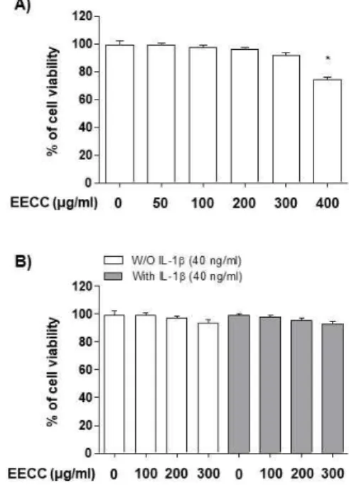
Effect of EECC on SW1353 chondrocyte cytotox-icity
The cytotoxic effects of EECC on the SW1353 chon-drocytes were determined by the MTT assay. As shown in Fig. 1A, EECC at the concentration range of 50–300 μg/ml did not show a cytotoxic effect on the SW1353 cells, but significant cytotoxicity was observed in the 400 μg/ml EECC treated group. Subsequent experi-ments did not show any adverse effect on cell viability when EECC was administered to the 40 ng/ml IL-1β -treated SW1353 cells at a concentration of 300 μg/ml or less (Fig. 1B). Thus, EECC concentrations up to 300 μg/ml were used in the following experiments.
EECC inhibited the IL-1β-induced NO and PGE2
production in the SW1353 chondrocytes
The effects of EECC on the levels of released in-flammatory mediators (NO and PGE2) were detected to assess the anti-inflammatory effects of EECC. The SW1353 cells were pretreated with various concen-trations of EECC for 1 h before stimulation with 40 ng/ml IL-1β for 24 h. Then, the NO concentration in the cell suspension was assayed by the Griess reaction, and the level of PGE2 was measured using an ELISA kit. As shown in the results in Fig. 2, IL-1β stimulation increased the release of NO and PGE2 compared with the non-stimulated control, but NO and PGE2 pro-duction was significantly decreased in the EECC-pre-treated cells in a concentration-dependent manner.
Figure 1. Effect of EECC and IL-1β on the cell viability
of the SW1353 chondrocytes. The cells were treated with various concentrations of EECC alone for 24 h (A) or pre-treated with or without EECC for 1 h before 40 ng/ml IL-1β stimulation for 24 h (B). Cell viability was analyzed using an MTT assay (*p<0.05 in comparison with the control group).
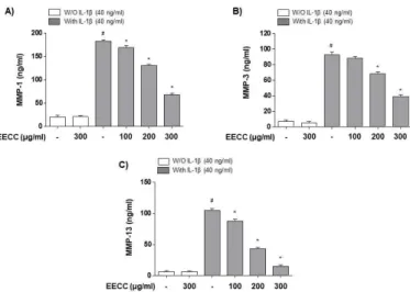
EECC attenuated the IL-1β-mediated increase in MMP-1, -3, and -13 production in the SW1353 chon-drocytes
pathogenesis, it was investigated whether EECC could avoid the generation of MMPs having an essential part in cartilage degradation. The results showed that the amount of MMPs (MMP-1, -3, and -13) released into the culture supernatant after stimulation with IL-1β was significantly increased. However, the enhanced pro-duction of these MMPs by IL-1β was suppressed by EECC pretreatment, and this effect was dependent on the EECC treatment concentration (Fig. 3).
Figure 2. Inhibition of the IL-1β-induced NO and PGE2
pro-duction by EECC in the SW1353 chondrocytes. The cells were pretreated with various concentrations of EECC for 1 h before 40 ng/ml IL-1β stimulation for 24 h. (A) The NO concentration in the culture medium was determined by the Griess reaction. (B) The PGE2 concentration was determined
by a commercial ELISA kit (#p<0.05 in comparison with the
control group; *p<0.05 in comparison with the IL-1β group).
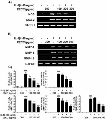
EECC decreased the expression of iNOS, COX-2 and MMP proteins increased by IL-1β in the SW1353 chondrocytes
I next examined whether EECC could inhibit the ex-pression of iNOS and COX-2 by IL-1β. According to the RT-PCR results, the increased expression of iNOS and COX- mRNA by IL-1β was concentration-depend-ently decreased in the cells cultured under the condition
of EECC pretreatment (Fig. 4A). Subsequently, it was also investigated whether the inhibition of MMP pro-duction by EECC in the IL-1β-treated SW1353 chon-drocytes was associated with the decreased expression of these genes. As shown in Fig. 4B, the expression of the three MMPs mRNAs was greatly enhanced by the IL-1β treatment, but their expression was dramati-cally reduced in the cells cultured under the condition of EECC pretreatment.
Figure 3. Inhibition of the IL-1β-induced MMPs production
by EECC in the SW1353 chondrocytes. The cells were pre-treated with various concentrations of EECC for 1 h before 40 ng/ml IL-1β stimulation for 24 h. Culture supernatants were then isolated, and the amounts of MMP-1 (A), -3 (B), and -13 (C) production were determined using commercial ELISA kits (#p<0.05 in comparison with the control group;
*p<0.05 in comparison with the IL-1β group).
EECC alleviated the IL-1β-mediated generation of ROS in the SW1353 chondrocytes
Because oxidative stress also plays an important role in the degradation of ECM and in the induction of chondrocyte apoptosis, I investigated whether EECC could suppress IL-1β-induced oxidative stress using the DCF-DA probe. The flow cytometry results showed that the level of the intracellular ROS contents gradu-ally increased with the stimulation of IL-1β, peaked at 2 h, and decreased thereafter (data not shown). However, the increase in ROS content in the SW1353 cells treated with IL-1β was dramatically reduced by
the addition of EECC. Moreover, as expected, the pre-treatment of a ROS scavenger, NAC, completely pro-tected the ROS production by IL-1β (Fig. 5A). Consistent with the results from the flow cytometry, the increase in the fluorescence intensity of DCF-DA observed in the cells treated with IL-1β was weakened by the pretreatment of EECC, as shown in Fig. 5B.
Figure 4. Inhibition of the iNOS, COX-2 (A) and MMPs (B)
mRNA expression by EECC in the IL-1β-stimulated SW1353 chondrocytes. The cells were pretreated with various concen-trations of EECC for 1 h and incubated with 40 ng/ml IL-1β for 24 h. RT-PCR was performed using the indicated primers and GAPDH was used as the internal control for the RT-PCR. (C) The relative density was calculated using ImageJ soft-ware, and normalized to GAPDH. The results are the average of three independent experiments and expressed as ± SD of the median. ###p<0.001 vs. the untreated group; ⁎⁎⁎p<0.001, ⁎⁎
p<0.01, and ⁎p<0.05 vs. the IL-1β-treated group.
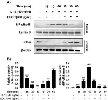
EECC suppressed nuclear translocation of NF-κB in IL-1β-stimulated the SW1353 chondrocytes
It was further investigated whether EECC could in-hibit the IL-1β-mediated activation of NF-κB because it is a critical mediator of IL-1β signaling in
chon-drocytes and the key factor controlling the transcription of inflammatory mediators and MMPs. As shown in Fig. 6, when the SW1353 cells were stimulated with IL-1β, the translocation of NF-κB p65 to the nucleus was significantly increased compared with the control group and peaked at 1 h. By contrast, the level of IκB-α in the cytoplasm was gradually decreased by the treat-ment of IL-1β. However, EECC significantly reduced the nuclear translocation of NF-κB p65 and the degra-dation of IκB induced by IL-1β (Fig. 6).
Figure 5. Effects of EECC on the IL-1β-induced ROS
gen-eration in the SW1353 chondrocytes. The cells were pre-treated with 300 μg/ml EECC or 10 mM NAC for 1 h and then treatment with 40 ng/ml IL-1β for 2 h. (A) The DCF-DA-stained cells were collected, and then DCF fluo-rescence was calculated by a flow cytometry. The results shown are the average of two independent experiments. (B) The images were obtained using a fluorescence microscope and presented from one experiment.
Discussion
In this study, I investigated the effect of the ethanol extract of C. costata (EECC) on the IL-1β-induced in-flammatory response in the SW1353 chondrocytes. According to the results, EECC remarkably reversed the production of IL-1β-mediated inflammatory media-tors, such as NO and PGE2, and MMP-1, -3, and -13
in the SW1353 chondrocytes. Moreover, EECC sig-nificantly attenuated the IL-1β-induced ROS gen-eration and of NF-κB activation.
Figure 6. Inhibition of the NF-κB nuclear translocation by
EECC in the IL-1β-stimulated SW1353 chondrocytes. The cells were pretreated with 300 μg/ml EECC for 1 h before the 40 ng/ml IL-1β treatment for the indicated times. (A) Nuclear and cytoplasmic proteins isolated from IL-1β-treated cells were isolated and the expression levels of NF-κB and IκB-α were determined by Western blotting. Lamin B and β-actin were presented as internal controls for each protein fraction. (B) Bands were quantified using ImageJ and normal-ized to lamin B and β-actin, respectively, and the ratio was determined. Data are expressed as mean ± SD. All experi-ments were repeated three times. ###p<0.001 vs. the untreated
group; ⁎⁎⁎p<0.001 vs. the IL-1β-treated group.
Inflammation has emerged as a major cause of OA pathogenesis. In particular, IL-1β acts as a key media-tor of articular cartilage degradation through the stim-ulation of inflammatory mediators, such as iNOS and COX-2 [6,8]. These mediators can accelerate the devel-opment of OA, and inhibiting their expression may im-pair OA development. In this study, the effects of EECC on the secretion of NO and PGE2 were inves-tigated to examine the inhibitory effect of EECC on the IL-1β-mediated inflammatory response. It was found that EECC significantly reduced the expression
of iNOS and COX-2 mRNA induced by IL-1β, which was associated with decreased NO and PGE2 production. Therefore, the data demonstrate that the an-ti-inflammatory effect of EECC is due at least to the reduced expression of proteins involved in the gen-eration of NO and PGE2. Many previous studies showed that MMPs, the major proteases increased by chondrocytes according to IL-1β, contribute to the for-mation of the most important proteolytic system in the degradation of ECM [5,6]. In addition, their expression is increased in the cartilage and synovial tissue of most OA patients [3,4]. Therefore, the application of candi-date substances that block the activity of MMPs could be applied as a new treatment strategy for OA. In the current study, MMPs such as MMP-1, -3, and -13, which are important enzymes for cartilage degradation, were significantly increased in the IL-1β-exposed chondrocytes. However, the elevated levels of the MMPs mRNA by IL-1β were obviously inhibited by the EECC treatment. These results indicate that EECC can protect the ECM degradation and chondrocytes by inhibiting the expression of MMPs.
The ROS levels are significantly greater in OA carti-lage than in normal carticarti-lage, and this has been reported in many clinical studies [14,23]. High levels of ROS in chondrocytes promote ECM degradation, cell dys-function, and death. In addition, ROS stimulates the production of inflammatory cytokines and alters the function of critical cell signaling pathways [14,15], suggesting that oxidative stress acts as a risk factor for the pathogenesis of OA. As a result of examining the influence of EECC on oxidative stress by IL-1β, it was showed that EECC effectively repressed the production of ROS by IL-1β. Although the antioxidant potential of C. costata extract was reported in previous studies [24], the results of this study support the use of EECC as an antioxidant for OA management.
As noted in many studies, NF-κB plays a critical role in the control of inducible ant-inflammatory mediators, cytokines, and MMPs expression in OA pathogenesis [13,25]. Typically, NF-κB forms a complex with the inhibitory subunit IκB-α and remains inactive in the
cytoplasm. When IκB-α is phosphorylated and de-graded through the upstream signaling systems by in-flammatory stimulation, NF-κB migrates to the nu-cleus, triggering transcriptional activation of in-flammation-inducing genes as well as catabolic en-zymes [26,27]. Therefore, the efficacy of EECC on IL-1β-induced NF-κB activation was further evaluated because blocking the activity of NF-κB could be effec-tive in treating OA. The current results showed that the translocation of NF-κB from the cytoplasm to the nucleus and the degradation of IκB-α were increased in IL-1β-treated SW1353 chondrocytes, but EECC ef-fectively blocked the nuclear translocation of NF-κB, an essential step for NF-κB activation. Therefore, the inhibitory effect of EECC on the increased expression of iNOS, COX-2 and MMP in IL-1β-treated chon-drocytes is due to blocking of the nuclear translocation of NF-κB.
However, further studies are needed to determine the role of other cellular signaling pathways that may be involved in the anti-inflammatory activity of EECC oth-er than the NF-κB signaling pathway, and to detoth-ermine the direct relationship with NF-κB signaling. In addi-tion, the identification and role of other intracellular or-ganelles in cells that are involved in ROS production by EECC should be continuously identified. It is also necessary to identify the anti-inflammatory active in-gredients contained in EECC and re-confirm the an-ti-cancer efficacy of EECC through animal experiments.
Conclusion
In conclusion, the current study clearly indicates that EECC has the potential to inhibit the IL-1β-induced inflammatory response in chondrocytes, and that its an-ti-inflammatory effect is associated with the sup-pression of the NF-κB signaling pathway. Therefore, I suggest that EECC may have cartilage protection po-tential and can be used as a candidate for OA treatment.
Acknowledgements
This work was supported by Dong-eui University Grant (202003410001).
References
1. Brittberg, M., Gomoll, A. H., Canseco, J. A., Far, J., Lind, M. and Hui, J. 2016. Cartilage repair in the degenerative ageing knee. Acta. Orthop. 87, S26-38.
2. Andriacchi, T. P., Favre, J., Erhart-Hledik, J. C. and Chu, C. R. 2015. A systems view of risk factors for knee osteo-arthritis reveals insights into the pathogenesis of the disease. Ann. Biomed. Eng. 43, 376-387.
3. Son, Y. O. and Chun, J. S. 2018. Estrogen-related receptor γ is a novel catabolic regulator of osteoarthritis pathogenesis. BMB Rep. 51, 165-166.
4. Hochberg, M., Chevalier, X., Henrotin, Y., Hunter, D. J. and Uebelhart, D. 2013. Symptom and structure mod-ification in osteoarthritis with pharmaceutical-grade chon-droitin sulfate: what's the evidence? Curr. Med. Res. Opin.
29, 259-267.
5. Martel-Pelletier, J., Boileau, C., Pelletier, J. P. and Roughley, P. J. 2008. Cartilage in normal and osteoarthritis conditions. Best Pract. Res. Clin. Rheumatol. 22, 351-384. 6. Blasioli, D. J. and Kaplan, D. L. 2014. The roles of catabo-lic factors in the development of osteoarthritis. Tissue Eng.
Part B. Rev. 20, 355-363.
7. Jotanovic, Z., Mihelic, R., Sestan, B. and Dembic, Z. 2012. Role of interleukin-1 inhibitors in osteoarthritis: an evi-dence-based review. Drugs Aging 29, 343-358. 8. Rai, M. F. and Sandell, L. J. 2011. Inflammatory
media-tors: tracing links between obesity and osteoarthritis. Crit.
Rev. Eukaryot. Gene Expr. 21, 131-142.
9. Panina, S. B., Krolevets, I. V., Milyutina, N. P., Sagakyants, A. B., Kornienko, I. V., Ananyan, A. A., Zabrodin, M. A., Plotnikov, A. A. and Vnukov, V. V. 2017. Circulating levels of proinflammatory mediators as potential biomarkers of post-traumatic knee osteoarthritis development. J. Orthop. Traumatol. 18, 349-357. 10. Ramonda, R., Lorenzin, M., Modesti, V., Campana, C.,
Ortolan, A., Frallonardo, P. and Punzi, L. 2013. Serological markers of erosive hand osteoarthritis. Eur.
J. Intern. Med. 24, 11-15.
11. Ding, Q. H., Cheng, Y., Chen, W. P., Zhong, H. M. and Wang, X. H. 2013. Celastrol, an inhibitor of heat shock protein 90β potently suppresses the expression of matrix metalloproteinases, inducible nitric oxide synthase and
cyclooxygenase-2 in primary human osteoarthritic chondrocytes. Eur. J. Pharmacol. 708, 1-7.
12. Goldring, M. B. and Otero, M. 2011. Inflammation in osteoarthritis. Curr. Opin. Rheumatol. 23, 471-478. 13. Roman-Blas, J. A. and Jimenez, S. A. 2006. NF-kappaB
as a potential therapeutic target in osteoarthritis and rheu-matoid arthritis. Osteoarthritis Cartilage 14, 839-848. 14. Lepetsos, P. and Papavassiliou, A. G. 2016. ROS/oxida-tive stress signaling in osteoarthritis. Biochim. Biophys.
Acta. 1862, 576-591.
15. Li, D., Xie, G. and Wang, W. 2012. Reactive oxygen species: the 2-edged sword of osteoarthritis. Am. J. Med.
Sci. 344, 486-490.
16. Klinge, S. A. and Sawyer, G. A. 2013. Effectiveness and safety of topical versus oral nonsteroidal anti-in-flammatory drugs: a comprehensive review. Phys.
Sportsmed. 41, 64-74.
17. Herndon, C. M. 2012. Topical delivery of nonsteroidal anti-inflammatory drugs for osteoarthritis. J. Pain Palliat.
Care Pharmacother. 26, 18-23.
18. Fitton, J. H. 2011. Therapies from fucoidan; multifunc-tional marine polymers. Mar. Drugs 9, 1731-1760. 19. Myers, S. P., O'Connor, J., Fitton, J. H., Brooks, L., Rolfe,
M., Connellan, P., Wohlmuth, H., Cheras, P. A. and Morris, C. 2010. A combined phase I and II open label study on the effects of a seaweed extract nutrient complex on osteoarthritis. Biologics 4, 33-44.
20. Shin, H. C., Hwang, H. J., Kang, K. J. and Lee, B. H. 2006. An antioxidative and anti-inflammatory agent for potential treatment of osteoarthritis from Ecklonia cava.
Arch. Pharm. Res. 29, 165-171.
21. Lee, G. H., Jin, S. W., Kim, S. J., Pham, T. H., Choi, J. H. and Jeong, H. G. 2019. Tetrabromobisphenol A in-duces MMP-9 expression via NADPH oxidase and the activation of ROS, MAPK, and Akt pathways in human breast cancer MCF-7 cells. Toxicol. Res. 35, 93-101. 22. Shin, S. K., Kim, J. H., Lee, J. H., Son, Y. H., Lee,
M. W., Kim, H. J., Noh, S. A., Kim, K. P., Kim, I. G. and Lee, M. J. 2017. Docosahexaenoic acid-mediated protein aggregates may reduce proteasome activity and delay myotube degradation during muscle atrophy in
vitro. Exp. Mol. Med. 49, e287.
23. Ziskoven, C., Jäger, M., Zilkens, C., Bloch, W., Brixius, K. and Krauspe, R. 2010. Oxidative stress in secondary osteoarthritis: from cartilage destruction to clinical pre-sentation? Orthop. Rev. (Pavia) 2, e23.
24. Zheng, J., Hewage, S. R., Piao, M. J., Kang, K. A., Han, X., Kang, H. K., Yoo, E. S., Koh, Y. S., Lee, N. H., Ko, C. S., Lee, J. C., Ko, M. H. and Hyun, J. W. 2016. Photoprotective effect of Carpomitra costata extract against ultraviolet B-induced oxidative damage in human keratinocytes. J. Environ. Pathol. Toxicol. Oncol. 35, 11-28.
25. Rigoglou, S. and Papavassiliou, A. G. 2013. The NF-κB signalling pathway in osteoarthritis. Int. J. Biochem. Cell
Biol. 45, 2580-2584.
26. Schuliga, M. 2015. NF-kappaB signaling in chronic in-flammatory airway disease. Biomolecules 5, 1266-1283. 27. O'Dea, E. and Hoffmann, A. 2010. The regulatory logic of the NF-kappaB signaling system. Cold Spring Harb