Imaging of Oral Cavity Cancer
Department of Radiology, Hanyang University Medical School, Seoul, Korea Dong Woo Park, M.D.
Introduction
The oral cavity is the most anteriorly located structure of the head and neck, which is separated from the oropharynx by an imaginary line drawn across the circumvallate papillae, ante-rior tonsillar pillars, and junction of the hard and soft palates. Carcinomas in the oral cavity and oropharynx are different in both presentation and prognosis. The remaining boundaries of the oral cavity include the lips anteriorly; the cheeks laterally; the hard palate, superior alveolar ridge, and teeth superiorly; and the inferior alveolar ridge, teeth, and the mylohyoid mu-scle inferiorly. So, oral cavity consists of the lips, including the vermillion border, anterior two thirds of the oral tongue, floor of mouth (FOM), buccal mucosa, upper and lower gingiva, hard palate, and retromolar trigone.
Although oral cavity cancer can arise from any of the tiss-ues within the oral cavity, which accounts for 2% of new cases of all cancer in the Korea (2002), the most common site of oral cavity cancer is the tongue (25.0%), and then the gums (15.4%) and the tonsils (13.5%). Most oral cancers are squa-mous cell carcinomas (SCCs) comprising 90% of all oral ma-lignant lesions. Additional carcinomas include salivary gland tumors such as adenoid cystic or mucoepidermoid carcinomas comprising approximately 3% of tumors, and then lymphoma, melanoma, and rare unusual sarcomas or bone tumors. Most patients have been usually diagnosed at advanced stages, III or IV, resulting in poor stage-related survival rate, despite of me-dical and public health development.
Imaging of Oral Cavity Cancer
Although oral cancer screening may be done on clinical examination, imaging study plays a critical role in determining tumor especially its deep margins, staging and planning of treatment. Common imaging modalities for the oral cavity in-clude CT, MRI, ultrasonography (US), plain radiography, nu-clear medicine scintigraphy, and PET. CT and MRI have been
the primary studies for evaluation of oral cavity malignan-cies. CT is very sensitive for detecting small areas of cortical bone invasion, while MRI is better for evaluating the extent of marrow invasion by tumor. MR imaging is the initial study of choice for tumors confined to the oral tongue and those with possible perineural spread. Neither enlargement nor contrast enhancement of a nerve is pathognomonic of perineural spread, as it may also occur secondary to edema and inflammation. Fortunately, false positive perineural sp-read is rare on MR imaging. Recently introduced perfusion CT can assess tumor vascularity, which helps to detect both primary and recurrent cancer. There are many pre-malignant lesions including the various grades of dysplasia which may mimic oral SCC, which remain superficial and may not re-quire imaging. Tumor thickness is a significant independent prognostic factor, which is most delineated on contrast-en-hanced T1-weighted fat-suppressed image. Tumors under 3 mm have a low incidence of local-regional recurrence and excellent disease-free survival, whereas tumor thicknesses over 9 mm carry a 24% of local recurrence and 66% of 5-year disease-free survival. A critical assessment of the lingual arteries, veins, nerves (including the hypoglossal) is manda-ted in oral and oropharyngeal cancers. The relationship of oral tongue and FOM cancer to the neurovascular bundles, both ipsilateral and contralateral, is essential since its loca-tion adjacent to these structures is highly suggestive of neu-rovascular invasion. FOM and ventral tongue tumors with aggressive margins greater than 2cm in size that deeply in-vade the sublingual space, likely involve the neurovascular bundle. The relationship of the tumor to the lingual septum is important since its involvement indicates spread across the midline. The orifices of submandibular and sublingual ducts frequently become obstructed and dilated with or without associated inflammatory involvement of the salivary glands. A stranded “dirty” appearance of parapharyngeal space fat indicates cancer spread into this space and possible further extension. For oral tongue cancer, which most commonly oc-curs on the ventrolateral surface, imaging is primarily used
to evaluate the depth of invasion and to ascertain the pre-sence of pathologic adenopathy. For this, MR imaging is the modality of choice. Sublingual space fat and the mucosal in-terface stripe of the lateral tongue are normally preserved and symmetrical, whereas their asymmetry or effacement frequ-ently reveals small tumors.
Denervation atrophy of the mylohyoid and anterior belly of digastric or tongue muscles occurs due to injury to the man-dibular or hypoglossal nerves, respectively. These injuries are sometimes caused by cancer, but probably more frequen-tly by aggressive treatment. Denervation atrophy is seen as decreased size and hypodensity of the involved muscles, si-milar to fat on CT. The appearance of denervation atrophy on MR changes over time, the muscles are generally hyperin-tense on both T1 and T2-weighted images and may show post-contrast enhancement.
It is important to closely evaluate the LNs, particularly levels I-III in any oral cancer. Although skip lesions to the level IV LNs are uncommon, this level should always be evaluated at imaging. The most widely accepted criteria for metastatic disease state that submandibular (level IB) and jugulodigastric level II nodes generally should not exceed 15 mm, and other nodal stations should not exceed 10 mm in the longest diameter. Imaging criteria for extracapsular tumor spread include spiky, irregular margins, loss of fat cleavage plane around the node, thickening of adjacent fascia, and apparent invasion of adjacent structures. A 5-mm node with central necrosis and peripheral enhancement or other abnor-mal enhancement pattern is almost certainly metastatic. Also, round nodes are more worrisome than elongated, bean-sha-ped ones. Ultrasmall superparamagnetic iron oxide (SPIO) nanoparticles, new MRI contrast agent, tends to accumulate in the reticuloendothelial system of LNs, which are taken up by macrophages within normal functioning nodes, reducing their signal on post-contrast MR because of the magnetic susceptibility effects of iron oxide. Metastatic nodes, on the other hand, remain hyperintense on post-contrast T2*-weigh-ted gradient echo images.
In oral cavity cancers, the site of origin is a key factor in the propensity of the tumor to spread in certain directions and the adjacent critical structures affected. 5 major findings on imaging study play a key prognostic role in oral cavity cancer, namely, tumor 1) size and 2) thickness, 3) perineural invasion, 4) bone marrow invasion, and 5) LN involvement with extracapsular spread. Multiple studies demonstrated in-creased tumor size is associated with dein-creased overall sur-vival, thus, the important T factor on the AJCC chart. The T
factor affects both the 5-year survival rates and rates of po-sitive cervical lymphadenopathy. In the oral tongue, tumor thickness is an important prognostic factor for local recur-rence, nodal metastasis, and overall patient morbidity and mortality. Perineural invasion is strongly associated with de-creased survival and inde-creased local recurrence requiring aggressive treatment. Bone marrow invasion is a finding on the AJCC chart that upstages a lip carcinoma to a T4. False-positive results may occur with loss of normal fatty marrow and cortical margins due to chemical shift artifact arising from bone marrow fat obscuring the cortical margin or from inflammation surrounding the tumor in the inferior alveolar canal. The presence or absence of lymphadenopathy is often considered one of the most important prognostic factors.
1. Lip carcinoma
Lip carcinomas usually arise from the vermillion border and may spread to involve adjacent skin or underlying mus-cles of the mouth such as the orbicularis oris. Early small lesions are often easier to detect on clinical examination. The goal of imaging is to determine the full extent of the lesion in advanced or indeterminate margined lesion, also including possible perineural spread especially of mental nerve and the integrity of the adjacent bone. If the tumor has invaded the bone, the lip carcinoma becomes upstaged to a T4, which is a contraindication for surgery.
2. FOM carcinoma
Most FOM SCCs arise near the anterior midline FOM and have a propensity for lateral spread with involvement of the adjacent mandible or submucosally involving the ipsilateral or contralateral lingual neurovascular bundle. Assessment as to whether the tumor has invaded through the midline lingual septum or involved the contralateral neurovascular bundles is a determining factor between a hemiglossectomy or total glossectomy, with many institutions avoiding the total glos-sectomy secondary to the morbidity associated with this pro-cedure. Additional critical factors that may upstage an FOM cancer to a stage 4 include bone involvement, extension to the base of tongue, and beyond the posterior edge of the mylohyoid muscle into the neck. Additional findings include secondary dilatation of the salivary ducts from tumoral ob-struction. LN drainage pathways that need to be assessed in-clude levels I and II, submental, submandibular, and internal jugular LNs. Enhancement within anterior FOM tumors can be difficult to identify on CT due to the adjacent dense mandi-ble and artifact from dental amalgam.
3. Alveolus, gingiva and buccal mucosa carcinoma
Although tumors of the gingiva account for less than 10% of oral cavity cancers, the proximity of these tumors to bony cortical margins is worrisome for potential bony intramedul-lary and perineural extension. Buccal carcinomas most com-monly originate along the lateral margins. The most common spread patterns of gingival and buccal cancers are laterally and submucosally along the buccinator muscle to the ptery-gomandibular raphe and subsequent involvement of the un-derlying bone. These may be seen to better advantage using the “puffed cheek” technique at CT, as this technique may provide a clearer and more detailed evaluation of the buccal mucosal surface. The common route of entry into the mandi-ble is often the occlusal surface, especially in edentulous patients. The mucosal extent of both gingival and hard palate tumors is often underestimated on imaging study, and they are better assessed with endoscopy. It is important to use T1-weighted pre- and post-gadolinium MR images to detect pe-rineural spread along the greater and lesser palatine nerves into the pterygopalatine fossa. Obliteration of the fat below a foramen, enhancement of the nerve, or enlargement of a nerve is considered evidence of tumor involvement.
4. Tongue carcinoma
The oral tongue is attached to the FOM and is mobile, which makes to evaluate easier in sagittal and coronal planes and more difficult on axial images. MRI well demonstrates the intrinsic and extrinsic muscle anatomy and the interdigi-tated fat. The styloglossus muscle extends from the styloid through the parapharyngeal fat and can be identified as it ex-tends into the posterior tongue and subsequently interweaves into the hyoglossus muscle. The adjacent tissues around the styloglossus are possible pathways of tumor spread from the FOM and tongue to the skull base. In addition, inferior tu-moral extension allows tumor access to the mylohyoid and geniohyoid muscles. Most oral tongue SCCs arise from the antero-lateral undersurface of the tongue, imaging study is primarily used to evaluate T and N staging including tumor thickness. Sublingual space fat and the mucosal interface st-ripe of the lateral tongue are normally preserved and sym-metrical, whereas their asymmetry or effacement frequently reveals small tumors.
5. Hard palate carcinoma
Carcinomas of the hard palate are especially required to be assessed for the degree of bone invasion and for potential perineural spread. Adenoid cystic tumors in this area have a propensity for perineural tumoral spread along the greater
and lesser palatine nerves into the pterygopalatine fossa. Pe-rineural tumoral spread into the pterygopalatine fossa allows for multiple routes of tumoral spread, including along CN V2 into foramen rotundum or anteriorly into the infraorbital foramen, and also along the vidian nerve in the vidian canal. The vidian nerve is a combination of the greater superficial petrosal nerve (GSPN) and deep petrosal nerve. The GSPN exits from the first genu of CN VII, exiting the petrous tem-poral bone via the greater petrosal foramen to enter the mid-dle cranial fossa. The nerve then passes below the trigeminal ganglion to reach foramen lacerum, which acts like an eleva-tor shaft, with the GSPN extending along its lateral margins until it arrives at the vidian canal where it subsequently joins the deep petrosal nerve containing the sympathetic fibers from the plexus surrounding the internal carotid artery, be-coming the vidian nerve. All of these nerves serve as routes for potential perineural tumoral spread into skull base and cavernous sinus.
6. Retromolar trigone carcinoma
Retromolar trigone tumors account for 7% of tumors af-fecting the oral cavity. The retromolar trigone is situated be-hind the posterior molars. This unique location, which is a junction point between the oral cavity, oropharynx, and na-sopharynx, allows for complex spread of tumors. Multiple muscles, including the buccinator, orbicularis oris, and su-perior constrictor muscles insert into the retromolar trigone, allowing different routes of tumor growth. Beneath the mu-cosal surface of the retromolar trigone is the pterygoman-dibular raphe that attaches superiorly to the hook of the ha-mulus arising from the medial pterygoid plate and inferiorly to the posterior mylohyoid line of the mandible. This raphe allows tumor spread into the medial pterygoid muscle po-steriorly, mylohyoid muscle inferiorly, and buccinator muscle anterolaterally. Spread of retromolar trigone tumors along the pterygomandibular raphe superiorly to the pterygoid plates may allow access to the pterygopalatine fossa, and from there the lesion may extend cephalad to the skull base and caver-nous sinus. Alternatively, involvement of the pterygoid mus-cles in the masticator space leads to perineural spread along the mandibular nerve and proximally to the skull base. The spread of tumors of the retromolar trigone can be difficult to detect clinically, so imaging study plays an important role in mapping the extent of tumor. Tumoral spread is assessed by obliteration of the normal fat planes.
7. Other imaging modalities
toring tumor recurrence, especially if US-guided fine-needle aspiration (FNA) is indicated. Technetium-99m labeled scin-tigraphy can be helpful for detecting early bone invasion re-lated to increased osteoblastic activity, but this technique is nonspecific because tumors, infarction, and bone infection may all show tracer uptake. 18-FDG PET scanning plays as a functional tumor detection modality, particularly for clini-cally occult primary lesions, nodal staging, treatment response, second primary lesions, distant metastases, clinically negative neck (Accuracy;up to 100%), and differentiation of recur-rent tumor from post-therapy changes. However, 18-FDG PET is not without its limitations because physiological up-take may occur in lymphoid tissue, nasal mucosa, salivary glands, muscle activity, and in irradiated and postoperative tissues. On diffusion weighted image, apparent diffusion co-efficient (ADC) values of lymphoma are lower than those of SCC. ADC values of benign solid lesions were significantly higher than malignant tumors. Choline (Cho)/creatine (Cr)
ratios can be used as metabolic markers distinguishing SCC from normal muscle with higher in SCC and also benign tumors than in muscle in the extracranial head and neck. Recurrent SCC of the head and neck may elevate Cho/Cr ratios and increase signals from either lipid or lactic acid.
Conclusions
In conclusions, the unique anatomy of the oral cavity al-lows it to be clinically inspected. However, cross-sectional im-aging studies, especially CT and MRI, play an important role in delineating tumor size, tumor thickness, perineural invasion, bone marrow invasion, and LN involvement with extracap-sular spread. These findings are important prognostic indica-tors for patient morbidity and mortality. Ultrasonography (US), FDG-PET/CT and other imaging studies may often play a complementary role for the evaluation of oral cavity cancer, such as the assessment for lymphadenopathy,
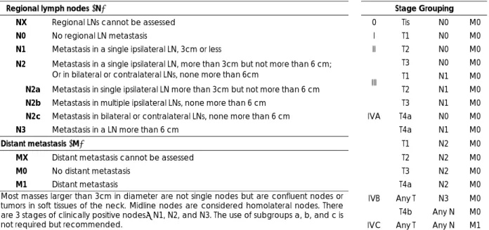
metas-Regional lymph nodes (N) Stage Grouping
NX Regional LNs cannot be assessed 0 Tis N0 M0
N0 No regional LN metastasis I T1 N0 M0
N1 Metastasis in a single ipsilateral LN, 3cm or less II T2 N0 M0
T3 N0 M0
N2 Metastasis in a single ipsilateral LN, more than 3cm but not more than 6 cm;
Or in bilateral or contralateral LNs, none more than 6cm T1 N1 M0 N2a Metastasis in single ipsilateral LN more than 3cm but not more than 6 cm T2 N1 M0 N2b Metastasis in multiple ipsilateral LNs, none more than 6 cm
III
T3 N1 M0
N2c Metastasis in bilateral or contralateral LNs, none more than 6 cm IVA T4a N0 M0
N3 Metastasis in a LN more than 6 cm T4a N1 M0
Distant metastasis (M) T1 N2 M0
MX Distant metastasis cannot be assessed T2 N2 M0
M0 No distant metastasis T3 N2 M0
M1 Distant metastasis T4a N2 M0
IVB Any T N3 M0 T4b Any N M0 Most masses larger than 3cm in diameter are not single nodes but are confluent nodes or
tumors in soft tissues of the neck. Midline nodes are considered homolateral nodes. There are 3 stages of clinically positive nodes:N1, N2, and N3. The use of subgroups a, b, and c is
not required but recommended. IVC Any T Any N M1
Table 1. AJCC staging of lip and oral cavity cancer Primary tumor (T)
TX Primary tumor cannot be assessed T0 No evidence of primary tumor Tis Carcinoma in situ
T1 Tumor 2cm or less (in greatest dimension) T2 Tumor more than 2cm but not more than 4cm T3 Tumor more than 4cm
T4 (Lip) Tumor invades adjacent structures; through cortical bone, inferior alveolar nerve, floor of mouth, or skin of face, i.e., chin or nose
(not T4 by superficial erosion alone of bone/tooth socket by gingival primary)
T4a0 (Oral cavity) Tumor invades adjacent structures; through cortical bone, into deep (extrinsic) muscle of tongue (GG, HG, PG, SG), maxillary sinus, or skin of face
tatic disease and perineural or bone involvement.
References
________________________________
1) Rumboldt Z, Day TA, Michel M. Imaging of oral cavity cancer. Oral Oncol. 2006;42:854-865.
2) Kirsch C. Oral cavity cancer. Top Magn Reson Imaging. 2007; 18:269-80.
3) Chong V. Oral cavity cancer. Cancer Imaging. 2005;5 Spec No A:S49YS52.
4) Dammann F, Horger M, Mueller-Berg M, et al. Rational diag-nosis of squamous cell carcinoma of the head and neck region: comparative evaluation of CT, MRI, and 18FDG PET. AJR Am J Roentgenol. 2005;184:1326-1331.
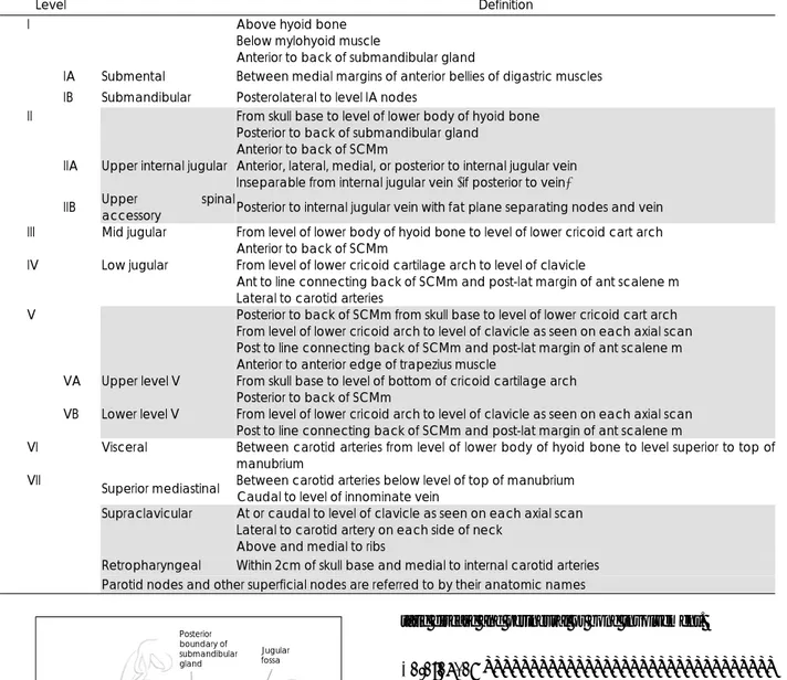
Table 2. Summary of the imaging-based nodal classification
Level Definition
I Above hyoid bone
Below mylohyoid muscle
Anterior to back of submandibular gland
IA Submental Between medial margins of anterior bellies of digastric muscles IB Submandibular Posterolateral to level IA nodes
II From skull base to level of lower body of hyoid bone Posterior to back of submandibular gland
Anterior to back of SCMm
IIA Upper internal jugular Anterior, lateral, medial, or posterior to internal jugular vein Inseparable from internal jugular vein (if posterior to vein)
IIB Upper spinal accessory Posterior to internal jugular vein with fat plane separating nodes and vein III Mid jugular From level of lower body of hyoid bone to level of lower cricoid cart arch
Anterior to back of SCMm
IV Low jugular From level of lower cricoid cartilage arch to level of clavicle
Ant to line connecting back of SCMm and post-lat margin of ant scalene m Lateral to carotid arteries
V Posterior to back of SCMm from skull base to level of lower cricoid cart arch From level of lower cricoid arch to level of clavicle as seen on each axial scan Post to line connecting back of SCMm and post-lat margin of ant scalene m Anterior to anterior edge of trapezius muscle
VA Upper level V From skull base to level of bottom of cricoid cartilage arch Posterior to back of SCMm
VB Lower level V From level of lower cricoid arch to level of clavicle as seen on each axial scan Post to line connecting back of SCMm and post-lat margin of ant scalene m
VI Visceral Between carotid arteries from level of lower body of hyoid bone to level superior to top of manubrium
VII
Superior mediastinal Between carotid arteries below level of top of manubrium Caudal to level of innominate vein Supraclavicular At or caudal to level of clavicle as seen on each axial scan
Lateral to carotid artery on each side of neck Above and medial to ribs
Retropharyngeal Within 2cm of skull base and medial to internal carotid arteries Parotid nodes and other superficial nodes are referred to by their anatomic names
Posterior boundary of submandibular gland Jugular fossa Lower border of hyoid Lower margin of cricoid cartilage Left common carotid artery Top of manubrium Internal jugular vein