저작자표시-비영리-변경금지 2.0 대한민국 이용자는 아래의 조건을 따르는 경우에 한하여 자유롭게 l 이 저작물을 복제, 배포, 전송, 전시, 공연 및 방송할 수 있습니다. 다음과 같은 조건을 따라야 합니다: l 귀하는, 이 저작물의 재이용이나 배포의 경우, 이 저작물에 적용된 이용허락조건 을 명확하게 나타내어야 합니다. l 저작권자로부터 별도의 허가를 받으면 이러한 조건들은 적용되지 않습니다. 저작권법에 따른 이용자의 권리는 위의 내용에 의하여 영향을 받지 않습니다. 이것은 이용허락규약(Legal Code)을 이해하기 쉽게 요약한 것입니다. Disclaimer 저작자표시. 귀하는 원저작자를 표시하여야 합니다. 비영리. 귀하는 이 저작물을 영리 목적으로 이용할 수 없습니다. 변경금지. 귀하는 이 저작물을 개작, 변형 또는 가공할 수 없습니다.
의학 석사학위 논문
Effect of Size and Location of
Nevi on Postoperative Pain and
Emergence Agitation in Children
Undergoing Nevi Excision
아 주 대 학 교 대 학 원
의 학 과
Effect of Size and Location of
Nevi on Postoperative Pain and
Emergence Agitation in Children
Undergoing Nevi Excision
지도교수 김 지 은
이 논문을 의학 석사학위 논문으로 제출함.
2020년 2월
아 주 대 학 교 대 학 원
의 학 과
석 수 현
석수현의 의학 석사학위 논문을 인준함.
심사위원장 김 지 은 인
심 사 위 원 김 종 엽 인
심 사 위 원 박 성 용 인
아 주 대 학 교 대 학 원
2020년 1월 9일
Abstract
Congenital melanocytic nevi need surgical excision. However, the effect of size and location of nevi on pain and emergence agitation have yet to be studied. The objective of this study was to evaluate (1) the ideal parameter of nevus size and (2) the effects of size and location of nevus on pain and emergence agitation. This observational study enrolled 100 children scheduled for excision of nevus under sevoflurane anesthesia. Parameters of nevus size included long diameter, area before resection, area of resection, and proportion (area of resection/total body surface). Nevus locations included trunk, face, scalp, and extremities.
Proportion of nevi was the most ideal parameter in evaluating the pain and emergence agitation. A large size showed higher emergence agitation than a small size (median [range]; 6 [0-20] in small group vs. 12.5 [0-20] in large groups, p = 0.021). However, the pain was comparable. Nevus location did not influence pain or emergence agitation. In multivariate regression analysis, younger age and extensive excision were associated with higher pain and emergence agitation.
In conclusion, large nevi induced more severe emergence agitation. However, nevus location did not affect the outcome. In addition, younger age was associated with pain and emergence agitation. Clinicians need to consider the proportion of nevi when managing children undergoing nevus excision.
차 례
Abstract...i
I. Introduction...1
II. Materilas and Methods...3
III. Results...7
IV. Discussion...12
V. Conclusions...17
References...18
그림목차
Figure 1. Intraoperative concentration of sevoflurane...8 Figure 2. The (A) heart rate and (B) mean arterial pressure during perioperative period...9
표목차
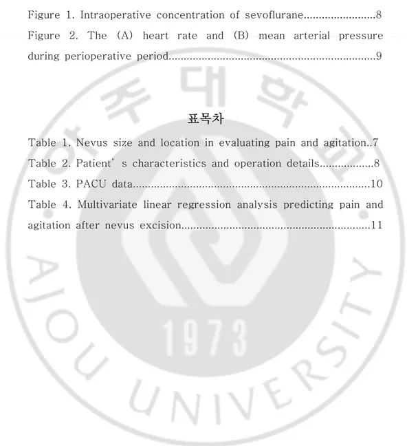
Table 1. Nevus size and location in evaluating pain and agitation..7 Table 2. Patient’s characteristics and operation details...8 Table 3. PACU data...10 Table 4. Multivariate linear regression analysis predicting pain and agitation after nevus excision...11
I. Introduction
Congenital melanocytic nevi (CMN) are defined as benign melanocytic nevi that are present at birth. CMN is found in 1% to 6% of neonates. However, nevi being identical CMN are reported to show a prevalence of more than 15% in older children [1]. CMN tend to extend deeper into dermis and subcutaneous tissues compared with acquired melanocytic nevi, and large CMN are associated with a higher risk for melanoma than smaller CMN, although the exact risk is unknown. Because of the potential for malignant changes, enlargement in proportion to the child’s growth, and cosmetic appearance, prophylactic surgical excision is usually advocated early in life, although there is no consensus about the risk of melanoma and optimal management practices in CMN [2].
Effective pain management in children remains a challenge. A large proportion of children are reported to receive insufficient analgesic
medication after surgery [3], and thus, suffer from
moderate-to-severe postoperative pain up to 44% [4]. Skin incision alone similar to nevus excision is sufficient to induce mechanical or heat hyperalgesia, although cutaneous combined with deep tissue incision trigger prolonged hyperalgesia [5]. In addition, sensitivity to painful stimuli is reported to be affected by the specific body region [6]. Although disputed, the sensitivity in the facial area was higher compared with that in other body regions.
Emergence agitation in children occurs generally during recovery from sevoflurane anesthesia. Although emergence agitation resolves spontaneously, it increases the risk of patient injury, surgical site damage, and parental dissatisfaction with any extra treatments. Emergence agitation has been attributed to multiple factors including age, temperament, pain, rapid anesthetic emergence, anesthetic and surgical types.
Turkmen et al [7] compared the CMNs based on dimension, total area, and total area-to-body surface area, and proposed the total nevus area for determining the risk of malignant transformation. However, no reliable parameter based on nevus size is available for estimation of the postoperative pain and emergence agitation after nevus excision. In addition, no studies have investigated the association between the size or body region of nevus and the degrees of pain and emergence agitation.
Therefore, the aims of this study were to evaluate (1) the adequate parameter of nevus size and (2) the effects of size and location of nevus on postoperative pain and emergence agitation in pre-school children undergoing nevus excision.
II. Materials and Methods
This single-center prospective observational study was approved by the Ajou University Hospital Institutional Review Board (protocol number: AJIRB-MED-OBS-16-533) and was registered at http://clinicaltrials.gov (registration number: NCT03094754). Between March 2017 and January 2018, 100 children who were scheduled to undergo general anesthesia for excision of nevi were enrolled. All parents provided written informed consent. Inclusion criteria were American Society of Anesthesiologists physical status 1 and 2, age between 1 and 7 years, previously healthy or with mild systemic disease, excision warranting general anesthesia, and
elective surgery. Exclusion criteria were developmental,
psychiatric, and neuromuscular diseases, excision of nevi involving more than 3 sites, and giant nevi.
All children had intravenous (IV) access and were not treated with any medication previously. On arrival at the operating room, standard monitors including electrocardiogram, pulse oximeter, and application of noninvasive blood pressure were used. Anesthesia was induced with 5 mg/kg of thiopental and 0.3 mcg/kg of fentanyl. After confirming the absence of response to eye stimulus, 0.6 mg/kg of rocuronium was administered. Ventilation was conducted with a mask using 100% oxygen and sevoflurane 5 vol%, followed by orotracheal intubation. Anesthesia was maintained with the end-tidal concentration of sevoflurane 2.0–2.5
vol% during surgery. We did not perform the regional block or premedication. All lesions were excised totally or in stages, together with the underlying subcutaneous tissue, and tension-free closure, regardless of melanoma suspicion. Surgical methods comprised direct primary closure and excision with local flap, but did not include excision with skin graft, use of tissue expander, and dermabrasion. After surgery, children were extubated after neuromuscular reversal and transferred to postanesthesia care unit (PACU). If the Modified Aldrete score was ≥ 8, children were transferred to ward.
Parameters related to size of nevi included long diameter, area before resection, area of resection, and proportion. Long diameter was measured in centimeters using a ruler. Area was measured by tracing the total nevus area onto a transparent film, OPSITE ELEXIGRID (Smith & Nephew, Inc. USA); to accurately assess the size of the nevus in cm2, the tracing was transposed onto paper
divided into millimeters, which is considered to be accurate up to 1 mm. “Area before resection” was measured at time between the anesthesia induction and the start of surgery. “Area after resection” was measured at time between the end of surgery and the surgical dressing by adding a semipermeable polyurethane membrane (Tegaderm) under the transparent film. The “area of resection” was calculated as area before resection minus area after resection. The total body surface (TBS) area was estimated for each patient using the formula of TBS (cm2) = 0.007184 ×
[weight (kg)0.425]× [height (cm)0.725]. The proportion as area of
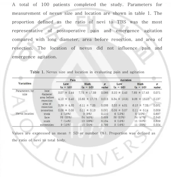
resection / TBS area was calculated as a percentage. Regions of nevi were defined as trunk, face, scalp, and extremities. Postoperative pain was assessed using the Face, Legs, Activity, Cry, Consolidation (FLACC) scale (0 = no pain, 1–3 = mild discomfort, 4–6 = moderate pain, 7–10 = severe discomfort or pain), which is a measurement tool used to assess pain among children between the ages of 2 months and 7 years or in individuals who are unable to communicate their pain. Emergence agitation was rated with Pediatric Agitation and Emergence Delirium (PAED) scale. Children were defined as having pain (if FALCC score ≥ 4) or emergence agitation (if PAED score ≥ 10). Pain and emergence agitation was evaluated upon PACU arrival, at 10 min, 30 min, 1 hr, and 2 hr after surgery. Fentanyl 0.5 mcg/kg was administered in patients with pain ≥ FLACC scale 4. Hemodynamic data was recorded before induction (baseline), after intubation, the start of surgery, the end of surgery, and after extubation. A single anesthesiologist observed and assessed the children throughout the study. For analysis, the children were divided into 2 groups according to the median value of proportion: a small group with less than 0.042 cm in proportion (n = 50), and a large group as 0.042 or greater in proportion (n = 50).
Results were expressed as mean ± standard deviation, median (range), or the number of patients (%). Normality of distribution was assessed with the Shapiro–Wilk test and the parametric and
nonparametric data were analyzed using the independent t-test and the Mann–Whitney U-test, respectively. Categorical variables were evaluated by chi-square test or Fisher’s exact test when appropriate. Repeatedly measured data were analyzed with a linear mixed model. A multivariate linear regression analysis was performed to assess the independent factors predicting pain and agitation. A value of p < 0.05 was considered statistically significant. The statistical analyses were conducted using IBM SPSS Statistics ver. 23.0 (IBM Corp., Armonk, NY, USA)
III. Results
A total of 100 patients completed the study. Parameters for measurement of nevus size and location are shown in table 1. The proportion defined as the ratio of nevi to TBS was the most representative of postoperative pain and emergence agitation compared with long diameter, area before resection, and area of resection. The location of nevus did not influence pain and emergence agitation.
Table 1. Nevus size and location in evaluating pain and agitation
Values are expressed as mean ± SD or number (%). Proportion was defined as the ratio of nevi to total body.
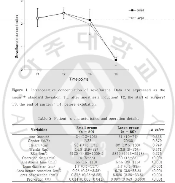
Figure 1. Intraoperative concentration of sevoflurane. Data are expressed as the mean ± standard deviation. T1, after anesthesia induction; T2, the start of surgery; T3, the end of surgery; T4, before extubation.
Table 2. Patient’s characteristics and operation details.
Values are expressed as median (min-max) or number (%). Proportion was defined as the ratio of nevi to total body surface; BSA, body surface area
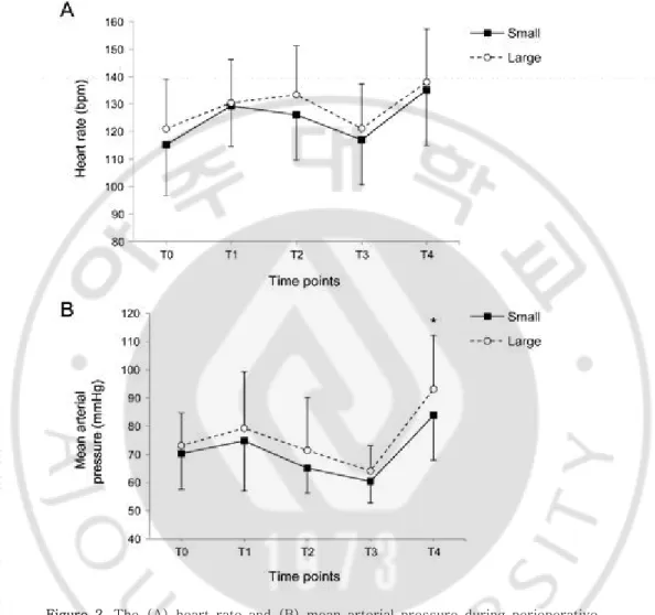
Figure 2. The (A) heart rate and (B) mean arterial pressure during perioperative period. Data are expressed as the mean ± standard deviation. T0, before
anesthesia induction (baseline); T1, after intubation; T2, the start of surgery; T3, the end of surgery; T4, after extubation.
Among the 100 children, the incidence of emergence agitation was 51%, and incidence of pain was 66%. Classification into two groups according to the median value of proportion showed that the
patient’s characteristics were comparable between the two groups (Table 2). However, operation time, anesthesia time, and nevus size were significantly higher in the large group. The intraoperative end-tidal concentration of sevoflurane was constantly maintained during surgery within 2.5 vol%, without any significant differences between the two groups (Figure 1). Perioperative HR was similar; however, MAP after extubation was higher in the large group compared with the small group (p = 0.007, Figure 2)
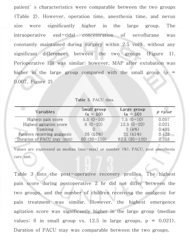
Table 3. PACU data.
Values are expressed as median (min-max) or number (%). PACU, post anesthesia care unit.
Table 3 lists the post-operative recovery profiles. The highest pain score during postoperative 2 hr did not differ between the two groups, and the number of children receiving the analgesic for pain treatment was similar. However, the highest emergence agitation score was significantly higher in the large group (median values; 6 in small group vs. 12.5 in large groups, p = 0.021). Duration of PACU stay was comparable between the two groups.
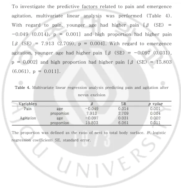
To investigate the predictive factors related to pain and emergence agitation, multivariate linear analysis was performed (Table 4). With regard to pain, younger age had higher pain [β (SE) = -0.049 (0.014), p = 0.001] and high proportion had higher pain [β (SE) = 7.913 (2.709), p = 0.004]. With regard to emergence agitation, younger age had higher pain [β (SE) = -0.097 (0.031), p = 0.002] and high proportion had higher pain [β (SE) = 15.803 (6.061), p = 0.011].
Table 4. Multivariate linear regression analysis predicting pain and agitation after nevus excision
The proportion was defined as the ratio of nevi to total body surface. Β, logistic regression coefficient; SE, standard error.
IV. Discussion
The primary endpoints included the effects of size and location of nevus on postoperative pain and emergence agitation. The proportion was the most ideal parameter when defining the role of nevus size in pain and emergence agitation. The large-sized group was significantly associated with higher emergence agitation following sevoflurane anesthesia compared with the small-sized group. However, the postoperative pain was comparable between the two groups. The specific location of nevus excision had no role in the differences in pain and emergence agitation associated with other areas of body. In multivariate linear regression analysis, younger age and large-sized excision were associated with increased pain and emergence agitation.
Emergence agitation is a postoperative behavioral disturbance observed after anesthesia in children, with a reported incidence of 70% following sevoflurane anesthesia [8]. Despite unclear mechanism, sevoflurane may exert an irritable effect on the central nervous system [9]. Several possible risk factors of emergence agitation were observed in this study. First, the large nevi triggered higher emergence agitation, although postoperative pain was comparable between large and small groups. Prolonged anesthesia time in the large group may increase the exposure to sevoflurane (55 min and 67.5 min in small and large groups, respectively), which may affect the development of emergence
agitation. The relationship between postoperative pain and agitation is still disputed. Emergence agitation was found in 50% of children
undergoing magnetic resonance imaging under sevoflurane
anesthesia without surgery [10]. Elimination of postoperative pain via scalp nerve block failed to reduce the occurrence of emergence agitation following sevoflurane anesthesia in children
undergoing excision of scalp nevi [11]. Conversely, a
meta-analysis found that propofol, alpha2-adrenoceptors,
ketamine, fentanyl, and perioperative analgesia induce a prophylactic effect against emergence agitation [12]. However, the authors concluded that the analgesic properties of these treatments do not play a role and reduced anesthetic requirements via potentiation of anesthesia might be involved [12]. Second, as another possible risk factor in this study, younger age was associated with emergence agitation. In 1997, Aono et al [13] showed a higher rate of emergence agitation in preschool boys anesthetized with sevoflurane compared with school boys. Since then, several studies confirmed that young age was a factor
associated with emergence agitation [14, 15]. In
pharmacodynamics, the minimal alveolar concentrations are very high in infants and decrease with increasing age [16]. In addition, in the very young subjects, blood solubility is reduced due to changing lipid and protein profiles, leading to a more rapid induction [16]. Thus, rapid awakening from sevoflurane in younger age may partly explain the phenomenon, although rapid awakening
is disputed [15, 17].
In this study, 0.3 mcg/kg of fentanyl was administered during anesthetic induction. Although fentanyl as adjuvant was reported to reduce the emergence agitation from sevoflurane anesthesia [18], the incidence of emergence agitation in this study was similar to that of Kim’s study (51% vs. 55%, respectively) [11]. First, fentanyl as rescue analgesic in the PACU was administered immediately on arrival at the PACU without pre-assessment of emergence agitation in Kim’s study. However, in this study, pain and emergence agitation scores were evaluated immediately on arrival at the PACU, and fentanyl administration was decided based upon the scores. The foregoing discussion suggests that the pain treatment in PACU does not influence the incidence of emergence agitation. Second, emergence agitation was rated using PAED scale in this study compared with Watch scale in Kim’s study. These two scales correlated reasonably well with each other, although the PAED scale showed greater sensitivity in detecting emergence compared with Watch scale [19].
In this study, the relationship between nevus size and postoperative pain varied when the two groups were compared or in multivariate analysis. Only 38% of pediatric patients receiving excision for scalp nevi required rescue analgesics in PACU, which was consistent with the results of the study by Xu et al [5], in which simple skin incision produced less severe and transient pain than in incision of skin combined with deep tissue. In the
development of pain, the extent of deep tissue injury appears to be more meaningful than the length of skin excision. When two approaches with the same length of skin incision (20 cm) and different degrees of deep muscle tissue injury were compared, the approach entailing incision of deep tissue induced greater pain at rest and morphine use [20]. In contrast, when two approaches with different lengths of skin incision (9 vs. 16 cm) and the same degree of deep muscle tissue injury were compared, no difference was found in pains at rest and following morphine use [21]. However, a previous case reported unusual pain after nevus excision, which transformed into complex regional pain syndrome [22].
In an observational study with Turkish children, CMN showed differential localization according to body region, predominantly in head and neck, or back [23]. During the study, nevus excision in the facial area was suspected to involve greater pain and emergence agitation than in other areas; however, in a final analysis, body location did not influence the pain or agitation. Although quantification and comparison of pain according to body location is important, no consensus is available regarding the outcomes [24]. The sensitivity of facial area to pain was found to be either higher [25] or similar to other body locations [26]. Distal parts were equally sensitive [27] or less sensitive than the proximal parts of the limb [25, 26]. However, because the degree of sensitivity to noxious pain is uniform across the body, the
comparison of pain sensitivity according to body location needs to be evaluated in a large sample size.
V. Conclusions
A large-sized nevus triggered more severe emergence agitation in children following sevoflurane anesthesia; however, the nevus location had no effect on the pain or emergence agitation. In addition, younger age was associated with pain and emergence agitation. Clinicians need to consider the proportion as size parameter when managing pain and emergence agitation in children undergoing nevus excision.
References
1. Price, H. N., and J. V. Schaffer. "Congenital Melanocytic
Nevi-When to Worry and How to Treat: Facts and
Controversies." Clin Dermatol 28, no. 3 (2010): 293-302.
2. Roldan, F. A., A. B. Hernando, A. Cuadrado, C. C. Blanco, R. S. Fernandez, J. M. Hermosa, and P. L. Ochaita. "Small and Medium-Sized Congenital Nevi in Children: A Comparison of the Costs of Excision and Long-Term Follow-Up." Dermatol Surg 35, no. 12 (2009): 1867-72.
3. Fortier, M. A., J. E. MacLaren, S. R. Martin, D. Perret-Karimi, and Z. N. Kain. "Pediatric Pain after Ambulatory Surgery: Where's the Medication?" Pediatrics 124, no. 4 (2009): e588-95.
4. Groenewald, C. B., J. A. Rabbitts, D. R. Schroeder, and T. E. Harrison. "Prevalence of Moderate-Severe Pain in Hospitalized Children." Paediatr Anaesth 22, no. 7 (2012): 661-8.
5. Xu, J., and T. J. Brennan. "Guarding Pain and Spontaneous Activity of Nociceptors after Skin Versus Skin Plus Deep Tissue Incision." Anesthesiology 112, no. 1 (2010): 153-64. 6. Meier, P. M., C. B. Berde, J. DiCanzio, D. Zurakowski, and N. F.
Sethna. "Quantitative Assessment of Cutaneous Thermal and Vibration Sensation and Thermal Pain Detection Thresholds in Healthy Children and Adolescents." Muscle Nerve 24, no. 10 (2001): 1339-45.
7. Turkmen, A., D. Isik, and M. Bekerecioglu. "Comparison of Classification Systems for Congenital Melanocytic Nevi." Dermatol Surg 36, no. 10 (2010): 1554-62.
8. Guler, G., A. Akin, Z. Tosun, S. Ors, A. Esmaoglu, and A. Boyaci. "Single-Dose Dexmedetomidine Reduces Agitation and Provides Smooth Extubation after Pediatric Adenotonsillectomy." Paediatr Anaesth 15, no. 9 (2005): 762-6.
9. Woodforth, I. J., R. G. Hicks, M. R. Crawford, J. P. Stephen, and D. J. Burke. "Electroencephalographic Evidence of Seizure Activity under Deep Sevoflurane Anesthesia in a Nonepileptic Patient." Anesthesiology 87, no. 6 (1997): 1579-82.
10. Isik, B., M. Arslan, A. D. Tunga, and O. Kurtipek. "Dexmedetomidine Decreases Emergence Agitation in Pediatric Patients after Sevoflurane Anesthesia without Surgery." Paediatr Anaesth 16, no. 7 (2006): 748-53.
11. Kim, J. S., G. W. Kim, D. H. Park, H. E. Ahn, M. Y. Chang, and J. Y. Kim. "Effects of Scalp Nerve Block on Pain and Emergence Agitation after Paediatric Nevus Surgery: A Clinical Trial." Acta Anaesthesiol Scand 61, no. 8 (2017): 935-41. 12. Dahmani, S., I. Stany, C. Brasher, C. Lejeune, B. Bruneau, C.
Wood, Y. Nivoche, I. Constant, and I. Murat. "Pharmacological Prevention of Sevoflurane- and Desflurane-Related Emergence Agitation in Children: A Meta-Analysis of Published Studies." Br J Anaesth 104, no. 2 (2010): 216-23.
"Greater Incidence of Delirium During Recovery from Sevoflurane Anesthesia in Preschool Boys." Anesthesiology 87, no. 6 (1997): 1298-300.
14. Voepel-Lewis, T., S. Malviya, and A. R. Tait. "A Prospective Cohort Study of Emergence Agitation in the Pediatric Postanesthesia Care Unit." Anesth Analg 96, no. 6 (2003): 1625-30, table of contents.
15. Dahmani, S., H. Delivet, and J. Hilly. "Emergence Delirium in Children: An Update." Curr Opin Anaesthesiol 27, no. 3 (2014): 309-15.
16. Johr, M., and T. M. Berger. "Paediatric Anaesthesia and Inhalation Agents." Best Pract Res Clin Anaesthesiol 19, no. 3 (2005): 501-22.
17. Cohen, I. T., J. C. Finkel, R. S. Hannallah, K. A. Hummer, and K. M. Patel. "Rapid Emergence Does Not Explain Agitation Following Sevoflurane Anaesthesia in Infants and Children: A Comparison with Propofol." Paediatr Anaesth 13, no. 1 (2003): 63-7.
18. Costi, D., A. M. Cyna, S. Ahmed, K. Stephens, P. Strickland, J. Ellwood, J. N. Larsson, C. Chooi, L. L. Burgoyne, and P. Middleton. "Effects of Sevoflurane Versus Other General Anaesthesia on Emergence Agitation in Children." Cochrane Database Syst Rev, no. 9 (2014): Cd007084.
19. Bajwa, S. A., D. Costi, and A. M. Cyna. "A Comparison of Emergence Delirium Scales Following General Anesthesia in
Children." Paediatr Anaesth 20, no. 8 (2010): 704-11.
20. Dorr, L. D., A. V. Maheshwari, W. T. Long, Z. Wan, and L. E. Sirianni. "Early Pain Relief and Function after Posterior Minimally Invasive and Conventional Total Hip Arthroplasty. A Prospective, Randomized, Blinded Study." J Bone Joint Surg Am 89, no. 6 (2007): 1153-60.
21. Ogonda, L., R. Wilson, P. Archbold, M. Lawlor, P. Humphreys, S. O'Brien, and D. Beverland. "A Minimal-Incision Technique in Total Hip Arthroplasty Does Not Improve Early Postoperative Outcomes. A Prospective, Randomized, Controlled Trial." J Bone Joint Surg Am 87, no. 4 (2005): 701-10.
22. Prager, J. P., and M. Csete. "An Unusual Cause of Pain after Nevus Excision: Complex Regional Pain Syndrome." J Am Acad Dermatol 37, no. 4 (1997): 652-3.
23. Oztas, P., M. N. Ilhan, M. Polat, and N. Alli. "Clinical and Dermoscopic Characteristics of Melanocytic Nevi in Turkish Children and Their Relationship with Environmental and Constitutional Factors." Dermatol Surg 33, no. 5 (2007): 607-13.
24. Defrin, R., M. Shachal-Shiffer, M. Hadgadg, and C. Peretz. "Quantitative Somatosensory Testing of Warm and Heat-Pain Thresholds: The Effect of Body Region and Testing Method." Clin J Pain 22, no. 2 (2006): 130-6.
25. Meh, D., and M. Denislic. "Quantitative Assessment of Thermal and Pain Sensitivity." J Neurol Sci 127, no. 2 (1994): 164-9.
26. Dyck, P. J., I. Zimmerman, D. A. Gillen, D. Johnson, J. L. Karnes, and P. C. O'Brien. "Cool, Warm, and Heat-Pain Detection Thresholds: Testing Methods and Inferences About Anatomic Distribution of Receptors." Neurology 43, no. 8 (1993): 1500-8.
27. Hagander, L. G., H. A. Midani, M. A. Kuskowski, and G. J. Parry. "Quantitative Sensory Testing: Effect of Site and Skin Temperature on Thermal Thresholds." Clin Neurophysiol 111, no. 1 (2000): 17-22