저작자표시-비영리-변경금지 2.0 대한민국 이용자는 아래의 조건을 따르는 경우에 한하여 자유롭게 l 이 저작물을 복제, 배포, 전송, 전시, 공연 및 방송할 수 있습니다. 다음과 같은 조건을 따라야 합니다: l 귀하는, 이 저작물의 재이용이나 배포의 경우, 이 저작물에 적용된 이용허락조건 을 명확하게 나타내어야 합니다. l 저작권자로부터 별도의 허가를 받으면 이러한 조건들은 적용되지 않습니다. 저작권법에 따른 이용자의 권리는 위의 내용에 의하여 영향을 받지 않습니다. 이것은 이용허락규약(Legal Code)을 이해하기 쉽게 요약한 것입니다. Disclaimer 저작자표시. 귀하는 원저작자를 표시하여야 합니다. 비영리. 귀하는 이 저작물을 영리 목적으로 이용할 수 없습니다. 변경금지. 귀하는 이 저작물을 개작, 변형 또는 가공할 수 없습니다.
碩士學位論文
병개암나무 잎으로부터 항산화,
항염 및 미백 활성 성분 규명
濟州大學校 大學院
化 學 科
李 容 範
병개암나무 잎으로부터 항산화,
항염 및 미백 활성 성분 규명
指導敎授 李 南 昊
李
容
範
이 論文을 理學 碩士學位 論文으로 提出함
2015年 2月
李容範의 理學 碩士學位 論文을 認准함
審査委員長
委
員
委
員
濟州大學校 大學院
印 印 印Identification of Anti-oxidative,
Anti-inflammatory and Skin Whitening
Constituents from the Leaves of
Corylus hallaisanensis
Yong Bum Lee
(Supervised by Professor Nam Ho Lee)
A thesis submitted in partial fulfillment of the requirement
for the degree of Master of Science
2015. 2.
This thesis has been examined and approved
Committee Chairman,
Dissertation Committee,
Dissertation Committee,
DateDepartment of Chemistry
GRADUATE SCHOOL
JEJU NATIONAL UNIVERSITY
목 차
List of Schemes ···ⅳ List of Tables ···ⅳ List of Figures ···ⅴ List of Abbreviations ···ⅶ Abstract ···ⅷ I. 서론 ···1 1. 항산화 ···1 2. 항염 ···2 3. 미백 ···3 II. 재료 및 방법 ···5 1. 시약 및 기기 ···5 2. 재료 ···6 3. 추출, 분획 및 분리 ···7 3-1. 병개암나무 잎의 추출 및 분획 ···73-2. Ethyl acetate layer의 활성 성분 분리 ···8
4. 활성실험 ···11
4-1. 항산화 활성 실험 ···11
4-1-1. 총 폴리페놀 함량 측정 ···11
4-1-2. DPPH radical 소거 활성 실험 ···11
4-1-3. ABTS radical cation 소거 활성 실험 ···13
4-2. RAW 264.7 cell을 이용한 항염 활성 실험 ···15
4-2-1. 세포배양 ···15
4-2-3-1. MTT assay ···16 4-2-3-2. LDH assay ···17 4-3. B16F10 melanoma cell을 이용한 미백 활성 실험 ···18 4-3-1. 세포배양 ···18 4-3-2. Melanogenesis 저해효과 측정 실험 ···18 4-3-3. 세포독성 평가 실험 ···19 4-3-3-1. MTT assay ···19 4-3-3-2. LDH assay ···19 III. 결과 및 고찰 ···20 1. 화합물의 구조 결정 ···20 1-1. Compound 1의 구조해석 ···20 1-2. Compound 2의 구조해석 ···22 1-3. Compound 3의 구조해석 ···25 1-4. Compound 4의 구조해석 ···28 1-5. Compound 5의 구조해석 ···31 2. 항산화 활성 실험 결과 ···37 2-1. 총 폴리페놀 함량 측정 ···37 2-2. 추출물 및 분획물의 DPPH radical 소거 활성 ···39
2-3. 추출물 및 분획물의 ABTS radical cation 소거 활성 ···40
3. RAW 264.7 cell을 이용한 항염 활성 실험 결과 ···41
3-1. 추출물 및 분획물의 nitric oxide 생성 억제 및 세포 독성 평가 ···41
3-2. 염증성 cytokine IL-6 생성 억제 평가 ···43
4. B16F10 murine melanoma cell을 이용한 미백 활성 실험 결과 ···44
4-1. 추출물 및 분획물의 melanin 생성 억제 및 세포 독성 평가 ···44
4-2. Intracellular tyrosinase 측정 결과 ···47
List of Schemes
Scheme 1. Procedure of extraction and solvent fractionation from
C. hallaisanenesis···7
Scheme 2. Procedure of isolation of compound 1 from C. hallaisanensis···8
Scheme 3. Procedure of isolation of compounds 2, 3 from C. hallaisanensis···9
Scheme 4. Procedure of isolation of compound 4 from C. hallaisanensis···10
Scheme 5. Procedure of isolation of compound 5 from C. hallaisanensis···10
List of Tables
Table 1. Elution conditions of MPLC ···8
Table 2. 1H and 13C NMR data of compound 1 (400 and 100 MHz, CD3OD)
···21
Table 3. 1H and 13C NMR data of compound 2 (500 and 125 MHz, DMSO-d6)
···23
Table 4. 1H and 13C NMR data of compound 3 (500 and 125 MHz, CD3OD)
···26
Table 5. 1H and 13C NMR data of compound 4 (400 and 100 MHz, CD3OD)
···29
Table 6. 1H and 13C NMR data of compound 5 (500 and 125 MHz, pyridine-d6)
List of Figures
Figure 1. Pictures of Corylus hallaisanensis···6
Figure 2. Scavenging of the DPPH radical by an anti-oxidant ···12
Figure 3. Scavenging of ABTS radical cation by an anti-oxidant ···13
Figure 4. Principle of MTT assay ···16
Figure 5. Principle of INT reagent ···17
Figure 6. Chemical structure of compound 1 ···20
Figure 7. 1H-NMR spectrum of compound 1 in CD3OD ···21
Figure 8. 13C-NMR spectrum of compound 1 in CD3OD ···21
Figure 9. Chemical structure of compound 2 ···22
Figure 10. 1H-NMR spectrum of compound 2 in DMSO-d6···24
Figure 11. 13C-NMR spectrum of compound 2 in DMSO-d6···24
Figure 12. Chemical structure of compound 3 ···25
Figure 13. 1H-NMR spectrum of compound 3 in CD3OD ···27
Figure 14. 13C-NMR spectrum of compound 3 in CD 3OD ···27
Figure 15. Chemical structure of compound 4 ···28
Figure 16. 1H-NMR spectrum of compound 4 in CD 3OD ···30
Figure 17. 13C-NMR spectrum of compound 4 in CD 3OD ···30
Figure 18. Chemical structure of compound 5 ···32
Figure 19. 1H-NMR spectrum of compound 5 in pyridine-d 6···34
Figure 20. 13C-NMR spectrum of compound 5 in pyridine-d 6···34
Figure 21. DEPT-135 spectrum of compound 5 in pyridine-d6···35
Figure 22. HMQC spectrum of compound 5 in pyridine-d6···35
Figure 23. HMBC spectrum of compound 5 in pyridine-d6···36
Figure 24. Calibration curve for quantification of total phenolic compounds ·· 37
Figure 26. DPPH radical scavenging activities of extract and solvent layers ···39
Figure 27. SC50 values of DPPH radical scavenging activities of extract and
solvent layers ···39 Figure 28. ABTS radical cation scavenging activities of extract and solvent layers ···40
Figure 29. SC50 values of ABTS radical cation scavenging activities of extract
and solvent layers ···40 Figure 30. NO production inhibitory activities of extract and solvent layers · 41 Figure 31. Cell viabilities and cytotoxicities of extract and solvent layers in RAW 264.7 cell ···42 Figure 32. NO production inhibitory activities of EtOAc layer ···42 Figure 33. Cell viabilities and cytotoxicities of EtOAc layer in
RAW 264.7 cell ···43 Figure 34. Inhibition of LPS-induced secretion of pro-inflammatory cytokine IL-6 of EtOAc layer ···43 Figure 35. Melanin contents of extract and solvent layers ···44 Figure 36. Cell viabilities and cytotoxicities of extract and solvent layers in B16F10 murine melanoma cell ···45 Figure 37. Melanin contents of EtOAc layer ···45 Figure 38. Cell viabilities and cytotoxicities of EtOAc layer in
B16F10 murine melanoma cell ···46 Figure 39. Intracellular tyrosinase inhibition of EtOAc layer ···47
List of Abbreviations
ABTS 2,2'-Azino-bis(3-ethylbenzothiazoline-6-sulfonic acid)
C. hallaisanensis Corylus hallaisanensis
CC Column chromatography
CD3OD Methanol-d4
CHCl3 Chloroform
d Doublet
DEPT Distorionless enhancement by polarization transfer
DMSO Dimethylsulfoxide
DPPH 2,2-Diphenyl-1-picryhydrazyl
EtOAc Ethyl acetate
EtOH Ethanol
Fr. Fraction
HMBC Heteronuclear multiple bond correlation
HMQC Heteronuclear multiple quantum correlation
int Integration IL-6 Interleukin-6 J Coupling constant (Hz) LPS Lipopolysaccharide m Multiplet MeOH Methanol
MTT Dimethyl thiazolyl diphenyl tetrazolium salt
NMR Nuclear magnetic resornance
n-BuOH n-Butanol
n-Hex n-Hexane
s Singlet
SC50 Scavenging concentration of 50%
sepha Sephadex LH-20 CC sublayer
TLC Thin layer chromatography
Abstract
Identification of Anti-oxidative, Anti-inflammatory and Skin Whitening Constituents from the Leaves of
Corylus hallaisanensis
Yong Bum Lee
Department of Chemistry and cosmetics, Graduate School Jeju National University, Korea
Supervised by Professor Nam Ho Lee
Corylus hallaisanensis Nakai, a deciduous shrub growing in Jeju island, was investigated for skin whitening, anti-oxidative and anti-inflammatory
activities. In skin whitening screenings, ethanol extract, n-Hex, EtOAc and
n-BuOH layers showed melanin production inhibitory activity in α-MSH
induced B16F10 murine melanoma cell. For the anti-oxidation tests, the
ethanol extract, EtOAc, and n-BuOH layers showed good DPPH and ABTS+
radical scavenging activities. The total phenolic contents for the extract and
solvent layers (n-Hex, EtOAc, n-BuOH, water) were estimated as 184, 64,
425, 347 and 83 μg GAE/1 mg respectively. In anti-inflammatory screenings,
n-Hex and EtOAc layers showed nitric oxide production inhibitory activity in
LPS-induced RAW 264.7 cell.
Phytochemical studies were conducted for the EtOAc layer, which led to
isolation of four constituents such as gallic acid (1), kaempferol 3-O-β
-D-xylopyranoside (2), kaempferol 3-O-α-L-rhamnopyranoside (3) and
apigenin (4).
This study demonstrated the potential of Corylus hallaisanensis extract as
I. 서론
1. 항산화활성산소란 산소가 가지는 화학적 특성으로 인하여 생성되는 free radical 및 이로부터 유래된 산소화합물을 일컬으며 대부분 불안정하여 전자를 잃거나 얻어
서 보다 안정된 상태로 가려는 성질이 있다.1) 산소는 각종 공해물질, 물리 화학
적, 환경적 요인 등에 의해 superoxide radical (O·-2), hydroxy radical (HO·),
hydrogen peroxide (H2O2), singlet oxygen (1O2)와 같은 반응성이 매우 큰 free
radical로 전환되면서 생체에 치명적인 산소독성을 일으키는 양면성이 있다.2)
그러나 인체는 산화적 손상을 예방하고 차단하기 위한 항산화 방어망을 구축 하고 있는데 superoxide dismutase (SOD), glutathione peroxidase (GSHPx), glutathione S-transferase (GST), catalase (CAT) 등의 효소가 중요 방어 역할 을 하고 있다. 이러한 효소적 항산화제 외에 β-carotenes, flavonoids, L-ascorbic acid, α-tocopherol 등의 성분 역시 항산화제로 작용을 하게 된다. 이들은 reactive oxygen species (ROS)를 직접 소거하거나 또는 ROS에 의한 연쇄반응 을 차단하여 세포 및 피부의 생리성분이 산화적 손상을 받는 것으로부터 보호한 다.3)
지금까지 free radical을 제거하기 위해 butylated hydroxyaniso (BHT) 및 butylate hydroxy toluene (BHT) 등의 합성 항산화제가 개발되어 식품, 화장품 등에 산화방지제로 많이 사용되어 왔으나 안전성에 논란이 있어 허용대상 식품 이나 사용량이 엄격히 규제되고 있으며,4) 기존에 개발된 식물 유래 천연 항산화 제의 경우 항산화 효과가 비교적 낮고 원료 수급의 문제, 낮은 수율 및 안정성 등의 문제점이 있으며, 또한 항산화 효소는 단백질로 구성되어 온도나 pH에 영 향을 많이 받아 산업적 이용에 어려움이 있다.5-7) 그러므로 현재에는 보다 안전하고 강한 항산화제의 개발이 요구되고 있는 실 정이다. 따라서 최근에는 각종 생약 추출물 등에서 보다 안전하고 항산화 효과가 뛰어난 천연 항산화제를 개발하기 위한 많은 연구가 활발히 이루어지고 있다.8)
2. 항염 염증(inflammation)은 상처를 줄 수 있는 자극에 대한 생체의 방어 반응으로, 염증반응에서는 미소혈관의 확장, 혈액성분의 조직간극으로의 유출, 백혈구의 염 증 부위로의 유주 및 조직의 변성과 섬유화 등이 연쇄적으로 일어나게 된다. 임 상적으로는 발적, 발열, 종창, 동통, 기능 장애등의 다양한 증상이 나타난다.9) 염증을 일으키는 요인 중에서 세균에 의한 것이 가장 많으며, 일단 자극이 가
해지면 국소적으로 histamine, serotonine, bradykinin, prostaglandin,
hydroxyeicosatetraenoic acid (HETE), leukotriene과 같은 혈관생성 물질이 유리 되어 혈관 투과성이 증대되면서 염증이 유발된다. 그런데 지속적인 염증반응은 도리어 점막손상을 촉진하고, 그 결과 일부에서는 암 발생 등과 같은 질환의 요
인이 되기도 한다.10)
NO (nitric oxide)는 체내 방어기능, 신호전달기능, 신경독성, 혈관확장 등의 다 양한 생리 기능을 가지고 있다. 일반적인 NO의 형성은 박테리아를 죽이거나 종 양을 제거시키는 중요한 역할을 하지만, 염증상태에서 inducible nitric oxide synthase (iNOS)에 의해 과잉 생성된 NO는 혈관 투과성, 부종 등의 염증반응을 촉진시킬 뿐만 아니라 염증매개체의 생합성을 촉진하여 염증을 심화시키는 것으 로 알려져 있다.11-14) 다양한 염증 유도 인자 중 NO는 iNOS에 의해 생성되며, 자극을 받지 않는 세 포에서는 발현되지 않지만, cytokines이나 감염 등에 의해 대식세포, 간세포, 근 육세포 등에서 발현될 수 있다. 특히, 대식세포의 경우 iNOS의 발현을 위해 Lipopolysaccharide (LPS)가 요구된다.15-17) LPS는 대식세포 표면의 TLR4를 자극하여 하부 세포신호전달경로인
mitogen-activated protein kinase (MAPK)의 활성화를 유도한다.18)
활성화된 대식세포에서는 interleukin (IL)-1b, IL-6, IL-10, IL-12 와 TNF-α와
같은 pro-inflammatory cytokine과 PGE2 등을 생성하게 된다.19-20)
염증에 관여하는 염증 매개 물질의 형성은 phospholipase A2의 활성으로 인하
및 cyclooxygenase-2 (COX-2)에 의해 형성된다. COX-2는 염증반응, 면역반응 에 깊이 관여하고 있는 PGE2를 형성시킨다. 이와 같은 염증 매개인자들의 발현 을 조절할 수 있는 물질이 염증질환의 예방 및 치료제로서 주목을 받고 있 다.21-23) 3. 미백 일반적으로 피부색은 멜라닌(melanin)의 함량, 분포 등에 따라 색이 결정된다. 세포내 멜라노사이트(melanocyte)에서 생성된 후 세포 외부로 방출되는 멜라노좀 (melanosome)의 수와 분포에 연관되어 있다.24) 이러한 멜라닌의 생합성 과정에서 작용하는 주요 효소가 tyrosinase이다. Tyrosinase는 polyphenol oxidase의 일종이며 구리를 함유한 효소로써 색소 세포 에서 tyrosine을 L-3,4-dihydroxyphenylalanine (DOPA)으로 변환하고 효소적 산 화 반응에 의한 단계를 거쳐 dopaquinone, dopachrome으로 변환하여 멜라닌을 생합성한다.25-27) 멜라닌은 인체의 피부에 존재하며 자외선에 대항하는 기능을 수행한다. 또한 아민, 유리기, 금속이온 등과 같은 세포독성물질에 대한 제거제로 작용하여 세포 를 보호하는 작용을 한다. 반면에 체내, 외적인 여러 요인에 의해 멜라닌의 생성 이 증가하여 다량의 멜라닌이 각질형성세포에 전달되고 피부 상피층에 축적하여 과색소 침착 현상이 나타난다. 이런 멜라닌의 과잉생산은 인체에 기미, 주근깨를 형성하고 피부노화를 촉진하며 피부암의 유발에 관여하는 것으로 알려졌다. 피부에서 색소의 침착방지는 주로 다음의 네 가지 관점에서 연구되어 왔다. 첫째 로, 멜라닌 합성의 주효소인 tyrosinase 활성을 조절하기 위하여 tyrosinase 합성 저해물질이나, tyrosinase의 기질에 대한 길항물질(antagonist)을 개발한다. 둘째 로, 동물의 멜라닌 생합성 장소인 melanocyte의 기능을 저하시키기 위해 melanocyte에 독성을 나타내는 물질을 개발한다. 셋째로, DOPA의 산화방지를 위해 DOPA 환원 물질을 개발한다. 마지막으로 멜라닌 생성기구인 제1효소 tyrosinase와 DOPA chrome에서 DHICA로의 변환을 촉매하는 제2효소인 DOPA
indole-5,6-quinone-2-carboxylic acid로의 변환을 촉매하는 제3의 효소의 활성을
동시에 감소시킨다.28) 이러한 작용기전 외에 melanocyte에서 합성된 melanin이
melanosome의 과립형 입자로 keratinocyte를 통해 피부 표면으로 이동하는 과정 에서 melanocyte와 keratinocyte 간의 melanosome transfer를 차단하는 작용 역
시 미백효과를 나타낼 수 있다.29)
최근 멜라닌 생합성 대사에 대한 연구가 arbutin,30) kojic aicd,31)
oxyresveratrol,32) hydroquinone33) 등에 대해 이루어져 화장품 산업에서 미백제,
의약품 산업에서 피부 질환 치료제 등으로 사용되고 있다. 그러나 미백 화장품의
원료로 사용되었던 kojic acid와 hydroquinone에서 부작용이 보고되면서,33) 최근
에는 안전성을 고려하여 천연물을 이용한 미백연구가 활발히 이루어지고 있 다.34-35)
따라서 본 연구에서는 제주에서 자생하고 있는 특산식물인 병개암나무에서 유 효성분을 분리·정제하여 안정화된 생리활성 물질 탐색에 목적이 있다.
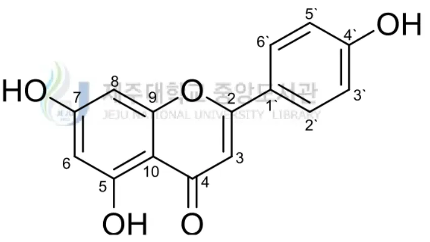
병개암나무(Corylus hallaisanensis)는 자작나무과(Betulaceae)의 낙엽활엽 관
목으로 높이는 3 m에 달하고 어린 가지에 잔털이 있다. 잎은 호생하고 넓은 도 란형 또는 난상 타원형으로 길이 4 ~ 9.7 cm, 너비 2 ~ 5.3 cm이며 끝이 뾰족 하고 겹 톱니가 있으며 측맥은 8 ~ 13쌍이고 맥 사이에 잔털이 있으며 뒷면 맥 위와 엽병에도 잔털이 있다. 꽃은 자웅일가로 암꽃이삭은 두상이며 포는 서로 겹 쳐지고 각각 2개의 암꽃이 달리며 봄에 핀다. 과실은 견과로 난형이며 숙존성인 총포로 싸여 도토리 모양을 하고 10월에 성숙한다. 본종은 참개암나무에 비해 총 포는 열매 전체를 싸나 끝이 길어지지 않는다. 과실은 식용 또는 약용한다. 한국 특산종으로 제주도 한라산 1,300 ~ 1,800 m의 산지 양지바른 곳에 난다.36) 현재 병개암나무에 관한 연구로는 가지로부터 화합물이 분리 동정되어 보고되 어있다.37) 본 연구에서는 병개암나무 잎 추출물 및 분획물의 다양한 생리활성을 검색하 고 병개암나무 잎로부터 분리된 화합물들의 구조를 동정하였다.
II. 재료 및 방법
1. 시약 및 기기
본 연구에서 시료의 추출, 용매분획 및 분리에 사용된 용매들은 Merk, Junsei 의 제품을 사용하였다. MPLC (midium pump liquid chromatography)에는 reversed phase silica gel (C18HS 12+M1946-3, Biotage Co.), normal-phase column chromatography (CC)에는 silica gel (95-110 μm, Merk Co.)이 사용되었 고, 겔 여과 크로마토그래피에는 Sephadex LH-20 (0.1-0.025 mm, GE Healthcare Life Sciences)이 사용되었다. 분리 과정에 사용된 thin-layer chromatography (TLC)는 precoated silica gel aluminium sheet (Silica gel 60
F254, 2.0 mm, Merk Co.)를 사용하였다. TLC 상에서 분리된 물질들을 확인하여
위하여 UV lamp (254 nm)를 사용하거나, visualizing agent에 침적시킨 후
heat-gun을 이용하여 건조시켰다. Visualizing agent로는 KMnO4 수용액(3 %
KMnO4, 20 % K2CO3, 0.25 % NaOH) 및 1 % anisaldehyde-MeOH를 필요에 따
라 사용하였다.
분리된 화합물의 생리활성 연구를 위한 흡광도 측정에는 SunriseTM (Tecan
Co.)을 이용하였다.
구조분석에 이용된 NMR (nuclear magnetic resonance spectrometer)은 JNM-ECX 400 (FT-NMR system, JEOL)과 AVANCE Ⅲ (Bruker Co., 500
MHz)을 이용하였으며, NMR 측정 용매는 Merk의 NMR 전용용매로 CD3OD,
Figure 1. Pictures of Corylus hallaisanensis.
2. 재료
실험에 사용된 병개암나무(Corylus hallaisanensis) 잎(시료번호 : 321)은 2011
년 6월에 제주 한라수목원에서 채집하였다. 채집한 병개암나무 잎은 실온 및 음 지에서 완전히 자연 건조 하였으며 분쇄하여 사용하였다(Figure 1).
Dried leaves of C. hallaisanensis 301.0 g
Extract 51.6 g (17.1%)
70% EtOH, stirring, 24 hr, 3 times.
Extract 40.0 g
n-Hexane layer 2.8 g (7.0%)
Suspend with 1 L water and add 1 L n-hexane, 3 times.
Add 1 L ethyl acetate, 3 times.
Ethyl acetate layer
6.4 g (16.0%) Add 1 L n-butanol, 3 times. n-Butanol layer 7.4 g (18.5%) Water layer 22.2 g (55.5%)
Scheme 1. Procedure of extraction and solvent fractionation from
C. hallaisanensis. 3. 추출, 분획 및 분리 3-1. 병개암나무 잎의 추출 및 분획 건조된 병개암나무 잎 301.0 g을 분쇄한 후, 이를 70 % 에탄올 6 L에 넣고 실 온에서 24시간 교반하였다. 침출시킨 시료를 감압 여과 장치를 이용하여 여액만 취하였으며, 이와 같은 방법으로 분리한 잔사에 대하여 동일한 조건으로 2회 더 반복 실시하였다. 이렇게 여과하여 얻어진 여액은 40 ℃ 이하의 수욕 상에서 진 공농축기(rotary vacuum evaporator)로 농축하여 70 % 에탄올 추출물 51.6 g을 얻었다. 이 중 추출물 40.0 g을 증류수 2 L에 현탁시키고, 분별 깔때기를 이용
해 극성순서에 따라 순차적으로 분획하여 n-hexane layer, ethyl acetate layer,
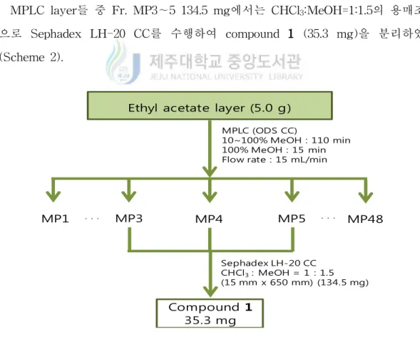
Ethyl acetate layer (5.0 g)
MPLC (ODS CC)
10~100% MeOH : 110 min 100% MeOH : 15 min Flow rate : 15 mL/min
MP1 ㆍ ㆍ ㆍ MP3 MP4 MP5 ㆍ ㆍ ㆍ MP48 Sephadex LH-20 CC CHCl3: MeOH = 1 : 1.5 (15 mm x 650 mm) (134.5 mg) Compound 1 35.3 mg
Scheme 2. Procedure of isolation of compound 1 from
C. hallaisanensis.
3-2. Ethyl acetate layer의 활성 성분 분리
용매분획 후 얻어진 각 분획 층들 중 ethyl acetate layer 5.0 g을 분획하기 위 하여 ODS (C-18) column을 이용하여 medium pressure liquid chromatography (MPLC)를 수행하였다. MPLC의 조건은 다음 아래의 표와 같으며 각 35 mL씩 용출하여 총 48의 layer을 얻었다(MP1∼MP48).
Table 1. Elution conditions of MPLC.
조건 시간
10∼100% MeOH 110 min
100% MeOH 15 min
Flow rate 15 mL/min
MPLC layer들 중 Fr. MP3∼5 134.5 mg에서는 CHCl3:MeOH=1:1.5의 용매조건
으로 Sephadex LH-20 CC를 수행하여 compound 1 (35.3 mg)을 분리하였다 (Scheme 2).
Ethyl acetate layer (5.0 g)
MPLC (ODS CC)
10~100% MeOH : 110 min 100% MeOH : 15 min Flow rate : 15 mL/min
MP1 ㆍ ㆍ ㆍ MP25 MP26 MP28 ㆍ ㆍ ㆍ MP48 Sephadex LH-20 CC CHCl3: MeOH = 5 : 1 (20 mm x 650 mm) (122 mg) Compound 3 6.2 mg MP27 Sephadex LH-20 CC CHCl3: MeOH = 2 : 1 (13 mm x 280 mm) MP22 MP23 MP24 Silica gel CC CHCl3: MeOH = 5 : 1 (18 mm x 670 mm) (231.9 mg) Compound 2 9.2 mg
Scheme 3. Procedure of isolation of compounds 2, 3 from
C. hallaisanensis. Fr. MP22∼24 231.9 mg에서는 CHCl3:MeOH=5:1의 용매조건으로 순상 silica gel CC를 수행하여 compound 2 (9.2 mg)을 분리하였다. 또한 Fr. MP25∼28 122 mg 에서는 CHCl3:MeOH=5:1의 용매조건으로 sephadex LH-20 CC를 수행 하여 얻어진 layer (30 mg)을 이용하여 다시 한번 CHCl3:MeOH=2:1의 용매조건 으로 sephadex LH-20 CC를 수행하여 compound 3 (6.2 mg)을 분리하였다 (Scheme 3).
Ethyl acetate layer (5.0 g)
MPLC (ODS CC)
10~100% MeOH : 110 min 100% MeOH : 15 min Flow rate : 15 mL/min
MP1 ㆍ ㆍ ㆍ MP10 MP29 ㆍ ㆍ ㆍ MP48 Compound 4 1.3 mg MP40 Silica gel CC n-Hex : EtOAc = 1.5 : 1 (15 mm x 650 mm) (41.8 mg) ㆍ ㆍ ㆍ ㆍ ㆍ ㆍ MeOH soluble
Scheme 4. Procedure of isolation of compound 4 from
C. hallaisanensis.
Fr. MP29를 감압농축한 후 MeOH를 첨가하였을 때 녹지 않고 가라앉는
powder를 제거 후 녹은 부분(41.8 mg)을 이용하여 n-Hex:EtOAc=1.5:1의 용매조
건으로 silica gel CC를 수행하여 compound 4 (1.3 mg)을 분리하였다(Scheme 4).
Fr. MP40∼44 148.0 mg에서는 CHCl3:MeOH=15:1의 용매조건으로 순상 silica
Ethyl acetate layer (5.0 g )
MPLC (ODS CC)
10~100% MeOH : 110 min 100% MeOH : 15 min Flow rate : 15 mL/min
MP1ㆍ ㆍ ㆍ MP40 MP41 MP44 ㆍ ㆍ ㆍMP48 Silica gel CC CHCl3: MeOH = 15 : 1 (15mm x 690 mm) (148 mg) Compound 5 11.1 mg MP43 MP42
Scheme 5. Procedure of isolation of compound 5 from
4. 활성실험
4-1. 항산화 활성 실험
4-1-1. 총 폴리페놀 함량 측정
폴리페놀 화합물의 함량은 Folin-Denis법38)을 약간 변형하여 실시하였다. 먼저
gallic acid 표준용액을 사용하여 폴리페놀 화합물의 정량을 위한 표준곡선으로 이용하였다. 표준곡선을 작성하기 위해 gallic acid 1 mg을 DMSO:EtOH=1:1 용 액 1 mL에 녹여 stock solution을 제조하고, 이를 0, 31.25, 62.5, 125, 250 및 500 μg/mL이 되도록 묽혀 표준곡선 작성에 사용하였다. 각 시료들은 1 mg/mL 농도로 용매에 녹인 후 실험에 이용하였다. 각각의 표준물질과 시료용액 100 μL 를 micro tube에 취하고 증류수 900 μL를 가하여 total volume이 1 mL가 되도 록 희석하였다. 여기에 100 μL Folin-ciocalteu's phenol reagent를 첨가하여 잘
혼합한 후 실온에 3분간 방치하였다. 이 용액에 7 % Na2CO3 용액 200 μL를 가
하여 혼합하고, 증류수 700 μL을 가하여 total volume이 2 mL이 되도록 희석하 여 실온에서 1시간 방치한 뒤, 상등액을 취하여 UV-Visible spectrophotometer로 700 nm에서 흡광도를 측정하고, 폴리페놀 화합물 함량을 환산하였다.
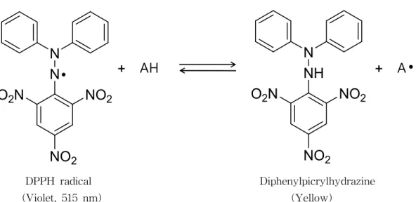
4-1-2. DPPH radical 소거 활성 실험
DPPH (2,2-diphenyl-1-picrylhydrazyl) radical 소거 활성 실험은 시료의 free radical 소거 능력이나 수소 공여 능력을 평가하는 방법 중에 하나이다. 대부분의 radicals는 반응성이 커서 매우 불안정하지만, DPPH radical은 안정한 free radical을 가진 물질로써 515 nm에서 강한 흡수를 나타내는 보라색의 화합물이 다. 하지만 free radical을 소거할 수 있는 항산화제로부터 수소를 공여 받아 2,2-diphenyl-1-picrylhydrazine (DPPH-H)이 되면 노란색으로 변화되어 515 nm 에서의 흡광도가 감소되므로, 이러한 원리를 이용하여 시료의 항산화 활성을 측
N
NO
2NO
2O
2N
N
+ AH
NH
NO
2NO
2O
2N
N
+ A
● ● DPPH radical Diphenylpicrylhydrazine (Violet, 515 nm) (Yellow)Figure 2. Scavenging of the DPPH radical by an anti-oxidant.
DPPH radical 소거 활성 실험은 Blois 방법39)을 응용하였고, 실험방법은 다음
과 같다.
96 well plate에 농도별로 희석한 sample 용액 20 μL (in EtOH)와 0.2 mM DPPH (in EtOH) 용액 180 μL를 혼합하여 상온에서 25분간 반응시킨 후, ELISA Reader를 이용하여 515 nm에서 흡광도를 측정하였다. 라디칼 소거율(%)은 다음의 식에 의해 계산되었으며, 각 시료의 소거활성 백 분율이 50 % 일 때의 시료 농도(SC50)를 구하였다. 양성대조군(positive control) 으로는 vitamin C를 사용하였다. Acontrol : 515 nm에서 DPPH의 흡광도 Asample : 515 nm에서 sample과 DPPH 반응액의 흡광도 Ablank : 515 nm에서 sample 자체의 흡광도
N S N HO3S N S N SO3H + AH ABTS+ radical (blue green, 700nm) + N S N HO3S N S N SO3H + A ABTS+ (colorless) + H
Figure 3. Scavenging of ABTS radical cation by an anti-oxidant.
4-1-3. ABTS radical cation 소거 활성 실험
ABTS [2,2'-Azino-bis(3-ethyl benzothiazoline-6-sulfonic acid)] radical 소거 활성 실험은 total antioxidant activity (TAC)의 측정방법 중의 한 가지로 무색
의 환원된 ABTS가 산화되면 특징적인 청록색을 띄는 ABTS + (ABTS radical
cation)이 형성되는 것을 이용한 방법이다. 이 청록색의 ABTS+은 산화될 수 있
는 물질과 반응하면 본래의 무색 ABTS로 환원되고 그와 반응한 물질의 산화가 일어나게 되어 흡광도의 감소가 일어나게 되면 항산화 능력을 측정할 수 있다 (Figure 3).
ABTS radical cation 소거 활성은 Re, Pellegrin 등의 방법40)을 응용하였고, 실 험방법은 다음과 같다.
ABTS radical cation을 만들기 위하여 7.0 mM ABTS (in D.W) 용액과 2.45 mM potassium persulfate (in D.W)용액을 1:1로 혼합하여 16시간동안 암소에서 반응시킨다. 반응 시킨 ABTS 용액과 ethanol을 1:60으로 희석하여 700 nm에서 흡광도가 0.78±0.002가 되도록 조절하여 실험에 사용하였다.
96 well plate에 농도별로 희석한 sample 용액 20 μL (in EtOH)와 ABTS 용 액 180 μL를 혼합하여 15분간 암소에서 반응시킨 후, ELISA Reader를 이용하여 700 nm에서 흡광도를 측정하였다. 라디칼 소거율(%)은 다음의 식에 의해 계산되었으며, 각 시료의 소거활성 백 분율이 50 % 일 때의 시료 농도(SC50)를 구하였다. 양성대조군(positive control) 으로는 vitamin C를 사용하였다. Acontrol : 700 nm에서 ABTS의 흡광도
Asample : 700 nm에서 sample과 ABTS 반응액의 흡광도 Ablank : 700 nm에서 sample 자체의 흡광도
4-2. RAW 264.7 cell을 이용한 항염 활성 실험 4-2-1. 세포배양
마우스 대식세포 계열(murine macrophage cell line)인 RAW 264.7 cell은 한국
세포주은행(KCLB; Seoul, Korea)으로부터 분양 받아 1 %
penicillin-streptomycin과 10 % fetal bovine serum (FBS)이 함유된 Dulbecco's Modified Eagle Medium (DMEM, GIBCO, Grand Island, NY, USA) 배지를 사용하여 37
℃, 5 % CO2 incubator 조건에서 배양하였으며, 2일 간격으로 계대 배양을 시행
하였다.
4-2-2. Nitric oxide 생성 억제 평가 실험
24 well plate에 RAW 264.7 cells를 2×10⁵ cells/mL로 분주하고 37 ℃, 5 %
CO2 incubator 조건하에서 18시간 배양한 후 실험에 사용하였다. 전 배양시킨
cells을 1 μg/mL LPS가 포함된 배지로 교환 후, sample을 농도별로 각각 첨가하 여 24시간 배양하였다. 생성된 NO의 양은 Griess 시약(1 % sulfanilamide, 0.1 % naphylethylenediamine in 2.5 % phosphoric acid)을 이용하여 세포배양액 중에
존재하는 NO2-의 형태로 측정하였다. 세포배양 상등액 100 μL와 Griess 시약
100 μL를 혼합하여 96 well plate에서 10분 동안 반응시킨 후 540 nm에서 흡광
도를 측정하였다. 생성된 NO의 양은 sodium nitrite (NaNO2)를 이용하여 검정곡
Cell viability (%) = × 100
A
sampleA
control N N N N S N Br yellow MTT mitochondrial reductase N N NH N S N purple formazanFigure 4. Principle of MTT assay.
4-2-3. 세포독성 평가 실험 4-2-3-1 MTT assay
MTT [3-(4,5-dimethyl-thiazol-2-yl)-2,5-diphenyl tetrazolium bromide] assay는
RAW 264.7 cell을 24 well plate에 2×10⁵ cells/mL로 분주하고 37 ℃, 5 % CO2
조건하에서 18시간 배양 후, 1 μg/mL LPS와 sample을 농도별로 각각 첨가하여 24시간 배양하였다. 24시간 배양 후, 500 μg/mL 농도로 MTT를 첨가하여 37 ℃ 에서 3시간 동안 반응시킨 후, 상층액을 제거하였다. 여기에 DMSO를 가하여 살 아있는 세포와 반응하여 생긴 formazan 침전물을 용해시킨 다음, 이를 96 well plate에 옮긴 후 570 nm에서 흡광도를 측정하였다. Cell viability는 다음의 식에 의해 계산되었다.
Acontrol : 570 nm에서 용매와 LPS를 처리한 control의 흡광도 Asample : 570 nm에서 시료와 LPS를 처리한 sample의 흡광도
Cell cytotoxity (%) = × 100
A
sample-A
controlA
highcontrol-A
control C N N N N I NO2 diapholase C N N N N I NO2 HInt (yellow) Formazan (red) 490 nm
Figure 5. Principle of INT reagent. 4-2-3-2 LDH assay
LDH는 세포막이 손상되면 세포외, 즉 배지 중으로 방출된다. LDH는 젖산의 탈수소화반응의 촉매작용을 하여 pyruvate와 NADH를 생성하는데 이때 NADH 는 diapholase라는 탈수소효소에 의해 노란색의 INT (2-(4-Iodophenyl)-3-(4-nitrophenyl)-5-phenyltetrazolium chloride)을 환원시켜 490 nm의 흡수를 갖는 적색의 formazan 색소를 형성한다. LDH assay는 위의 원리를 이용한 cytotoxicity detection kit로 측정하였다. 먼저 RAW264.7 cell을 24 well plate에
2×10⁵ cells/mL로 분주하고 37 ℃, 5 % CO2 조건하에서 18시간 배양 후,
1 μg/mL LPS와 sample을 농도별로 각각 첨가하여 24시간 배양하였다. 24시간
배양 후, 배양액 100 μL와 diapholase, NAD+ 및 INT 혼합액 100 μL를 혼합한
후 암실에서 5분간 반응시켜 ELISA reader에서 490 nm에서 흡광도를 측정하였 다. Cell cytotoxyty는 다음의 식에 의해 계산되었다.
Acontrol : 490 nm에서 용매와 LPS를 처리한 control의 흡광도 Asample : 490 nm에서 시료와 LPS를 처리한 sample의 흡광도 Ahighcontol : 490 nm에서 tripton X100을 처리한 control의 흡광도
4-3. B16F10 murine melanoma cell을 이용한 미백 활성 실험 4-3-1. 세포배양
Murine B16F10 melanoma cell은 한국세포주은행(KCLB; Seoul, Korea)으로부 터 분양 받아 1 % penicillin-streptomycin과 10 % fetal bovine serum (FBS)이 함유된 Dulbecco's Modified Eagle Medium (DMEM, GIBCO, Grand Island,
NY, USA) 배지를 사용하여 37 ℃, 5 % CO2 incubator 조건에서 배양하였으며, 3
일 간격으로 계대 배양을 시행하였다.
4-3-2. Melanogenesis 저해효과 측정 실험
6 well plate에 5×10⁴ cells/mL로 세포를 분주하고 37 ℃, 5 % CO2 incubator
조건하에서 24시간 배양한 후 실험에 사용하였다. 전 배양시킨 cells을 1 μg/mL
α-MSH를 포함하는 배지로 교환 후, sample을 농도별로 각각 첨가하여 72시간
배양하였다. 배양이 끝나면 배지를 제거하고 PBS buffer로 세척한 후 trypsin-EDTA를 처리하여 세포를 회수하여 원심분리 시켜 pellet만을 취한다. Pellet에 sonication buffer (1 % triton X-100 and 0.2 mM PMSF (Phenylmethyl sulfonyl fluoride in sodium phosphate buffer)를 넣고 세포를 sonication해 준 후 에 원심분리 시켜 분리한 pellet에는 1 N NaOH를 300 μL 첨가하여 ELISA reader를 이용하여 405 nm에서 melanin contents를 확인하고, 상등액은 L-DOPA와 반응시켜 ELISA reader로 475 nm에서 cell에 있던 tyrosinase를 확 인하였다. 대조군으로는 melasolv를 사용하였다.
4-3-3. 세포독성 평가 실험 4-3-3-1 MTT assay
MTT [3-(4,5-dimethyl-thiazol-2-yl)-2,5-diphenyl tetrazolium bromide] assay를
이용하였다. B16F10 murine melanoma cell을 6 well plate에 5×10⁴ cells/mL로
분주하고 37 ℃, 5 % CO2 조건하에서 24시간 배양 후, 1 μg/mL α-MSH와 sample을 농도별로 각각 첨가하여 72시간 배양하였다. 72시간 배양 후, 500 μ g/mL 농도로 MTT를 첨가하여 37 ℃에서 3시간 동안 반응시킨 후, 상층액을 제 거하였다. 여기에 DMSO를 가하여 살아있는 세포와 반응하여 생긴 formazan 침 전물을 용해시킨 다음, 이를 96 well plate에 옮긴 후 570 nm에서 흡광도를 측정 하였다. 4-3-3-2 LDH assay LDH는 세포막이 손상되면 세포외, 즉 배지 중으로 방출된다. LDH는 젖산의 탈수소화반응의 촉매작용을 하여 pyruvate와 NADH를 생성하는데 이때
NADH는 diapholase라는 탈수소효소에 의해 노란색의 INT (2-(4-Iodophenyl)-3 -(4-nitrophenyl)-5-phenyltetrazolium chloride)을 환원시켜 490 nm의 흡수를 갖 는 적색의 formazan 색소를 형성한다. LDH assay는 위의 원리를 이용한 cytotoxicity detection kit로 측정하였다. 먼저 B16F10 murine melanoma cell을
6 well plate에 5×10⁴ cells/mL로 분주하고 37 ℃, 5 % CO
2 조건하에서 24시간 배
양 후, 1 μg/mL α-MSH와 sample을 농도별로 각각 첨가하여 72시간 배양하였
다. 72시간 배양 후, 배양액 100 μL와 diapholase, NAD+ 및 INT 혼합액 100 μL
를 혼합한 후 암실에서 5분간 반응시켜 ELISA reader를 이용하여 490 nm에서 흡광도를 측정하였다.
OH
OH
HO
HO
O
1
2
3
4
5
6
Figure 6. Chemical structure of compound 1.
III. 결과 및 고찰
1. 화합물의 구조 결정
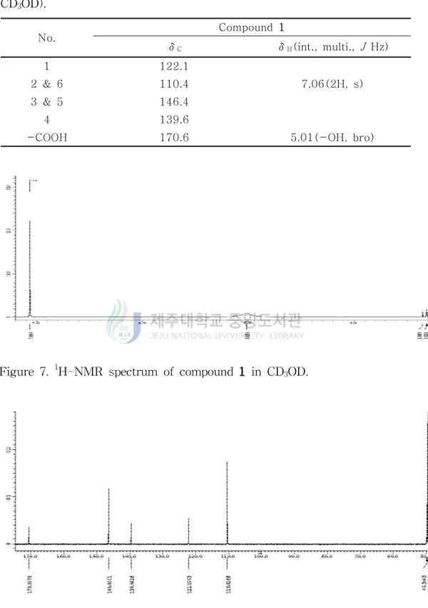
1-1. Compound 1의 구조해석
Compound 1은 1H-NMR spectrum 분석 결과 δ 7.06(2H, s)의 signal로부터
sp2혼성을 갖는 aromatic ring의 대칭구조의 proton을 예상하였다. 13C-NMR
spectrum 분석결과 탄소 수는 5개 이상으로 예측되며, 170.6의 signal은 carbonyl carbon임을 예상하였고, 146.4, 139.6, 122.1, 110.4의 signal들은 aromatic ring의
signal임을 확인하였다. 이를 바탕으로 compound 1은 문헌41)과 비교하여 gallic
Figure 8. 13C-NMR spectrum of compound 1 in CD3OD.
Figure 7. 1H-NMR spectrum of compound 1 in CD3OD.
No. Compound 1 δC δH(int., multi., J Hz) 1 122.1 2 & 6 110.4 7.06(2H, s) 3 & 5 146.4 4 139.6
-COOH 170.6 5.01(-OH, bro)
Table 2. 1H and 13C NMR data of compound 1 (400 and 100 MHz,
O
OH
HO
O
O
OH
O
HO
OH
OH
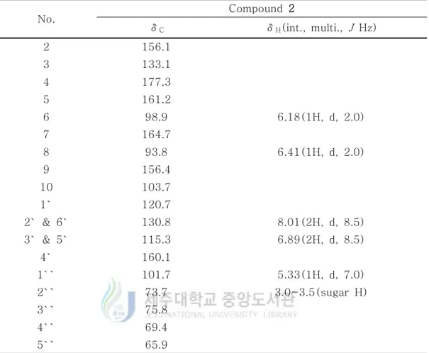
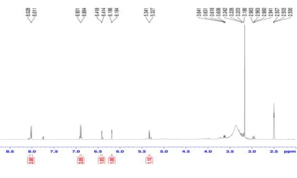
2 3 4 5 6 7 8 9 10 1` 6` 5` 4` 3` 2` 1`` 2`` 3`` 4`` 5`` 1-2. Compound 2의 구조해석 Compound 2는 1H-NMR spectrum 분석 결과 δ 6.89(2H, d, J = 8.5 Hz)와 δ8.02(2H, d, J = 8.5 Hz)의 signal은 aromatic ring의 proton으로 적분 값과
coupling constant 값을 통하여 ortho-coupling을 하는 대칭구조임을 예상할 수
있었고, δ 6.18(1H, d, J = 2 Hz)과 δ 6.41(1H, d, J = 2 Hz)의 signal은 coupling
constant 값을 통하여 meta-coupling을 하는 sp2 proton으로 예상하였다. 또한 δ
5.33(1H, d, J = 7 Hz) 및 δ 3.0~3.5 부근의 복잡한 signal로부터 당이 하나 치환 된 구조임을 예상하였다.
13C-NMR spectrum 분석 결과, 19개 이상의 carbon signal과 δ 156.1(C-2),
133.1(C-3), 177.3(C-4)의 carbon peak로부터 flavone 3-ol의 골격임을 알 수 있
었으며, 1H과 13C NMR data를 비교한 결과 aglycone은 kaempferol이며 3번 위
치에 당이 치환될 것으로 예상 하였다. 치환된 당의 signal은 δ 101.7, 73.7, 75.8,
69.4, 65.9이며 anomeric proton의 coupling constant 값(J = 7 Hz)을 통하여 문
헌42)과 비교하여 β-D-xylopyranoside임을 확인 하였다. 이를 바탕으로
compound 2은 kaempferol 3-O-β-D-xylopyranoside로 동정하였고 문헌과 일치
No. Compound 2 δC δH(int., multi., J Hz) 2 156.1 3 133.1 4 177.3 5 161.2 6 98.9 6.18(1H, d, 2.0) 7 164.7 8 93.8 6.41(1H, d, 2.0) 9 156.4 10 103.7 1` 120.7 2` & 6` 130.8 8.01(2H, d, 8.5) 3` & 5` 115.3 6.89(2H, d, 8.5) 4` 160.1 1`` 101.7 5.33(1H, d, 7.0) 2`` 73.7 3.0~3.5(sugar H) 3`` 75.8 4`` 69.4 5`` 65.9
Table 3. 1H and 13C NMR data of compound 2 (500 and 125 MHz,
Figure 10. 1H-NMR spectrum of compound 2 in DMSO-d6.
Figure 11. 13C-NMR spectrum of compound 2 in DMSO-d
O
OH
HO
O
O
OH
O
HO
OH
OH
CH
3
2 3 4 5 6 7 8 9 10 1` 6` 5` 4` 3` 2` 1`` 2`` 3`` 4`` 5`` 6``Figure 12. Chemical structure of compound 3.
1-3. Compound 3의 구조해석
Compound 3은 1H-NMR spectrum 분석 결과 δ 6.93(2H, d, J = 9.0 Hz)와 δ
7.76(2H, d, J = 9.0 Hz)의 signal은 aromatic ring의 proton으로 적분 값과
coupling constant 값을 통하여 ortho-coupling을 하는 대칭구조임을 예상할 수
있었고, δ 6.18(1H, d, J = 2 Hz)과 δ 6.36(1H, d, J = 2 Hz)의 signal은 coupling
constant 값을 통하여 meta-coupling을 하는 sp2 proton으로 예상하였다. 또한 δ
5.36(1H, d, J = 1.5 Hz) 및 δ 3.0~4.0 부근의 복잡한 signal로부터 당이 하나 치
환된 구조임을 알 수 있었는데, δ 0.91(3H, d, J = 5.5 Hz)에서 doublet으로 나타
나는 특징적인 methyl proton peak를 통하여 치환된 당이 rhamnose인 것으로 예상하였다.
13C-NMR spectrum 분석 결과, 19개 이상의 carbon signal과 δ 158.6(C-2),
136.3(C-3), 179.5(C-4)의 carbon peak로부터 flavone 3-ol의 골격임을 알 수 있
었으며, 1H과 13C NMR data를 비교한 결과 aglycone은 kaempferol이며 3번 위
치에 rhamnose가 치환되어 있는 것으로 예상 하였다. 치환된 당의 signal은 δ
103.6, 72.0 73.3, 72.2, 72.1, 17.8이며 anomeric proton의 coupling constant 값 (J
= 1.5 Hz)을 통하여 문헌43)과 비교하여 α-L-rhamnopyranoside임을 확인 하였다.
이를 바탕으로 compound 3은 kaempferol 3-O-α-L-rhamnopyranoside로 동정하
No. Compound 3 δC δH(int., multi., J Hz) 2 158.6 3 136.3 4 179.7 5 163.3 6 99.9 6.18(1H, d, 2.0) 7 166.0 8 94.8 6.36(1H, d, 2.0) 9 159.4 10 106.0 1` 122.7 2` & 6` 132.0 7.76(2H, d, 9.0) 3` & 5` 116.6 6.93(2H, d, 9.0) 4` 161.7 1`` 103.6 5.37(1H, d, 1.5) 2`` 72.0 4.0~4.5(sugar H) 3`` 72.1 4`` 73.3 5`` 72.2 6`` 17.8 0.92(3H, d. 5.5)
Table 4. 1H and 13C NMR data of compound 3 (500 and 125 MHz, CD
Figure 13. 1H-NMR spectrum of compound 3 in CD3OD.
O
O
OH
OH
HO
2 3 4 5 6 7 8 9 10 1` 2` 3` 4` 5` 6`Figure 15. Chemical structure of compound 4.
1-4. Compound 4의 구조해석
Compound 4는 1H-NMR spectrum 분석 결과, δ 7.85(2H, d, J = 8.7 Hz),
6.92(2H, d, J = 8.7 Hz), 6.44(1H, d, J = 1.83 Hz), 6.19(1H, d, J = 1.83 Hz)의 sp2 혼성을 갖는 proton들로부터 coupling constant값을 통하여 proton들 간의
ortho-, meta- coupling을 하고 있는 2개의 aromatic ring이 있음을 예상하였다. 또한, 6.58(1H, s)의 signal을 통하여 주변에 전기음성도가 큰 산소가 결합해 있 는 proton으로 예상 할 수 있었다.
13C-NMR spectrum 분석 결과, 탄소수가 12개 이상임을 예측할 수 있었고,
proton과 carbon spectrum으로 전형적인 flavone형태의 구조를 예상할 수 있었 다. 이러한 결과를 바탕으로 표준품의 NMR data와 비교하여 compound 4는 apigenin으로 동정하였다.
No. Compound 4 δC δH(int., multi., J Hz) 2 167.0 3 103.8 6.58(1H, s) 4 183.9 5 163.3 6 100.5 6.44(1H, d, 1.83) 7 166.3 8 95.3 6.19(1H, d, 1.83) 9 159.6 10 105.1 1` 123.3 2` & 6` 129.5 6.92(2H, d, 8.7) 3` & 5` 117.1 7.85(2H, d, 8.7) 4` 163.0
Table 5. 1H and 13C NMR data of compound 4 (400 and 100 MHz, CD
Figure 16. 1H-NMR spectrum of compound 4 in CD3OD.
Figure 17. 13C-NMR spectrum of compound 4 in CD
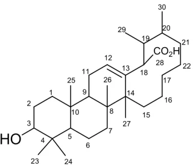
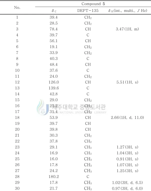
1-5. Compound 5의 구조해석 Compound 5는 1H-NMR spectrum 분석 결과 δ 3.47(1H, dd, J = 3.5, 6.5 Hz) signal로부터 sp3 혼성을 갖는 proton을 예상하였고, 전형적으로 1∼2 ppm대에 나타나는 proton보다 약간 deshielding 된 것으로 보아 전기음성도가 큰 산소가 치환되어 있을 것이라 예상하였다. 대부분 2 ppm이하에서 나오는 signal들은 aliphatic 탄소에 결합된 proton들 일 것이라 예상하였으며, 그 중 δ 1.27(3H, s), 1.25(3H, s), 1.07(3H, s), 1.04(3H, s), 1.02(3H, d, J = 6.5 Hz), 0.97(3H, d, J =
6.5 Hz), 0.91(3H, s)에서 총 7 개의 methyl proton signal을 확인하였다.
13C-NMR spectrum 분석 결과, δ 180.2의 carboxyl기의 carbon signal과 δ
139.6, 126.0은 olefin 구조의 carbon signal이라 예상하였다. 그리고 δ 78.4 signal 을 통해 전기음성도가 큰 원소가 인접하여 deshielding 된 methine carbon으로 1 개의 산소에 인접한 위치의 carbon이 있음을 예상하였다.
또한, 1H-NMR data와 마찬가지로 대부분의 signal이 δ 16.0, 16.9, 17.8, 17.8,
21.7, 24.2, 29.1에 분포하였으며 이 signal 또한 sp3혼성을 갖는 7개의 methyl기가
있음을 예상하였다.
13C-NMR data를 통해 화합물이 30개 이상의 탄소로 이루어졌음을 예상하였
고, DETP-135를 통해 6개의 4차 탄소, methine기가 7개, methylene이 10개 있음 을 확인하였다(figure 21). 1H-NMR, 13C-NMR, DEPT-135를 통해 예상한 부분구조의 정확한 구조 동정 을 위해 2D NMR을 분석하였다. HMQC를 통해 탄소에 직접 연결되어 있는 수 소를, HMBC를 통해서는 각 탄소에 대해 long-range결합을 하고 있는 수소를 확인하였다. 1D NMR과 2D NMR의 data를 기본으로 하여 문헌44)을 검색한 결 과 compound 5는 아래와 같은 구조로 자연계에서 처음 분리된 화합물일 것이라 예상하였다(figure 18-23).
HO
CO
2H
1 2 3 4 5 6 7 8 9 10 11 12 13 14 15 16 17 18 19 20 21 22 23 24 25 26 27 28 29 30No. Compound 5
δC DEPT-135 δH(int., multi., J Hz)
1 39.4 CH2 2 28.5 CH2 3 78.4 CH 3.47(1H, m) 4 39.7 C 5 56.1 CH 6 19.1 CH2 7 33.9 CH2 8 40.3 C 9 48.4 CH 10 37.6 C 11 24.0 CH2 12 126.0 CH 5.51(1H, s) 13 139.6 C 14 42.8 C 15 29.0 CH2 16 25.2 CH2 17 31.4 CH2 18 53.9 CH 2.66(1H, d, 11.0) 19 39.7 CH 20 39.8 CH 21 30.3 CH2 22 37.8 CH2 23 29.1 CH3 1.27(3H, s) 24 16.9 CH3 1.04(3H, s) 25 16.0 CH3 0.91(3H, s) 26 17.8 CH3 1.07(3H, s) 27 24.2 CH3 1.25(3H, s) 28 180.2 C 29 17.8 CH3 1.02(3H, d, 6.5) 30 21.7 CH3 0.97(3H, d, 6.0)
Table 6. 1H and 13C NMR data of compound 5 (500 and 125 MHz,
Figure 19. 1H-NMR spectrum of compound 5 in pyridine-d5.
Figure 20. 13C-NMR spectrum of compound 5 in pyridine-d
Figure 21. DEPT-135 spectrum of compound 5 in pyridine-d5.
y = 0.0008 x - 0.0070 R² = 0.9990 0.0 0.1 0.2 0.3 0.4 0.5 0 100 200 300 400 500 600 Ab so rb an ce
Gallic acid concentration (μg/mL)
Figure 24. Calibration curve for quantification of total phenolic compounds. 2. 항산화 활성 실험 결과 2-1. 총 polyphenolic compounds 함량 측정 Polyphenolic compounds는 식물계에 널리 분포되어 있는 2차 대사물의 하나로 다양한 구조와 분자량을 가지며 한 분자 내에 2개 이상의 phenolic hydroxyl기를 가지기 때문에 단백질 및 기타 거대 분자들과 결합하는 성질을 가지며 항암 및 항산화 효과와 같은 다양한 생리활성 기능을 가지는 것으로 알려져 있다.45)
Gallic acid 표준용액을 사용한 검정곡선을 작성하여(Figure 18) 병개암나무 가 지 70 % 에탄올 추출물 및 분획물의 총 polyphenolic compounds 함량을 측정하 였으며, 추출물 및 분획물의 중량 1 mg 당 함유하고 있는 gallic acid 의 양 (GAE)으로 환산하여 나타내었다. 실험 결과, 추출물에서 184 μg GAE/1 mg, 분
획물 중 EtOAc 및 n-BuOH layer에서 각각 425, 347 μg GAE/1 mg으로 비교적
높은 polyphenolic compounds 함량을 나타내었다(Figure 25). 이는 다른 항산화 실험 결과와도 관련이 있을 것이라 예상되었다.
0 20 40 60 80 100 120 DP PH ra dic al sc av en gin g a cti vit y ( %) 3.125 μg/mL 6.25 μg/mL 12.5 μg/mL 25 μg/mL 50 μg/mL 100 μg/mL
n-Hex layer EtOAc layer n-BuOH layer H2O layer
Extract Vit.C
Figure 26. DPPH radical scavenging activities of extract and solvent layers.
34.36 13.57 17.24 13.29 0 10 20 30 40 50
SC
50(μ
g/
m
L)
n-BuOH layer EtOAc layer Extract Vit.CFigure 27. SC50 values of DPPH radical scavenging activities of extract and
solvent layers.
2-2. 추출물 및 분획물의 DPPH radical 소거 활성
병개암나무 잎 70 % 에탄올 추출물 및 분획물의 DPPH radical 소거 활성을 측정하였다. 추출물과 각각의 분획물에 대하여 3.125, 6.25, 12.5, 25, 50, 100 μ
g/mL의 농도로 실험을 실시하여 SC50 값을 계산하였다.
그 결과, n-Hex layer, H2O layer를 제외하고, 추출물 34.36 μg/mL, EtOAc
layer 13.57 μg/mL, n-BuOH layer 17.24 μg/mL로 대조군인 vitamin C (13.29 μ
g/mL) 만큼의 비교적 좋은 DPPH radical 소거 활성을 나타내었다(Figure 26, 27).
0 20 40 60 80 100 120 AB TS ra di ca l c at io n sc av en gi ng a ct iv ity (% ) 3.125 μg/mL 6.25 μg/mL 12.5 μg/mL 25 μg/mL 50 μg/mL 100 μg/mL n-Hex layer EtOAc layer n-BuOH layer H2O layer
Extract Vit.C
Figure 28. ABTS radical cation scavenging activities of extract and solvent layers. 18.72 58.14 8.33 9.39 49.61 7.87 0 10 20 30 40 50 60 SC50 (μ g/ m L)
n-Hex layer EtOAc layer n-BuOH layer H2O layer
Extract Vit.C
Figure 29. SC50 values of ABTS radical cation scavenging activities of
2-3. 추출물 및 분획물의 ABTS radical cation 소거 활성
병개암나무 잎 70 % 에탄올 추출물 및 분획물의 ABTS radical cation 소거 활 성을 측정하였다. 추출물과 나머지 분획물에 대하여 3.125, 6.26, 12.5, 25, 50, 100
μg/mL의 농도로 실험을 실시하여 SC50 값을 계산하였다. 그 결과, 추출물에서
18.72 μg/mL, EtOAc, n-BuOH에서 각각 8.33, 9.39 μg/mL로 대조군인 vitamin
C (7.87 μg/mL)만큼 좋은 ABTS radical cation 소거 활성을 나타내었다(Figure 28, 29).
0 20 40 60 80 100 120 N O P ro d u ct ( % ) * sample concentration : 100 μg/mL * 2-amino-4-picoline : 10 μM LPS(-) LPS(+) 2-amino-4-picoline
n-Hex layer EtOAc layer n-BuOH layer H2O layer
Extract
Figure 30. NO production inhibitory activities of extract and solvent layers. 3. RAW 264.7 cell을 이용한 항염 활성 실험 결과
3-1. 추출물 및 분획물의 Nitric oxide 생성 억제 및 세포 독성 평가
RAW 264.7 cell을 이용하여 병개암나무 잎 70 % 에탄올 추출물 및 분획물에 대한 nitric oxide(NO) 생성 억제 및 세포독성을 측정하였다. 우선 sample 100 μ g/mL의 농도로 실험을 진행하여 활성이 있는지 확인하였다. 그 결과 LPS와 용 매만을 처리한 LPS(+)와 NO 생성량을 비교하였을 때, 70 % 에탄올 추출물과
n-BuOH layer에서는 각각 31.2 %, 34.4 %가 감소하였으며 n-Hex 및 EtOAc
layer에서 각각 67.5 %, 67.2 %로 큰 세포독성 없이 NO 생성 저해 활성을 나타내 었다(Figure 30, 31).
0 20 40 60 80 100 120 140 160 C e ll vi ab ili ty & c yt o to xi ci ty ( % ) * sample concentration : 100 μg/mL * 2-amino-4-picoline : 10 μM MTT LDH LPS(-) LPS(+) 2-amino-4-picoline n-Hex layer n-BuOH layer EtOAc layer H2O layer Extract
Figure 31. Cell viabilities and cytotoxicities of extract and solvent layers in RAW 264.7 cell. 0 20 40 60 80 100 120 N O P ro d u ct ( % ) * 2-amino-4-picoline : 10 μM LPS(-) LPS(+) 2-amino-4-picoline EtOAc layer 100 μg/mL EtOAc layer 50 μg/mL EtOAc layer 25 μg/mL
Figure 32. NO product inhibition activities of EtOAc layer.
위 결과를 바탕으로 100 μg/mL 농도에서 큰 세포독성 없이 좋은 활성을 보였 던 EtOAc layer에 대해 25, 50, 100 μg/mL의 농도로 실험을 더 진행하였다 (Figure 32, 33).
0 20 40 60 80 100 120 140 160 Ce ll v iab ilit y & cy to to xic ity (% ) * 2-amino-4-picoline : 10 μM MTT LDH LPS(-) LPS(+) 2-amino-4-picoline EtOAc layer 100 μg/mL EtOAc layer 50 μg/mL EtOAc layer 25 μg/mL
Figure 33. Cell viabilities and cytotoxicities of EtOAc layer in RAW 264.7 cell.
0 20 40 60 80 100 120 IL -6 Pr od uc t ( % ) * 2-amino-4-picoline : 10 μM LPS(-) LPS(+) 2-amino-4-picoline EtOAc layer 100 μg/mL EtOAc layer 50 μg/mL EtOAc layer 25 μg/mL
Figure 34. Inhibition of LPS-induced secretion of pro-inflammatory cytokine IL-6 of EtOAC layer.
그 결과 EtOAc layer에서 큰 세포독성 없이 25, 50, 100 μg/mL 농도에서 각각 23.8, 48.4, 67.3 %로 매우 좋은 NO 생성 저해 활성을 보이는 것을 확인하였다.
3-2. 염증성 cytokine IL-6 생성 억제 평가
초기 염증 반응 진행에 중요하게 작용하는 염증성 cytokine으로 알려진 IL-6의 생성량 측정 실험을 진행하였다. 먼저 병개암나무 잎 분획물 중 NO 저해 활성이
좋았던 n-Hex 및 EtOAc layer에 대하여 IL-6 양을 측정한 결과, LPS와 용매만
을 처리한 LPS(+)와 비교하였을 때 25, 50, 100 μg/mL 농도에서 각각 68.4, 58.4, 29.6 %로 IL-6 생성이 억제되는 것을 확인할 수 있었다(Figure 34).
0 20 40 60 80 100 120 M e la n in c o n te n ts ( % ) * sample concentration : 100 μg/mL * Melasolv : 20 μM
α-MSH(-) α-MSH(+) melasolv Extract n-Hex layer EtOAc layer n-BuOH layer H2O layer
Figure 35. melanin contents of extract and solvent layers. 4. B16F10 murine melanoma cell을 이용한 미백 활성 실험 결과
4-1. 추출물 및 분획물의 melanin 생성 억제 및 세포 독성 평가
B16F10 murine melanoma cell을 이용하여 병개암나무 잎 70 % 에탄올 추출물 및 분획물에 대한 melanin 생성 억제 활성 및 세포독성을 측정하였다. 우선 sample 100 μg/mL의 농도로 실험을 진행하여 활성이 있는지 확인하였다. 그 결 과 α-MSH와 용매만을 처리한 α-MSH(+)와 melanin 생성량을 비교하였을 때,
70 % 에탄올 추출물과 n-Hex, n-BuOH layer에서는 각각 65.0 %, 63.5 %, 58.2 %
가 감소하였다.
EtOAc layer에서는 79.2 %의 높은 저해율을 보였으나 세포 독성 실험에서 큰 독성이 확인 되었다. 이를 세포들이 죽었는지를 확인하기 위해 LDH 키트실험을 진행한 결과 세포들이 죽은 것이 아니라 생장억제 효과가 있었음을 알 수 있었 다(Figure 35, 36).
0 20 40 60 80 100 120 140 160 180 C e ll vi ab ili ty & c yt o to xi ci ty ( % ) * sample concentration : 100 μg/mL * Melasolv : 20 μM MTT LDH
α-MSH(-) α-MSH(+) melasolv n-Hex layer EtOAc layer n-BuOH layer
H2O layer Extract
Figure 36. Cell viabilities and cytotoxicities of extract and solvent layers in B16F10 murine melanoma cell.
0 20 40 60 80 100 120 M e la n in c o n te n ts ( % ) * melasolv : 20 μM α-MSH(-) α-MSH(+) melasolv EtOAc 50 μg/mL EtOAc 25 μg/mL EtOAc 12.5 μg/mL
Figure 37. Melanin contents of EtOAc layer.
위 결과를 바탕으로 100 μg/mL 농도에서 좋은 활성을 보였던 EtOAc layer에 대해 12.5, 25, 50 μg/mL의 농도로 실험을 더 진행하였다.
0 20 40 60 80 100 120 140 C e ll v ia b il it y & c y to to x ic it y ( % ) * melasolv : 20 μM MTT LDH α-MSH(-) α-MSH(+) melasolv EtOAc 50 μg/mL EtOAc 25 μg/mL EtOAc 12.5 μg/mL
Figure 38. Cell viabilities and cytotoxicities of EtOAc layer in B16F10 murine melanoma cell.
그 결과 EtOAc layer 12.5, 25, 50 μg/mL의 농도에서 각각 76.9 %, 47.5 %, 20.0 % melanin 생성을 저해함을 알 수 있었으며, 특히 25 μg/mL의 농도에서 세포독 성 그리고 세포생장 효과 없이 좋은 melanin 생성을 저해 하는 것을 확인하였다 (Figure 37, 38).
0 20 40 60 80 100 120 In tr ac e llu la r ty ro si n as e a ct iv it y (% ) * melasolv : 20 μM α-MSH(-) α-MSH(+) melasolv EtOAc 50 μg/mL EtOAc 25 μg/mL EtOAc 12.5 μg/mL
Figure 39. Intracellular tyrosinase inhibition of EtOAc layer. 4-2. Intracellular tyrosinase 측정 결과
B16F10 murine melanoma cell을 이용한 melanin contents 생성량과 세포독성 에서 좋은 활성을 보였던 EtOAc layer에 대해 intracellular tyrosinase 저해활성 을 확인한 결과 12.5, 25, 50 μg/mL의 농도에서 각각 77.8 %, 45.4 %, 6.7 %로 intracellular tyrosinase를 저해함을 알 수 있었으며, melanin contents와 마찬가 지로 EtOAc layer 25 μg/mL 농도에서 좋은 intracellular tyrosinase 저해 활성을 확인하였다(Figure 39).
IV. 요약 및 결론
본 논문은 제주도 한라산에 서식하는 제주 특산종인 병개암나무(Corylus hallaisanensis) 잎을 추출하여 그 추출물로부터 화합물들을 분리하고, 분리된 화 합물에 대해 항산화, 항염 및 미백 활성에 대해 천연 소재로 활용 가능성을 알아 보고자 연구를 진행하였다. 병개암나무 잎 70 % 에탄올 추출물을 용매의 극성순서에 따라 순차적으로 분획하여 n-hexane, ethyl acetate, n-butanol, water layer를 얻었다. 추출물과 각
분획물들에 대해 항산화, 항염 및 미백 활성을 진행하였다.
추출물 및 분획물들의 항산화 활성실험 결과, 총 폴리페놀 함량 측정에서는
EtOAc, n-BuOH layer에서 425 μg, 347 μg GAE/1mg 가장 높게 나왔다. DPPH
라디칼 소거능 실험에서는 n-Hex, H2O layer를 제외한 나머지 70 % 에탄올 추
출물, EtOAc, n-BuOH layer에서 positive control인 vitamin C (SC50 13.3 μ
g/mL)와 비교했을 때 각각 34.4, 13.6, 17.2 μg/mL로 좋은 라디칼 소거 활성을 보였다. 또한, 추출물 및 분획물의 ABTS radical cation 소거 활성결과, 70 % 에
탄올 추출물에서 18.7 μg/mL, EtOAc, n-BuOH layer에서 각각 8.3, 9.4 μg/mL로
대조군인 vitamin C (7.9 μg/mL)만큼 좋은 ABTS radical cation 소거 활성을 나 타내었다.
추출물 및 분획물들의 항염 활성실험 결과는 NO 생성량을 비교하여 측정하였
는데, n-Hex, EtOAc layer에서 용매만을 처리한 LPS(+)와 비교하였을 때 세포
독성 없이 농도 의존적으로 NO의 생성량을 저해시키는 것을 확인 할 수 있었다. Cytokine 측정 실험에서도 EtOAc layer에서 IL-6의 양을 농도 의존적으로 저해 시켰다.
그리고 추출물 및 분획물들의 미백 활성실험 결과는 melanin 생성량을 비교하
여 측정하였는데, H2O layer를 제외하고 용매만을 처리한 α-MSH(+)와 비교하였
알 수 있었으며, 추가적으로 EtOAc를 농도별로 실험한 결과 25 μg/mL에서 세포 독성 그리고 세포생장억제 없이 미백활성이 나타남을 알 수 있었다.
이 같은 결과를 바탕으로 EtOAc layer에서 화합물 분리를 진행하였고, ODS (C-18) column을 이용한 medium pressure liquid chromatography (MPLC), 순 상 silica gel chromatography, sephadex LH-20 chromatography를 수행하여 화 합물을 분리하였다. 분리된 화합물의 구조는 NMR을 이용하여 확인하였고, 문헌 과 비교하여 총 4개의 화합물을 분리 동정하였다. 분리된 화합물은 gallic acid
(1), kaempferol 3-O-β-D-xylopyranoside (2), kaempferol 3-O
-α-L-rhamnpy-ranoside (3), apigenin (4)로 확인되었다.
위의 분리된 화합물 중 compound 146-49)과 compound 450-54)은 문헌조사를 통
하여 항산화, 항염 및 미백 활성 연구가 되어있는 것을 확인하였다.
본 연구의 실험결과를 바탕으로 병개암나무 잎을 이용한 천연 항산화, 항염 및 미백 활성을 갖는 화장품 소재로서의 개발 가능성을 확인할 수 있었다.
V. 참고문헌
1) Aniya, Y.; Naito, A. “Oxidative stress-induced activation of microsomal
glutathione S-transferase in isolated rat liver.“ Biochem Pharmacol, 1993,
45(1), 37-42
2) Haber, F.; Weiss, J. “The catalytic decomposition of hydrogen peroxide by
salt“ Proc. Roy. Soc. Ser. A. 1984, 147, 332-351
3) Packer, L. New comprehensive biochemistry, In CA. Rice-Evans & RH.
Burdon (Eds.). “Free radical damage and its control“ Elsevier Science,
Amsterdam. 1994, 239
4) Choe, S. Y.; Yang, K. H. “Toxicological studies of antioxidants, butylated
hydroxytoluene (BHT) and butylated hydroxyanisole (BHA)“ Korean J. Food
Sci. Technol., 1982, 12, 283
5) Cort, W. M. “Antioxidant activity of tocopherols, ascorbyl palmitate, and
ascorbic acid and their mode of action.“ J. Am. Oil Chem. Soc. 1974, 51,
321-325
6) Masaki, H.,; Sakaki, S.,; Atsumi, T.,; Sakurai, H. “Active-oxygen
scavenging activity of plant extracts.“ Biol. Pharm. Bull. 1995, 18: 162-166
7) Kim, K. B.,; Yoo, K. H.,; Park, H. Y.,; Jeong, J. M. “Anti-oxidative
activities of commercial edible plant extracts distributed in Korea.“ J . Korean
8) Masaki, H.,; Sakaki, S.,; Atsumi, T.,; Sakurai, H. “Activeoxygen scavenging
activity of plants extracts.“ Biol. Pharm. Bull, 1995, 18, 162-166
9) Storck, M.,; Schilling, M.,; Burkhardt, K.,; Prestel, R.,; Abendroth, D.,; Hammet, C. “Production of proinflammatory cytokines and adhesion molecules
in ex-vivo xenogeneic kidney perfusion.“ Transpl. Int. 1994, 7(suppll),
647-649
10) Willoughby, D. A. “Human arthritis applied to animal models towards a
better therapy” Annals of the Rheumatic Diseases, 1971, 34, 471-478
11) Moncada, S.,; Palmer, R. M.,; Higgs, E. A. “Nitric oxide: physiology,
pathophysiology, and pharmacology.“ Pharmacol. Rev., 1991, 43, 109-142
12) Weisz, A.,; Cicatiello, L.,; Esumi, H. “Regulation of the mouse inducible-type nitric oxide synthase gene promoter by interferon-γ, bacterial
lipopolysaccharide, and NG-monomethyl-L-arginine.“ Biochem. J., 1996, 316,
209-215
13) Ryu, J. H.,; Ahn, H.; Kim, J. Y.,; Kim, Y. K. “Inhibitory activity of plant
extracts on nitric oxide synthesis in LPS-activated macrophage.“ Phytother.
Res, 2003, 17, 485-489
14) Mu, M. M.,; Chakravortty, D.,; Sugiyama, T.,; Koide, N.,; Takahashi, K.,; Mori, I.,; Yoshida, T.,; Yokochi, T. “The inhibitory action of quercetin on lipopolysaccharide-induced nitric oxide production in RAW 264.7 macrophage
15) Yoon, W. J.; Ham, Y. M.; Kim, S. S.; Yoo, B. S.; Moon, J. Y.; Baik, J. S.; Lee, N. H.; Hyun, C. G. “Suppression of pro-inflammatory cytokines, iNOS, and COX-2 expression by brown algae Sargassum micracanthum in
RAW 264.7 macrophages, EurAsia J. BioSci., 2009, 3, 130-143
16) Aktan, F. “iNOS-mediated nitric oxide production and its regulation,” Life
Sciences, 2004, 75, 639-653
17) Farlic, M.; Reutterer, B.; Schindler, C.; Greten, F.; Vogl, C.; Muller, M.; Decker, T. “Conventional initiation complex assembly by STAT and NF-κB
transcription factors regulates nitric oxide synthase expression, Immunity,
2010, 33, 25-34
18) Akira, S.; Takeda, K. “Toll-like receptor signalling.” Nat. Rev. Immunol.,
2004, 4, 499-511
19) Horwood, N. J.; Page, T. H.; McDaid, J. P.; Palmer, C. D.; Campbell, J.; Mahon, T.; Brennan, F. M.; Webster, D.; Foxwell, B. N. “Bruton`s tyrosine kinase is required for TLR2 and TLR4-induced TNF, but not IL-6,
production.” J. Immunol., 2006, 176(6), 3635-3641
20) Hirohashi, N.; Morrison, D. C. “Low-dose Lipopolysaccharid(LPS) pretreatment of mouse macrophage modulates LPS-dependent interleukin-6
production in vitro.” Infect. Immun, 1996, 64(3), 1011
21) Shih, M. F.; Cheng, Y. D.; Shen, C. R.; Cherng, J. Y. “A molecular
pharmacology study into the anti-inflammatory actions of Euphorbia hirta L.0
22) Tsatsanis, C.; Androulidaki, A.; Venihaki, M. Margioris, A. N. “Signalling
networks regulating cyclooxygenase-2.“ Int. J. Biochem. Cell Biol, 2006, 38,
1654-1661
23) Kang, S. R.; Park, K. I.; Park, H. S.; Lee, D. H.; Kim, J. A.; Nagappan, A.; Kim, E. H.; Lee, W. S.; Shin, S. C.; Park, M. K.; Han, D. Y.; Kim, G. S.; “Anti-inflammatory effect of flavonoids isolated from Korea Citrus aurantium L. on lipopolysaccharide-induced mouse macrophage RAW 264.7 cells by blocking of nuclear factor-kappa B (NF-κB) and mitogen-activated protein
kinase (MAPK) signalling pathways.“ Food Chem, 2011, 129, 1721-1728
24) 최두영,; 안소영,; 이승기,; 한정선,; 김은철,; 이향복,; 신정현,; 김은기,; 노경호,
“홍경천에 포함된 미백성분의 분리 및 성능검사” korean J. Biotechnol. Bioeng.,
2004, 19(3), 169-173
25) Lerner, A. B.; Fitzpatrick, T. B. “Biochemistry of melanin formation.“
Physiological reviews, 1950, 30, 91-126
26) Swan, G. A. “Structure, chemistry, and biosynthesis of the melanins.“
Fortschritte Chemie. Organischer Naturstoffe, 1974, 31, 521-582
27) Korner, A.; Pawelek, J. “Mammalian tyrosinase catalyzes three reaction in
the biosynthesis of melanin.“ Science, 1982, 217, 1163-1165
28) 김은철,; 리광화,; 안소영,; 김은기,; 노경호, “중국산 천연물에서 미백성분의
29) 김다슬,; 김동현,; 오명진,; 이광근,; 국무창,; 박장서, “유자 부산물 에탄올 추
출물의 항노화 및 미백효과” J . Soc. Cosmet. Scientists Korea, 2010, 36(2),
137-143
30) Hori, I.; Nihei, K.; Kubo, I. “Structural criteria for depigmenting
mechanism of arbutin.” Phytother. Res., 2004, 18, 475-479
31) Canabas, J.; Chazarra, S.; Garcia-Carmona, F. “Kojic acid, a cosmetic skin whitening agent, is a slow-binding inhibitor of catecholase activity of
tyrosinase.“ J. Pharm. Pharmacol., 1994, 46, 982-985
32) Ikeda, T.; Tsutsumi, T. “Function and skin depigmental activity of crude
drugs.“ Fragrance J ., 1990, 18, 59-66
33) Elsner, P.; Maibach. H. I. “Cosmeceuticals: Drugs vs. Cosmetics.” Marcel
Dekker, Inc., 2000, 123-145.
34) Jang, D. I.; Lee, B. G.; Jeon, C. O.; Jo, N. S.; Park, J. H.; Cho, S. Y.;
Lee, H.; Koh, J. S. “Melanogenesis inhibitor from paper mulberry.“ Cosme. &
Toilet., 1997, 112, 59-62
35) Lee, S. H.; Park, J. S.; Kim, S. Y.; Kim, J. J.; Chung S. R. “The screening of the inhibitory compounds on tyrosinase activity for the natural
product“. Yakhak Haeji, 1997, 41, 456-461
36) 한국기준식물도감 “http://terms.naver.com/entry.nhn?docId=772214&cid =2694&categoryId=2699”
37) 김성천 “Identification of Anti-oxidative and Anti-inflammatory
Constituents from the Branches of Corylus hallaisanensis.” 제주국립대학교 대
학원 석사학위논문, 2014, 19-31
38) Folin, O.; Denis, W. “A colorimetric method for determination of
phenols(phenol dericatives) in urine” J. Biol. Chem., 1915, 22, 305-308
39) Blois, M. S. "Antioxidant determination by the use of a stable free
radical" Nature, 1958, 181, 1199-1200
40) Re, R.; Pellegrini, N.; Proteggente, A.; Pannala, A.; Yang, M.; Rice-Evans, C. "Antioxidant activity applying an improved ABTS radical cation
decolorzation assay" Free Radical Biology and Medicine, 1999, 26, 1231-1237
41) Zhijun, Liu1; Joshua, Schwimer; Dong, Liu; James, Lewis; Frank, L.; reenway,; David, A.; York and Eugene A.; oltering, W “Gallic Acid is Partially esponsible for the Antiangiogenic Activities of Rubus Leaf Extract.”
Phytother. Res., 2006, 20, 806-813
42) Monika, O.; Maria, W. “FLAVONOIDS FROM THE FLOWERS OF
PRUNUS SPINOSA l.” Acta Poloniae Pharmaceutica-Drug Reserch, 2001,
58(5) 367-372
43) 고하나 “Identification of Anti-oxidative and Anti-inflammatory
Constituents from the Branches of Carpinus turczaninowii.” 제주국립대학교 대