Gastric involvement of syphilis is rare manifestation of secondary and tertiary syphilis1). It has uncertain clinical, endoscopic, pathologic finding. And most of existing diagnostic tools were not so sensitive. So, diagnosis of gastric syphilis is very difficult.
Recently syphilis had increasing tendency, and related to human immunodeficiency virus (HIV) infection. So, gastric syphilis may become a more frequent clinical challenge2).
The diagnosis of gastric syphilis is depended on histology till now, in that, spirochetes organisms could be found. However in case of biopsy negative gastric syphilis, it is very difficult to diagnose the disease.
In these days, polymerase chain reaction (PCR) methods are widely used to diagnose several specific infectious
diseases such as tuberculosis, cytomegalovirus (CMV) in the endoscopic field. Likewise, PCR methods could be applied to diagnose gastric syphilis.
We reported a case of gastric syphilis diagnosed by PCR in the case of biopsy negative patient.
A 70-year-old man present with epigastric discomfort and anorexia for three weeks. He visited local clinic and underwent endoscopic examination with gastric biopsy. The endoscopic finding revealed diffuse fold thickening and multiple ulcers. Their pathologist reported only atypical cells and chronic inflammation with suggestion of lymphoepitheiliar lesion. Test for Helicobacter pylori was positive. For rule out mucosa associated lymphoid tissue (MALT) lymphoma and other malignancy condition, he was transferred to our hospital. He had mild epigastric tenderness. His blood pressure was 105/52 mmHg and heart rate was 72 beat/min. Body temperature was 36.7℃. Laboratory profiles were hemoglobin 11.5 g/dL, platelet 138,000 /mm3, albumin
PCR 방법으로 진단된 위매독 환자 1예
조재민, 김흥업, 허상택
제주대학교 의학전문대학원 내과학교실
(Received April 15, 2014; Revised April 22, 2014; Accepted April 29, 2014)
A Case of Gastric Syphilis Diagnosed by PCR Test: A Case Report
Jaemin Jo, Heung Up Kim, Sang Taek Heo
Department of Internal Medicine1, Pathology2, Jeju National University School of Medicine, Jeju, Korea
Syphilis is chronic and systemic inflammation caused by T. pallidum infection. Because the diagnosis is exclusively depended on histology, its diagnosis could be very difficult in the patients without positive biopsy finding. We experienced a case of gastric syphilis diagnosed by polymerase chain reaction (PCR) in case of biopsy negative patients. So we report this simple method to diagnose gastric syphilis that failed to show spirochetes on histology. A 70 year-old man visited our hospital via private clinic with suspicious gastric malignancy finding on gastroscopy. Our gastroscopy showed the characteristic shape of multiple nodular erosions and ulcers of gastric syphilis. Warthin-Starry silver stain failed to find spirochetes. Repeated gastroscopy was done to take biopsy specimen for T. pallidum PCR and proved positive. We confirmed gastric syphilis and treated with intravenous antibiotics. After 4 months of treatment, all gastric lesions were disappeared.(J Med Life Sci 2014;11(1):43-47)
Key Words
: Syphilis, Gastric; Polymerase Chain Reaction; DiagnosisIntroduction
Correspondence to : Heung Up Kim
Department of Internal Medicine, Jeju National University Hospital, 1753-3, Ara-1-dong, Jeju-Si, Jeju Special Self-Governing Province, 690-716, Korea
E-mail : [email protected]
Abstract
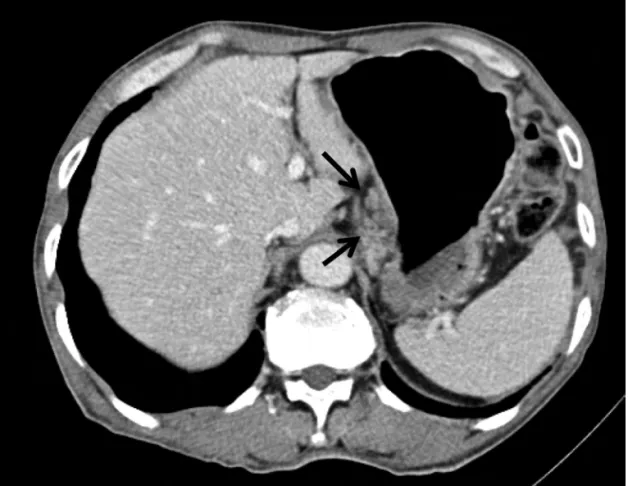
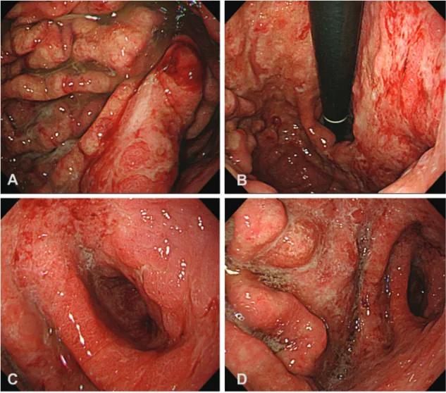
2.7 g/dL and high sensitive C-reactive protein 3.76 mg/dL. No stool occult blood was detected. Plain films of chest and abdomen were normal. Abdominal computed tomography showed diffuse gastric wall thickening and multiple perigastric lymph node enlargements (Fig. 1). Gastroscopic examination showed diffuse gastric wall thickening with multiple ulcers and erosions that were scattered in body and fundus and showed decreased air distensibility (Fig. 2). A Colonoscopic examination was normal. Biopsy showed erosions, chronic active inflammation with dense plasmocytic and neutrophilic infiltration, granuloma formation, glandular destruction (Fig. 3). No endarteritis obliterans were observed. Neither spirochete nor H. pylori were found even after Warthin-Starry silver stain. Serum rapid plasma regain (RPR) test was weakly positive (2.0 RPR unit). Further history taking was revealed his sexual contact history with prostitutes. Erythematous macules on the sole (Fig. 4A) and scrotal ulcers (Fig. 4B) were found on repeated physical
examination. The skin lesion of his sole and scrotum were corresponded to early secondary syphilis. We suspected the possibility of gastric syphilis based on his history, physical examination and endoscopic findings. Repeated gastroscopy to get a tissue specimen for real time PCR specific for pathogenic treponemes detection was done. The PCR (SeeplexⓇ STD4 ACE Detection; Seegene, Seoul, South Korea) was performed with newly acquired gastric specimen and finally proved T. pallidum. Tuberculosis and CMV PCR were also performed and proved negative. At this time, positive Treponema pallidum hemagglutination assay (TPHA) was also identified (318.47 titer unit). The patient was treated with ceftriaxone because he experienced penicillin shock after first injection of penicillin. Patient’s symptoms were resolved promptly during treatment. After 4 months, all gastric lesions were nearly disappreared on follow up gastroscopy (Fig. 5).
Figure 1. The abdominal computed tomography showed diffuse gastric wall thickening with
Figure 2. Gastroscopy showed multiple ulcers and erosions with diffuse fold thickening (A). The
mucosal lesion involved from cardia (B) to pyloric ring (C). Severe shortening of antrum was also noted.
Figure 3. Biopsy specimens revealed chronic inflammation with dense neutrophilic and plasmacytic
infiltration. Some focal glandular destructions (A) and granulation (B) were seen (Hematoxylin and eosin stain was used. Magnification, x200.)
Gastric syphilis is uncommon manifestation of secondary syphilis and it was first reported in 1834 by Andral3). Because of considerable overlap between stages of syphilis, it could be diagnosed at all stages of syphilis. Gastric syphilis most commonly affects young adults and have nonspecific symptoms including nausea, epigastric discomfort and weight loss. Gastrointestinal bleeding and obstruction were also reported1).
Gastric syphilis may exhibit variety of endoscopic and
radiologic appearance. Radiologic finding include fibrotic narrowing, rigidity of gastric wall, hypertrophic mucosal fold, nodule and mass lesion. Endoscopic finding include multiple ulceration, nodular mucosa, thickened fold, narrowing and mass. In some cases, characteristic shape of multiple ulcers and erosions with whitish exudate could be found1). Hypertrophic folds and nodular mucosa were more common in early disease, while fibrotic narrowing and rigidity were more common in late disease1).
Common histological finding include diffuse chronic inflammation with dense lymphocytic and especially Figure 4. Some erythematous macules were noted on his sole. It was typical finding of secondary syphilis. (A)
Painless and well marginated shallow ulcers with raised redish border were noted on his scrotum. (B)
Figure 5. Follow up gastroscopy showed mild mucosal atrophy with nearly healed mucosal lesions (A) and
improvement of fold thickening (B)
plasmacytic infiltration. Some represent granulomas and endarteritis obliterans4). Presence of abruptly end of microscopic lesion at the pyloric level and proliferative endarteritis obliterans should suggest the possibility of gastric syphilis4,5).
Diagnosis of gastric syphilis is commonly achieved based on combination of endoscopic, histologic findings and positive serology in patients at risk of sexually transmitted disease. Demonstration of T. pallidum in biopsy specimen provides confirmatory diagnosis.
Dark field microscopy test is simple and reliable test. However this method require fresh unfixed specimen and have low sensitivity.
Warthin-Starry silver stain is capable of demonstrating spirochetes and can be applied on fixed specimen. However detection of spirochetes by this method is difficult in a background of elastic and reticular fibers6).
Recently, Imunofluorescence staining method has been used to detect treponemes. Because of crossreactivity with other spirochets such as Borrelia burgdorferi was reported in initial methods7), monoclonal antibodies directed against the T. pallidum antigen were introduced and it has high specificity and sensitivity8). But this method has variable sensitivities and requires more experienced microscopist9).
More recently PCR method was used to detect T. pallidum6,10). PCR is considered more specific and sensitive than other traditional methods and immunofluorescence staining. (sensitive ranging from 94.7% in early disease to 80% in secondary stage)11). In addition, this method can achieve diagnosis earlier than immunofluorescence staining10).
In our case, we failed to prove spirochetes in Warthin-Starry silver stain but granulations were observed. Finally, we proved T. pallidum by PCR method with more high sensitivity. Prompt diagnosis resulted in successful treatment and reduced further unnecessary test and intervention. He had H.pylori in private clinic. But that was not found repeated endoscopic biopsy in our hospital. The patient said he took medication shortly though we failed to list the medication. So there was possibility he had take a kind of antibiotics or anti ulcer medication and that could be the cause of failed to find H. pylorior spirochetes on histology. In conclusion, gastric syphilis is very difficult to diagnosis, so it should be considered in a patients at risk of sexually transmitted disease who presented with non specific abdominal symptoms, unusual endoscopic finding unresponsive to standard anti-ulcer therapy. The endoscopic and histologic findings were relatively specific. So when such findings were observed, we must keep the possibility of
gastric syphilis in mind. Immediate use of PCR method for detect T. pallidum may be considerable in this condition and may be used to making definite diagnosis. Therefore, we cautiously suggest the use of T. pallidumPCR immediately when gastric syphilis was suspected on clinical and gastroscopic finding.
The authors have no financial conflicts of interest.
1) Mylona EE, Baraboutis IG, Papastamopoulos V, et al. Gastric syphilis: a systematic review of published cases of the last 50 years. Sex Transm Dis 2010;37:177-183. 2) Kim J, Kim WH, Cho C, et al. Evaluation of automated
architect syphilis TP as a diagnostic laboratory screening test for syphilis. Korean J Lab Med 2008;28:475-482. 3) Monod G. Syphilis of the Stomach. Proc R Soc Med
1922;15:1.
4) Kolb JC, Woodward LA. Gastric syphilis. Am J Emerg Med 1997;15:164-166.
5) Massironi S, Carmagnola S, Penagini R, Conte D. Gastric involvement in a patient with secondary syphilis. Dig Liver Dis 2005;37:368-371.
6) Inagaki H, Kawai T, Miyata M, Nagaya S, Tateyama H, Eimoto T. Gastric syphilis: polymerase chain reaction detection of treponemal DNA in pseudolymphomatous lesions. Hum Pathol 1996;27:761-765.
7) Magnarelli LA, Anderson JF, Johnson RC. Cross-reactivity in serological tests for Lyme disease and other spirochetal infections. J Infect Dis 1987;156:183-188. 8) Ito F, Hunter E, George R, Pope V, Larsen S. Specific
immunofluorescent staining of pathogenic treponemes with a monoclonal antibody. J Clin Microbiol 1992;30:831-838.
9) Jethwa HS, Schmitz JL, Dallabetta G, et al. Comparison of molecular and microscopic techniques for detection of Treponema pallidum in genital ulcers. J Clin Microbiol 1995;33:180-183.
10) Chen CY, Chi KH, George RW, et al. Diagnosis of gastric syphilis by direct immunofluorescence staining and real-time PCR testing. J Clin Microbiol 2006;44:3452-3456.
11) Palmer H, Higgins S, Herring A, Kingston M. Use of PCR in the diagnosis of early syphilis in the United Kingdom. Sex Transm Infect 2003;79:479-483.