I. INTRODUCTION
Behçet’s disease(BD) was named after the Turkish dermatologist Hulusi Behçet who first described the triple symptom-complex of recurrent oral aphthous ulcers, genital ulcers, and uveitis(Behçet, 1937). This complex, multisystemic disease includes involvement of the mucocutaneous, ocular, cardiovascular, renal, gastrointestinal, pulmonary, urologic, and central nervous systems as well as the joints, blood vessels, and lungs(Bøe et al, 1958; O’Duffy et al, 1971; Chajek et al, 1975).Although many studies have been conducted on the pathogenesis of the disease, exact mechanisms have not yet been fully understood. Until today, genetic, environmental, virological, bacterial and immunological factors have been discussed in the etiopathogenesis of the BD (O’Duffy, 1978; Griffin et al, 1982). The gastrointestinal(GI) tract is not infrequently involved in patients with BD. The frequency of GI involvement varies in different countries: there are several studies, mostly from Japan, reporting a high frequency(50-60%) of GI involvement in BD(Shimizu et al, 1979; Oshima et al, 1963), while a significant lower rate, less than 5%, is seen in Turkish patients(Yurdakul et al, 1996). GI involvement has been reported in 8% of patients in the United States(Balabanova et al, 1999). Any portion of the GI tract may be involved. The upper GI tract is less frequently affected, but ulcerations in the esophagus and stomach have been reported(Benamour, 1990). These patients commonly present with symptoms of hematemesis, melena, and epigastric pain.
The terminal ileum, cecum, and ascending colon are the sites more frequently affected, resulting in abdominal pain, diarrhea, and hematemesis.
Recurrent aphthous ulcer(RAU) represent a very common but poorly understood mucosal disorder. They occur in men and women of all ages, races and geographic regions. It is estimated that at least 1 in 5 individuals has at least once been afflicted with aphthous ulcers(Natah et al, 2004). The condition is classified as minor, major, and herpetiform on the basis of ulcer size and number. Attacks may be precipitated by local trauma, stress, food intake, drugs, hormonal changes and vitamin and trace element deficiencies(Natah et al, 2004). Local and systemic conditions, and genetic, immunological and microbial factors all may play a role in the pathogenesis of recurrent aphthous ulceration . However, to date, no principal cause has been discovered. Since the etiology is unknown, diagnosis is entirely based on history and clinical criteria and no laboratory procedures exist to confirm the diagnosis. Although RAU may be a marker of an underlying systemic illness such as celiac disease, or may present as one of the features of BD, in most cases no additional body systems are affected, and patients remain otherwise fit and well.
We often see patients of RAU with GI symptoms without any other symptoms of BD in out patient department. In many cases it is difficult to distinguish intestinal BD from RAU with gastrointestinal symptom. Because the pathognomic clinical features and tools are absent, the diagnosis of this two diseases relies on the characteristic clinical features and the judgement of the experienced physician.
Kim et al have reported that Anti-Saccharomyces cerevisiae Mannan antibody (ASCA) may be associated with Crohn’s disease and BD, whereas perinuclear antineutrophil cytoplasmic autoantibody (pANCA) with ulcerative colitis(Kim et al, 2002). A combination of both tests may aid the differential diagnosis of inflammatory bowel disease. Recently, Krause et al have reported the significantly higher ASCA values in BD patients compared with patients with RAU, Systemic lupus erythematosus or in healthy volunteers imply that ASCA, taken in appropriate clinical context, could become a useful diagnostic tool for BD(Krause et al, 2002).
The aim of this study was to help the differential diagnosis of intestinal BD and RAU with gastrointestinal symptoms through the comparison of clinical, endoscopic, histopathologic, and serologic findings.
II. MATERIALS AND METHODS
A. Subjects
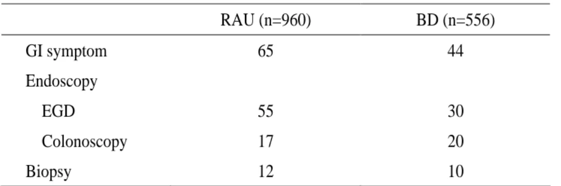
Sixty five of total 960 RAU patients and 44 of total 556 BD patients who visited the Depatment of Dermatology of Ajou University School of Medicine, Suwon, Korea, between January 1996 and December 2003 were invited to participate in this study and all of them had GI symptoms and were evaluated with esophagoduodenoscopy and colonoscopy (Table 1.). Sera were obtained from 12 patients with RAU, 16 patients with BD, 4 patients with healthy volunteers.
Table 1. Baseline data of the study group.
RAU (n=960) BD (n=556) GI symptom 65 44 Endoscopy EGD 55 30 Colonoscopy 17 20 Biopsy 12 10
RAU, recurrent aphthous ulcer; BD, Behçet’s disease GI, gastrointestinal; EGD, esophagogastroduodenosccopy
1. Clinical findings
The clinical records of these patients were reviewed. BD was diagnosed according to both the Japanese diagnostic criteria for BD revised in 1987 and to the International Study Group for Behçet’s Disease(ISGBG) criteria in 1990. We found out the patients of RAU didn’t have any other symptoms of BD. Review contents were age, sex, age of onset, first manifestation, gastrointesinal symptoms, other symptoms of BD.
2. Endoscopic findings
We investigated the endoscopic findings with respect to location, distribution, and morphology of the ulceration.
3. Histopathologic findings
A part of each specimen was fixed in 10% buffered neutral formalin and was embedded in paraffin. Sections were deparaffinized and rehydrated by sequential immersion in xylene, graded concentration of ethanol, and distilled water. Hematoxylin-eosin stain was done to observe the general histological changes.
We investigated all specimens with respect to presence of vasculitis and granuloma, infiltration type, dominant cell type and crypt architecture.
4. Serologic findings
(1) ASCA enzyme linked immunosorbent assay (ELISA)
Serum was obtained from 12 patients with RAU, 12 patients with BD, 4 patients with healthy volunteers. Each serum was collected in a sterile tube with EDTA coating. Then they were centrifuged at 4 and at 3000 rpm for 5 minutes. The s℃ erum was kept at -20 until the ℃ assay.
ASCA immunoglobin G (IgG) was evaluated in commercially available enzyme-linked immunosorbent assay (ELISA) kit (Inova Diagnostics, San Diego, California). ASCA ELISA was performed according to the manufacture’s manuals. The antigen consisted of phosphopeptidomannan extracted from Saccharomyces cerevisiae. All sera were tested in duplicate. Results were expressed as arbitrary units with a cut-off for positivity of 25 U/ml.
(2) pANCA indirect Immunofluorescence Assay
The ANCA indirect immunofluorescence assay was performed according to the following methods. In short, human peripheral blood neutrophils were smeared on 12-well Nutacon slides (Nutacon BV, Leimuiden, the Netherlands) and fixed in 96% ethanol (10 min at -20℃). Slides were incubated with 1 : 20 diluted patient’s serum (in phosphate buffered saline) and stained with rabbit anti-human IgG–fluorescein isothiocyanate (FITC) conjugate (Dako A/S, Glostrup, Denmark). The slides were evaluated by fluorescence microscopy. All sera were
tested in duplicate and scored by two well-trained observers who were unaware of the patients’ diagnoses. Depending on the brightness of the immunofluorescence staining pattern, the reactions were graded into negative (-), positive (+).
5. Statistics
Statistical analysis was done by Wilcoxon rank sum test and logistic regression. A p value of less than 0.05 was considered as statistically significant.
III. RESULTS
A. Clinical findings
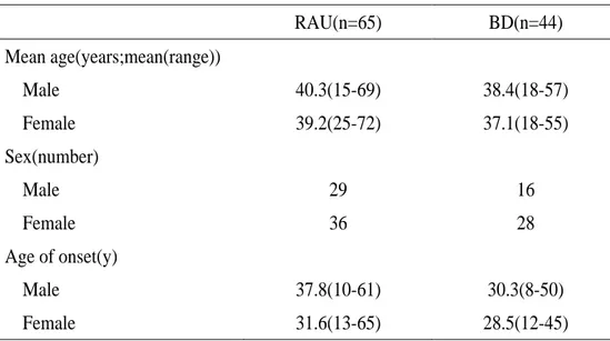
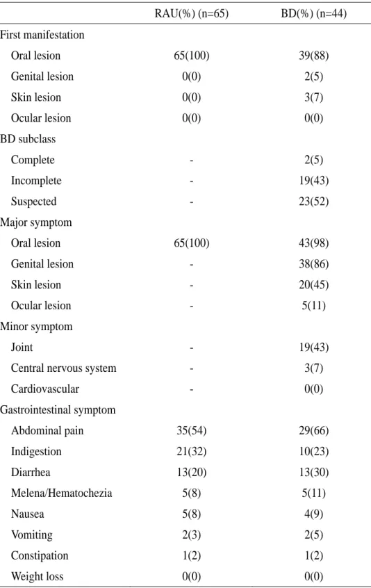
There were no significant differences between patients with RAU and BD by statistically analysis(Table 2.). Oral lesion was most common first manifestation of BD patients. Abdominal pain was most common gastrointestinal symptom of both group(Table 3.).
Table 2. Clinical data of the study group.
RAU(n=65) BD(n=44) Mean age(years;mean(range)) Male 40.3(15-69) 38.4(18-57) Female 39.2(25-72) 37.1(18-55) Sex(number) Male 29 16 Female 36 28 Age of onset(y) Male 37.8(10-61) 30.3(8-50) Female 31.6(13-65) 28.5(12-45)
Table 3. Clinical characteristic of patients with RAU and BD. RAU(%) (n=65) BD(%) (n=44) First manifestation Oral lesion 65(100) 39(88) Genital lesion 0(0) 2(5) Skin lesion 0(0) 3(7) Ocular lesion 0(0) 0(0) BD subclass Complete - 2(5) Incomplete - 19(43) Suspected - 23(52) Major symptom Oral lesion 65(100) 43(98) Genital lesion - 38(86) Skin lesion - 20(45) Ocular lesion - 5(11) Minor symptom Joint - 19(43)
Central nervous system - 3(7)
Cardiovascular - 0(0) Gastrointestinal symptom Abdominal pain 35(54) 29(66) Indigestion 21(32) 10(23) Diarrhea 13(20) 13(30) Melena/Hematochezia 5(8) 5(11) Nausea 5(8) 4(9) Vomiting 2(3) 2(5) Constipation 1(2) 1(2)
RAU, recurrent aphthous ulcer; BD, Behçet’s disease
Fig. 1. An aphthous ulcers on tongue in recurrent aphthous ulcer (A) and buccal mucosa in Behçet’s disease (B).
B. Endoscopic findings
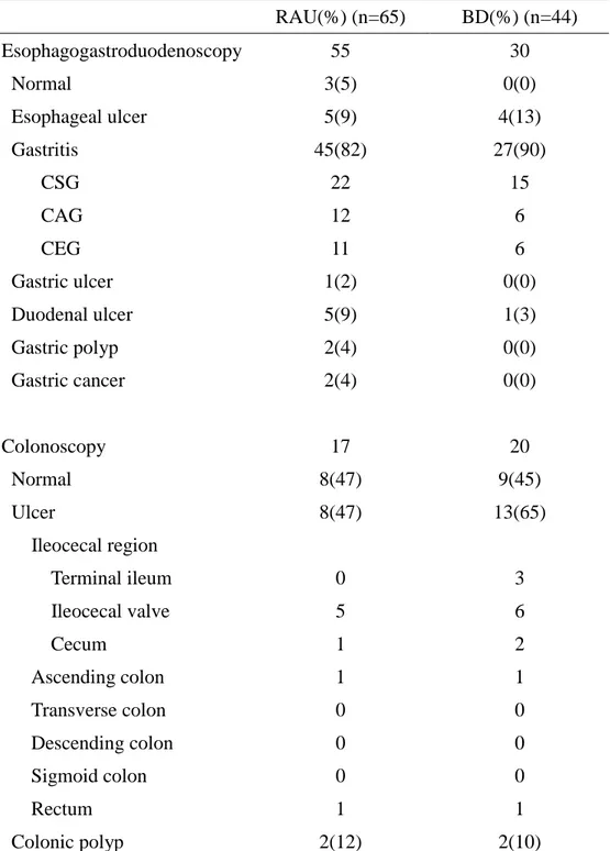
There were no significant differences in endoscopic characteristics between patients with RAU and BD(Table 4.). On esophagoduodenoscopy, ulcerations were observed 20% in RAU, 16% in BD. On colonoscopy, ulceration were observed 47% in RAU, 65% in BD. Ileocecal region was most common ulceration site of both two groups.
Table 4. Endoscopic characteristic of patients with RAU and BD. RAU(%) (n=65) BD(%) (n=44) Esophagogastroduodenoscopy 55 30 Normal 3(5) 0(0) Esophageal ulcer 5(9) 4(13) Gastritis 45(82) 27(90) CSG 22 15 CAG 12 6 CEG 11 6 Gastric ulcer 1(2) 0(0) Duodenal ulcer 5(9) 1(3) Gastric polyp 2(4) 0(0) Gastric cancer 2(4) 0(0) Colonoscopy 17 20 Normal 8(47) 9(45) Ulcer 8(47) 13(65) Ileocecal region Terminal ileum 0 3 Ileocecal valve 5 6 Cecum 1 2 Ascending colon 1 1 Transverse colon 0 0 Descending colon 0 0 Sigmoid colon 0 0 Rectum 1 1 Colonic polyp 2(12) 2(10)
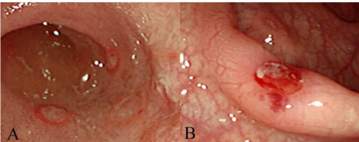
Fig. 2. Colonoscopic findings of recurrent aphthous ulcer and intestinal Behçet’s disease. Multiple, shallow, round and geographic ulcer on the ileocecal mucosa of recurrent aphthous ulcer (A) and Solitary, Deep, round, and punched-out ulcer on the ileocecal mucosa of Behçet’s disease (B).
There were no significant differences between patients with RAU and BD(Table 5.). But two cases of BD were perforation of ulcer.
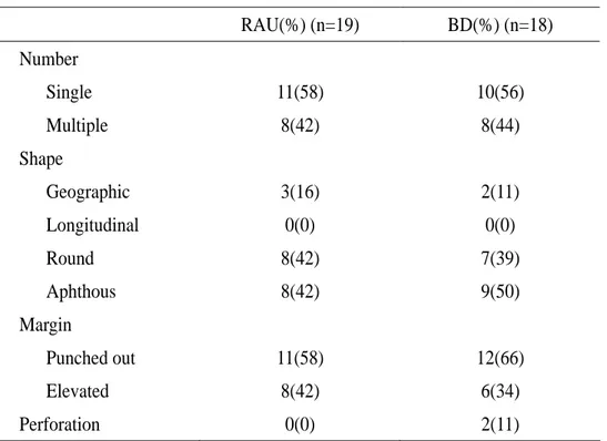
Table 5. Ulcer characteristic of patients with RAU and BD. RAU(%) (n=19) BD(%) (n=18) Number Single 11(58) 10(56) Multiple 8(42) 8(44) Shape Geographic 3(16) 2(11) Longitudinal 0(0) 0(0) Round 8(42) 7(39) Aphthous 8(42) 9(50) Margin Punched out 11(58) 12(66) Elevated 8(42) 6(34) Perforation 0(0) 2(11)
RAU, recurrent aphthous ulcer; BD, Behçet’s disease
C. Histopathologic findings
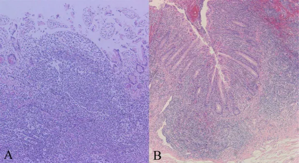
Most common histopathologic findings RAU and BD were vasculitis, patch mixed lymphocyte dominant infiltration(Table 6.). There were no significant differences between patients with RAU and BD.
Table 6. Histopathologic characteristic of patients with RAU and BD. RAU(%) (n=12) BD(%) (n=10) Histopathology Vasculitis 6(50) 7(70) Granuloma 0(0) 0(0) Lymphoid aggregation 6(50) 3(30) Infiltration type Patched, mixed 6(50) 8(80) Diffuse, mixed 3(25) 2(20) Mild, mixed 3(25) 0(0) Dominant cell Lymphocyte 12(100) 9(90) Plasma cell 0(0) 1(10) Crypt architecture Intact 12(100) 8(80) Damaged 0(0) 2(20)
RAU, recurrent aphthous ulcer; BD, Behçet’s disease
Fig. 4. Histopathologic findings of recurrent aphthous ulcer and cecal lesion in Behçet’s disease. Lymphocyte dominant, diffuse, mixed infiltration of recurrent aphthous ulcer (A) and lymphocyte dominant, patch, mixed infiltration with vasculitis and intact crypt of Behçet’s disease (B).
D. Serologic findings
1. ASCA enzyme linked immunosorbent assay (ELISA)
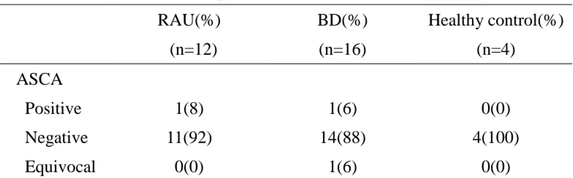
Table 7. shows that ASCA test result of patients with RAU, BD and healthy volunteers. Only one patient(6%) of intestinal Behçet’s disease and one patient(8%) of RAU was IgG ASCA positive(6%). Median level of ASCA was highest in serum of patient with BD. But
volunteers.
Table 7. ASCA test result of patients with RAU, BD and healthy control RAU(%) (n=12) BD(%) (n=16) Healthy control(%) (n=4) ASCA Positive 1(8) 1(6) 0(0) Negative 11(92) 14(88) 4(100) Equivocal 0(0) 1(6) 0(0)
RAU, recurrent aphthous ulcer; BD, Behçet’s disease; ASCA, Anti-Saccharomyces cerevisiae Mannan antibody; pANCA, perinuclear antineutrophil cytoplasmic autoantibody
Fig. 5. ASCA levels in patients with recurrent aphthous ulcer, Behçet’s disease, and healthy controls. Levels equal to or greater than 25 units are considered positive.
2. pANCA Indirect Immunofluorescence Assay
Table 8. shows that pANCA test result of patients with RAU, BD and healthy volunteers. Only one patient of intestinal BD was positive. There were no significant differences between patients with RAU, BD and healthy volunteers.
Table 8. pANCA test result of patients with RAU, BD and healthy control RAU(%) (n=12) BD(%) (n=16) Healthy control(%) (n=4) pANCA Positive 0(0) 1(6) 0(0) Negative 12(100) 15(94) 4(100)
RAU, recurrent aphthous ulcer; BD, Behçet’s disease; ASCA,Anti-Saccharomyces cerevisiae Mannan antibody; pANCA, perinuclear antineutrophil cytoplasmic autoantibody
IV. DISCUSSION
The clinical diagnosis of BD may pose considerable difficulties. Since the disease is multi-systemic and does not have any pathognomonic symptom or laboratory findings, the diagnosis is made solely on the basis of the criteria proposed by ISGBD in 1990, based on a cluster of clinical manifestations. Nonetheless, the diagnosis might be delayed for several years, until all ISG criteria have been fulfilled. Thus, the presence of a relatively specific laboratory marker can substantially facilitate the diagnosis of BD, and possibly support a diagnosis before all disease manifestations have occurred.
RAU is seen in most patients with BD; it commonly precedes other systemic features and can be of major, minor or herpetiform types. However, it is difficult to predict with any certainty those patients initially presenting with RAU who will subsequently proceed to develop multisystem involvement as part of BD. Two studies have addressed this problem; in one, a clinical comparison between 38 patients with BD and RAU-only controls showed an increased number of concurrent ulcers and involvement of the soft palate and oropharynx in those diagnosed with BD(Main and Chamberlain, 1992). No differences were detected with respect to duration, frequency, age of onset or family history. Scrutiny of clinical photographs taken of BD patients in this study suggested that both ‘herpetiform-type’ and ‘aphthous type’ ulcers appeared atypical(Main and Chamberlain, 1992). Clinical observations in oral medicine clinics suggest that aphthous ulcers in patients with BD appear to be associated with increased
tissue edema and appear to have an intensely erythematous border. The aphthae in BD often occur in the soft palate and oropharynx and have been observed on the hard palate, which is an unusual site for RAU in patients without BD(Main and Chamberlain, 1992). Patches of mucosal erythema may be observed in patients with the disease, indicating possible instability of the oral mucosa prior to ulceration. Patients may paradoxically report no painful symptoms during active disease, despite extensive oral ulceration being clinically evident. Bang et al examined the prognosis of the clinical relevance of recurrent oral ulceration in BD, and investigators found that approximately half the patients who were initially diagnosed as RAU-only developed other manifestations of BD at an average of 7.7 years after onset(Bang, 1995). They reported that highly recurrent RAU had appeared to be a warning signal for BD.
Many authors consider the onset to be the age at which the patient fulfilled the diagnostic criteria. The third decade is the most commonly reported age of onset for BD(Chajek and Fainaru, 1975; Bang et al, 1997; Yoo et al, 1997), and the fourth decade for intestinal BD(Kasahara et al,1981). In our study, intestinal BD occurred at a mean age of 29.0 years and RAU occurred at a mean age of 34.7 years. Althougt the mean age of RAU was higher than BD, no difference was found between the two groups in statistical analysis.
The male to female ratio in BD also differs geographically (Shimizu et al, 1979; Kaklamani et al, 1998); however, the gender ratio in terms of intestinal BD has not been well characterized in Mediterranean and Western countries, probably because of its relative rarity.
in Korea (0.7–1.3:1) (31,35,36) and Japan(1.7:1)(Kasahara et al, 1981). But there were no signicicant differences with BD and RAU(0.8:1).
It has been well known for many years that BD is often accompanied by such gastrointestinal symptoms as nausea, vomiting, and abdominal pain.(Lehner, 1977; Bayraktar, 2000). About 22 percent of patients with intestinal BD developed appendicitis-like symptoms during the clinical course(Kodama, 1977). This study showed that the most common presenting symptom in intestinal BD and RAU with GI symptoms is abdominal pain(66% and 54%). Diarrhea and indigestion were second common presenting symptoms in this two groups.
The pathologic hallmark of intestinal BD is known to be the presence of a few punched-out ulcers of variable size and appearance(Lee, 1986). The smaller ulcers (“aphthoid ulcers”) have been considered histologically to be similar to the oral aphthous ulcers of BD(Thach and Cummings, 1976). Larger ulcers usually have an oval or irregular configuration. The depth of penetration of the ulcers varies. Superficial ulcers occasionally have been shown to resolve(Smith et al, 1973; Empey, 1972; Lebwohl et al, 1977), but deeper ulcers, often with a narrow fissuring appearance, can extend through the bowel wall. Intestinal perforation is, therefore, a common complication(Shimizu et al, 1979; Kasahara et al, 1981; Ha et al,1998; Lee et al, 1997). Although intestinal BD is diagnosed radiologically or endoscopically in many cases, relatively few descriptions about the endoscopic characteristics of intestinal BD exist in the literature. Morphological characteristics of intestinal BD have been reported to be discrete
ulcerations that have a confluent and discrete border, appearing most commonly in the ileocecal region(Kasahara et al,1981; Bayraktar et al, 2000; Baba et al, 1976; Lee et al, 1997). In our series, most of the ulcers in intestinal BD and RAU on colonoscopy were found in the ileocecal areas, but they may be present at any site throughout the digestive system. Many of the patients had single or relatively few ulcers with localized distribution. The colonoscopic characteristics of intestinal BD could be summarized as large sized, round /oval or geographically shaped, deep and discrete ulcer with elevated margins. Because just we have examined the photography of endoscopy, we were difficult to find out differences of an ulcer including its shape, and location and the distribution pattern of lesions in intestinal BD and RAU.
The ulcers from the intestinal BD tend to be deep and penetrating and, thus, frequently required surgical interventions(Sayek et al, 1991; Ketch et al, 1980; Kim et al, 1994; Kim et al, 2000). Complications of GI ulceration in BD may include perforation and enterocutaneous fistula. Surgical treatment is often necessary, but postoperative recurrence is as high as 68%(Choi et al, 2000). In the current study, we found that two cases of intestinal BD were recurred after operation. Therefore, more attention should be paid in patients with perforating type of intestinal BD
The intestinal ulcer of BD is characterized not only by absence of the granulomatous formation of Crohn’s disease, but also by a deeper penetration of ulcer to a region nearer the
diagnos of BD. Of course, it is necessary to examine carefully the clinical symptoms and signs and the histopathological appearance, since in many cases the differential diagnosis is very difficult.
In our study, most common histopathologic findings of intestinal ulcers in BD were vasculitis, patched mixed lymphocyte dominant infiltration. There were no significant differences between patients with intestinal BD and RAU.
Since 1990, perinuclear anti-neutrophil cytoplasmatic antibodies (pANCAs), which show a specific staining on indirect immunofluorescence, have been consistently found in 40–80% of patients suffering from ulcerative colitis(Rump et al, 1990; Duerr et al, 1991; Oudkerk et al, 1993; Peeters et al, 2001). These are spontaneously produced by lamina propria and mesenteric node lymphocytes(Targan et al, 1995). In ulcerative colitis, the antigen recognized by pANCAs is located in the inner side of the nuclear periphery and is DNase sensitive, but the exact epitope remains as yet unknown. It is hypothesized that pANCAs are due to cross-reactivity with bacterial antigens(Seibold et al, 1998; Cohavy et al, 2000).
Antibodies to baker’s yeast and brewer’s yeast (ASCA) have been described in up to 65% of patients with Crohn’s disease(Main et al, 1988; McKenzie et al, 1990; Giaffer et al, 1992; Sendid et al,1996). It has been demonstrated that the specific antigen is a mannan localized in the yeast cell wall(Sendid et al,1996). The significance of ASCAs in Crohn’s disease is completely unknown, but one hypothesis links them to increased intestinal permeability(Vermeire et al, 2001). Due to this presumed break in the epithelial barrier,
increased exposure of the epithelium to common food antigens such as yeasts may result in an exaggerated antibody response.
Kim et al have reported that ASCA may be associated with Crohn’s disease and BD and pANCA with ulcerative colitis. Positivity for ASCA with pANCA negativity was associated with Crohn’s disesae and Behçet’s disease , whereas a positive pANCA in combination with a negative ASCA result was strongly correlated with ulcerative colitis. A combination of both tests may aid the differential diagnosis of inflammatory bowel disease(Kim et al, 2002). Recently, Krause et al have reported the significantly higher ASCA values in BD patients compared with patients with RAU, SLE or in healthy volunteers imply that ASCA, taken in appropriate clinical context, could become a useful diagnostic tool for BD(Krause et al, 2002). Monselise et al have reported ASCA titers were significantly higher in BD patients companied to their healthy family members(Monselise et al, 2002). ASCA seem to be strongly related to BD expression and are not associated with environmental or genetic factors.
But in our study, there were neither clinically, endoscopically, histopathologically nor serologically differences between patients with intestinal BD, RAU and healthy volunteers in ASCA and pANCA. Further prospective studies including more numbers of patients are needed to make a diagnosis of intestinal BD and RAU.
V. CONCLUSIONS
There were a wide range of clinical features for gastrointestinal involvement including abdominal pain, indigestion, diarrehea, melena, nausea, vomiting in the two groups, RAU with gastrointestinal symptoms and BD with gastrointestinal involvement.
In the two groups, localized ulcer was most common colonoscopic findings and most ulcers were discovered in ileocecal region. There are many hisopathologic findings of ulcer were lymphocyte dominant, mixed, patched infiltration and vasculitis.
Although median level of ASCA was highest in serum of patients with BD, there were no significant differences in clinical, endoscopic, histopathologic findings between the two groups. ASCA and pANCA levels were not different between the two groups unlike previous reports. Patient of RAU with gastrointestinal involvement need to be closed follow up.
REFERENCES
1. 유태욱: 대장에 발생한 Crohn 병과 Behcet 병의 형태학적 비교. 연세대학교, 1993
2. Aeberhard P, Berchtold W, Riedtmann HJ, Stadelmann G: Surgical recurrence of perforating and nonperforating Crohn’s disease: a study of 101 surgically treated patients. Dis Colon Rectum 39:80-87,1996
3. Baba S, Maruta M, Ando K: Intestinal Behçet’s disease: report of five cases. Dis Colon Rectum 19:428–440,1976
4. Balabanova M, Calamia KT, Perniciaro C, O'Duffy JD: A study of the cutaneous manifestations of Behcet's disease in patients from the United States. J Am Acad Dermatol 41:540-545,1999
5. Bang D, Yoon KH, Chung HG, Choi EH, Lee ES, Lee S: Epidemiological and clinical features of Behçet’s disease in Korea. Yonsei Med J 38: 428–436,1997
7. Behçet H: Über rezidivierende apthüse durch ein virus verusachte Geswüre am Mund, am Auge und an den Genitalien. Dermatol Wochenschr 105:1152–1157, 1937
8. Benamour S, Zeroual B, Bennis R, Amraoui A, Bettal S : Digestive manifestations of Behçet’s disease based on s series of 74 cases. Presse Med 6;19:1485-1489,1990
9. Bøe J, Dalgaard JB, Scott D: Mucocutaneous-ocular syndrome with intestinal involvement: a clinical and pathological study of four fatal cases. Am J Med 25:857–867, 1958
10. Chajek T, Fainaru M: Behçet’s disease: report of 41 cases and a review of the literature. Medicine 54:179-196, 1975
11. Choi IJ, Kim JS, Cha SD, Jung HC, Park JG, Song IS, Kim CY: Long-term clinical course and prognostic factors in intestinal Behçet’s disease. Dis Colon Rectum 43:692–700,2000
12. Cohavy O, Bruckner D, Gordon LK, Misra R, Wei B, Eggena ME, Targan SR, Braun J: Colonic bacteria express an ulcerative colitis pANCA-related protein epitope. Infect Immun 68: 1542-1548,2000
13. Duerr RH, Targan SR, Landers CJ, LaRusso NF, Lindsay KL, Wiesner RH, Shanahan F: Neutrophil cytoplasmic autoantibodies: a link between PSC and ulcerative colitis. Gastroenterology 100: 1385-1391,1991
14. Empey DW: Rectal and colonic ulceration in Behçet’s disease. Br J Surg 59:173–175,1972
15. Gamble CN, Wiesner KB, Shapiro RF, Boyer WJ: The immune complex pathogenesis of glomerulo-nephritis and pulmonary vasculitis in Behçet’s disease. Am J Med 66:1031–1039, 1979
16. Giaffer MH, Clark A, Holdsworth CD: Crohn's disease. Antibodies to Saccharomyces cerevisiae in patients with Crohn's disease and their possible pathogenic importance. Gut 33: 1071-1075,1992
17. Griffin JW, Harrison HB, Tedesco FJ, Mills LR 4th: Behçet’s disease with multiple sites of gastrointestinal involvement. South Med J 75:1405-1408,1982
Auh YH: Intestinal Behçet syndrome: CT features of patients with and patients without complications. Radiology 209:449–454,1998
19. Kaklamani VG, Vaiopoulos G, Kaklamanis PG: Behçet’s disease. Semin Arthr Rheum 27:197–217,1998
20. Kasahara Y, Tanaka S, Nishino M, Umemura H, Shiraha S, Kuyama T: Intestinal involvement in Behçet’s disease: review of 136 surgical cases in Japanese literature. Dis Colon Rectum 24:103–106,1981
21. Ketch LL, Buerk CA, Liechty RD: Surgical implications of Behçet’s disease. Arch Surg 115:759-760,1980
22. Kim BG, Kim YS, Kim JS, Jung HC, Song IS: Diagnostic role of anti-Saccharomyces cerevisiae mannan antibodies combined with antineutrophil cytoplasmic antibodies in
patients with inflammatory bowel disease Dis Colon Rectum 45:1062-1069, 2002
23. Kim JH, Choi BI, Han JK, Choo SW, Han MC: Colitis in Behçet’s disease;characteristics on double-contrast barium enema examination on 20 patient. Abdomen Imaging 19:132-136,1994
24. Kodama H: Intestinal Behcet’s. Surg Ther 37:51-8,1977
25. Kim JS, Lim SH, Choi IJ, Moon H, Jung HC, Song IS, Kim CY: Prediction of clinical course according to the macroscopic type by colonoscopy in Behçet’s colitis. Endoscopsy 32:635-640, 2000
26. Krause, Y Monselise, G Milo, A.Weinberger: Anti-Saccharomyces cerevisiae antibodies-A novel serologic marker for Behçet’s disease. Clin Exp Rrheumatol 20:S21-24,2002
27. Lebwohl O, Forde KA, Berdon WE, Morrison S, Challop R: Ulcerative esophagitis and colitis in a pediatric patient with Behçet’s syndrome. Response to steroid therapy. Am J Gastroenterol 68:550–555,1977
28. Lee CR, Kim WH, Cho YS, Kim MH, Kim JH, Park IS, Bang D: Colonoscopic findings in intestinal Behcet’s disease. Inflamm Bowel Dis 7(3):243-249,2001
29. Lee KS, Kim SJ, Lee BC, Yoon DS, Lee WJ, Chi HS: Surgical treatment of intestinal Behçet’s disease. Yonsei Med J 38:455–460,1997
31. Lehner T: Oral ulceration and Behçet’s syndrome. Gut 18:491–511, 1977
32. Main DM, Chamberlain MA: Clinical differentiation of oral ulceration in Behçet’s disease. Br J Rheumatol 31: 767–370, 1992
33. Main J, McKenzie H, Yeaman GR, Kerr MA, Robson D, Pennington CR, Parratt D: Antibody to Saccharomyces cerevisiae (baker's yeast) in Crohn's disease. BMJ 297: 1105-1106,1988
34. Mason RM, Barnes CG: Behçet’s syndrome with arthritis. Ann Rheum Dis 28:95–103,1969
35. McKenzie H, Main J, Pennington CR, Parratt D: Antibody to selected strains of Saccharomyces cerevisiae (baker's and brewer's yeast) and Candida albicans in Crohn's disease. Gut 31: 536-538, 1990
36. Monselise A, Krause I, Monselise Y, Weinberger A: Anti-saccharomyces cerevisiae antibodies among family members of Behçet’s disease patients. Clin Exp Rheumatol 20:S21-24, 2002
37. Natah SS, Konttinen YT, Enattah NS, Ashammakhi N, Sharkey KA, Hayrinen-Immonen R: Recurrent aphthous ulcers today: a review of the growing knowledge. Int J Oral Maxillofac Surg 33:221-234, 2004
38. O’Duffy JD, Carney JA, Deodhar S: Behçet’s disease: report of 10 cases, 3 with new manifestations. Ann Intern Med 75:561-570, 1971
39. O’Duffy JD: Summary of international symposium on Behçet’s disease. J Rheumatol 5:229-233, 1978
40. Oshima Y, Shimizu T, Yokohari R, Matsumoto T, Kano K, Kagami T, Nagaya H, Maruyama R.: Clinical studies on Behçet’s syndrome. Ann Rheum Dis 22:36–45,1963
41. Oudkerk Pool M, Ellerbroek PM, Ridwan BU, Goldschmeding R, von Blomberg BM, Pena AS, Dolman KM, Bril H, Dekker W, Nauta JJ: Serum antineutrophil cytoplasmic autoantibodies in inflammatory bowel disease are mainly associated with ulcerative colitis. A correlation study between perinuclear antineutrophil cytoplasmic autoantibodies and clinical parameters, medical, and surgical treatment. Gut 34: 46-50,1993
42. Peeters M, Joossens S, Vermeire S, Vlietinck R, Bossuyt X, Rutgeerts P. : Diagnostic value of anti-Saccharomyces cerevisiae and antineutrophil cytoplasmic autoantibodies in inflammatory bowel disease. Am J Gastroenterol 96: 730-734,2001
43. Rump JA, Schölmerich J, Gross V: A new type of perinuclear anti-neutrophil cytoplasmic antibody in active ulcerative colitis but not in Crohn's disease. Immunobiol 181: 406-413,1990
44. Sakane T, Takeno M, Suzuki N, Inaba G: Behçet’s disease. N Engl J Med 21:1284–1291,1999
45. Sayek I, Aran O, Uzunalimoglu B, Hersek E: Intestinal Behcet’s disease;surgical experience in seven cases. Hepatogastroenterology 38:81-83,1991
46. Seibold F, Brandwein S, Simpson S, Terhorst C, Elson CO: pANCA represents a cross-reactivity to enteric bacterial antigens. J Clin Immunol 1823: 153-160,1998
47. Sendid B, Colombel JF, Jacquinot PM, Faille C, Fruit J, Cortot A, Lucidarme D, Camus D, Poulain D: Specific antibody response to oligomannoside epitopes in Crohn's disease. Clin Diagn Lab Immunol 3: 219-226,1996
48. Shimizu T, Ehrlich GE, Inaba G: Behçet’s disease (Behçet’s syndrome). Semin Arthritis Rheum 8:223–260,1979
49. Smith GE, Kime LR, Pitcher JL: The colitis of Behçet’s disease: a separate entity? Colonoscopic findings and literature review. Am J Dig Dis 18:987–1000,1973
50. Targan SR, Landers CJ, Cobb L, MacDermott RP, Vidrich A: Perinuclear anti-neutrophil cytoplasmic antibodies are spontaneously produced by mucosal B cells of ulcerative colitis patients. J Immunol 155: 3262-3267,1995
51. Thach BT, Cummings NA: Behçet syndrome with “aphthous colitis”. Arch Intern Med 136:705–709,1976
52. Tsukada S, Yamazaki T, Iyo Sl: Neuro-Behçet’s syndrome: report of two cases and review of the literature. Saishin Igaku 19:1533–1541,1964
53. Vermeire S, Peeters M, Vlietinck R, Joossens S, Den Hond E, Bulteel V, Bossuyt X, Geypens B, Rutgeerts P: Anti-Saccharomyces cerevisiae antibodies (ASCA), phenotypes
8-15,2001
54. Yoo HM, Han KH, Kim BS: Clinical feature of intestinal Behçet’s disease and therapeutic effects of sulfasalazine. Korean J Gastroenterol 29:465–472,1997
55. Yurdakul S, Tuzuner N, Yokohari R: Gastrointestinal involvement in Behçet’s syndrome: a controlled study. Ann Rheum Dis 55:208-221,1996