Prevalence of Human Papilloma Virus Infections and Cervical
Cytological Abnormalities among Korean Women with Systemic
Lupus Erythematosus
We performed a multicenter cross-sectional study of 134 sexually active systemic lupus erythematosus (SLE) patients to investigate the prevalence of and risk factors for high risk human papilloma virus (HPV) infection and cervical cytological abnormalities among Korean women with SLE. In this multicenter cross-sectional study, HPV testing and routine cervical cytologic examination was performed. HPV was typed using a hybrid method or the polymerase chain reaction. Data on 4,595 healthy women were used for comparison. SLE patients had greater prevalence of high-risk HPV infection (24.6% vs. 7.9%, P<0.001, odds ratio 3.8, 95% confidence interval 2.5-5.7) and of abnormal cervical cytology (16.4 vs. 2.8%, P<0.001, OR 4.4, 95% CI 2.5-7.8) compared with controls. SLE itself was identified as independent risk factors for high risk HPV infection among Korean women (OR 3.8, 95% CI 2.5-5.7) along with ≥2 sexual partners (OR 8.5, 95% CI 1.2-61.6), and Pap smear
abnormalities (OR 97.3, 95% CI 6.5-1,456.7). High-risk HPV infection and cervical cytological abnormalities were more common among Korean women with SLE than controls. SLE itself may be a risk factor for HPV infection among Korean women, suggesting the importance of close monitoring of HPV infections and abnormal Pap smears in SLE patients.
Key Words: Systemic Lupus Erythematosus, Human Papilloma Virus; Cervical Cytological Abnormalities
You-Hyun Lee1, Jung-Yoon Choe2,
Sung-Hoon Park2, Yong-Wook Park3,
Shin-Seok Lee3, Young-Mo Kang4,
Eon-Jeong Nam4, Won Park5,
Seong-Ryul Kwon5, Sang-Cheol Bae6,
Yun-Jung Kim6, Chang-Hee Suh7,
Hyoun-Ah Kim7, Nam Wook Hur8,
and Jisoo Lee1
Division of Rheumatology, Department of Internal Medicine1, Ewha Womans University School of
Medicine, Seoul; Division of Rheumatology, Department of Internal Medicine2, Catholic
University of Daegu School of Medicine, Daegu; Division of Rheumatology, Department of Internal Medicine3, Chonnam National University Medical
School, Gwangju; Division of Rheumatology, Department of Internal Medicine4, Kyungpook
National University School of Medicine, Daegu; Division of Rheumatology, Department of Internal Medicine5, Inha University School of Medicine,
Incheon; Hanyang University Hospital for Rheumatic Diseases6, Seoul; Division of Rheumatology,
Department of Internal Medicine7, Ajou University
School of Medicine, Suwon; Biomedical Research Group Brain Korea 218, Hanyang University, Seoul,
Korea
Received: 11 February 2010 Accepted: 13 April 2010 Address for Correspondence: Jisoo Lee, M.D.
Division of Rheumatology, Department of Internal Medicine, Ewha Womans University School of Medicine, 204 Anyangcheon-gil, Yangcheon-gu, Seoul 158-710, Korea Tel: +82.2-2650-6164, Fax: +82.2-2650-2590 E-mail: [email protected]
This work was supported in part by the Korea Healthcare Technology R&D Project, Ministry for Health, Welfare and Family Affairs, republic of Korea (A080588).
DOI: 10.3346/jkms.2010.25.10.1431 • J Korean Med Sci 2010; 25: 1431-1437 Immunology, Allergic Disorders & Rheumatology
INTRODUCTION
Human papillomavirus (HPV) infection is thought to be princi-ple causal agent for cervical cancer worldwide (1). In particular, persistent infection with high-cancer-risk HPV type 16 and 18 is major risk factor for cervical cancer (2). In the case of immuno-compromised host, the risk of HPV infection was reported to be much greater than the general population due to high-load, per-sistent infection with oncogenic HPV genotypes (3-6). The
prev-alence of HPV infection was shown to increase in organ trans-plant recipients (3, 7), human immunodeficiency virus (HIV)-infected women (4-6), and patients with systemic autoimmune diseases such as systemic lupus erythematosus (SLE) (8, 9). Since SLE is a multi-system disease characterized by produc-tion of a vast array of autoantibodies against multiple tissues and organs, multiple factors including genetic and ethnic back-ground, immunological abnormalities, extent of organ damage, and behavioral patterns might account for the increased
sus-ceptibility of SLE patients to HPV infection and cervical cancer. The socioeconomic impact of the increased risk of cervical can-cer in Korean patients with SLE can be greater since it is known that there is an increase in prevalence of SLE in the Asian popu-lation (10). However, there is a paucity of data regarding the prev-alence of high risk HPV infection and cervical cytological ab-normalities in SLE patients of different ethnic background and regions. Moreover, disease related risk factors for HPV infections in SLE patients remain unclear. The purpose of our study was to investigate the prevalence of and risk factors for HPV infection, and cervical cytological abnormalities in Korean patients with SLE.
MATERIALS AND METHODS
Patients
All patients attending the rheumatology clinic at 7 tertiary hos-pitals in Korea who fulfilled the 1997 American College of Rheu-matology (ACR) criteria for SLE were invited to participate in this cross-sectional study. Participating hospitals were located throughout Korea including Seoul, Gyeonggi-do, Gyeongsang-do, and Jeolla-do regions including both urban and rural areas. Patients were eligible for the study if they had been sexually ac-tive. Patients who never had sexual intercourse and patients younger than 20 yr old were excluded. One hundred thirty four SLE patients who signed the informed consent out of total 1525 SLE patients were recruited between February 2006 and March 2007 through a survey in clinic. The study was approved by the local ethics committee of each institution. For comparison, data from a cross-sectional study of women attending the National Cervical Cancer Screening Program carried out by Oh et al. (11) on prevalence of HPV infection of 4595 women in Busan and Suwon, Korea were used. Women attending the National Cervi-cal Cancer Screening Program belonged to mostly low income social groups who visited government affiliated hospitals in both urban and rural areas. The data was supplemented by 113 wom-en aged 20-29 yr who visited local hospital for routine gyneco-logic care, since the National Cervical Cancer Screening Pro-gram included mostly women over 30 yr of age.
Clinical data collection
Patient interviews and chart reviews were performed at the time of the enrollment to obtain information regarding sexual be-haviors, obstetric and gynecological histories, and disease his-tory related to SLE. We obtained demographic information in-cluding age, marital status, duration of education, and tobacco smoking, and reproductive and sexual history including num-ber of gestations and parity, age at first intercourse, numnum-ber of sexual partners, and history of sexually transmitted diseases. Disease history related to SLE included disease duration, extent of organ involvement, and type of treatment received. Major
organ involvement was defined as involvement of one or more of the following organs: the central nervous system, lung, heart, kidney, intestine, and the hematologic system (hemolytic ane-mia, platelet <100,000/μL). Treatment with prednisolone >1 mg/ kg/day, azathioprine, cyclosporine, mycophenolate mofetil and/ or cyclophosphamide was defined as immunosuppressive ther-apy. Autoantibodies including antinuclear antibody (ANA), an-ti-double strand DNA (dsDNA), and systemic lupus erythema-tosus disease activity index (SLEDAI) were assessed at the time of gynecological evaluation.
Gynecological examination and specimen collection
All patients underwent gynecologic examinations, which in-cluded HPV testing and Pap smears, by gynecologists at each hospital. HPV DNA tests were performed on exfoliated cervical cells and examined by either Hybrid Capture II (HC-II) tech-nology (The digene HPV test, Gaithersburg, MD, USA) which detected 13 high-risk HPV types (16, 18, 31, 33, 35, 39, 45, 51, 52, 56, 58, 59, 68) or a polymerase chain reaction (PCR) based DNA microarray system (My HPV chip, Mygene, Seoul, Korea), which detect 16 types of high-risk HPV (16, 18, 31, 33, 35, 39, 45, 51, 52, 53, 54, 56, 58, 59, 66, 68) and 8 types of low-risk HPV (6, 11, 34, 40, 42, 43, 44, 70). Five of the 7 participating centers utilize the Hybrid Capture II technology and 2 utilized the PCR based DNA microarray system. The Hybrid Capture II technology system was used for the community control. Both HC-II and microar-rays were highly comparable in terms of sensitivity, specificity, and positive and negative predictive values for detecting HPV in cervical specimens (12). To perform hybrid capture technol-ogy, cervical specimens were combined with an extraction buf-fer to release and denature the HPV DNA. The released DNAs were combined with specific RNA probes to create the RNA-DNA hybrids, which were captured on a solid phase by an anti-body specific for the hybrids. The captured RNA-DNA hybrids were tagged with antibody reagent, and measured using a lumi-nometer. To perform PCR based DNA microarrays, target HPV DNA from cervical specimens were amplified by PCR using the primers of MY09/MY11 (5´-CGTCCMARRGGAWACTGATC-3´/5´-GCMCAGGGWCATAAYAATGG-3´) and GP5+/GP6+(5´- TTTGTTACTGTGGTAGATACTAC-3´/5´-Cy3-GAAAAATAAA-CTGTAAATCATATTC-3´). Primers that amplify β-globin were used as controls (5´-Cy3-CAACTTCATCCACGTTCACC-3´/5´-GAAGAGCCAAGGACAGGTAC-3´). The PCR product was hy-bridized onto the chip at 42°C for 4 hr and washed with buffer. Hybridized signals were visualized with a DNA chip scanner. If the binding of a high-risk HPV subtype was detected in hybrid capture and microarray tests, it was considered positive for HPV. In case of an abnormal Pap results, a histological examination was done. The interval between Pap smear and histological ex-amination was less than 1 month.
Statistical analysis
Clinical parameters are presented as mean±SD. Comparisons between the SLE patients and the controls were analyzed using the χ2-test. Odds ratios (ORs) with 95% confidence intervals (CIs) were used to estimate the association between each possible risk factor and HPV infection in SLE patients. Independent risk factors for HPV infection in SLE patients were determined using a multiple logistic regression equation. Statistical analysis was done using SPSS 12.0 (SPSS Inc, Chicago, IL, USA) software.
Ethics statement
The study was conducted according to the protocols approved by the institutional ethics committee of each institution in com-pliance with the Helsinki Declaration (IRB approval numbers: Ewha Womans University Mokdong Hospital; ECT 125-5, Daegu Catholic University Medical Center; CR-06-22-83, Chonnam National University Hospital; 06-nuh, Kyungpook National Uni-versity Hospital; 7405-1929-2009.07.07, Inha UniUni-versity Hospi-tal; 2006-580, Hanyang University Medical Center; 2006-29, Ajou University Hopital; AJIRB-CRO-06-109).
RESULTS
Characteristics of patients and controls
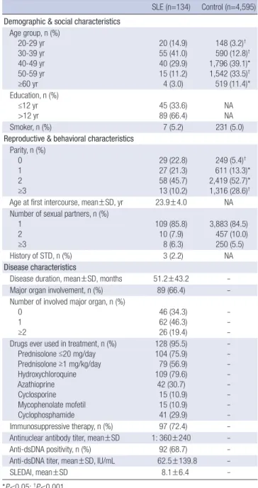
SLE patients and controls were all ethnically Koreans and de-mographically similar coming from both rural and urban areas, except that SLE patients included younger age group. The mean age of SLE patients was 39.5±9.4 (mean±SD) yr. Individuals younger than 39 yr of age accounted for 55.9% of the patient group, whereas 16.0% in the controls. Reproductive and behav-ioral characteristics between SLE patients and healthy controls were also similar with the exception of parity. SLE patients show-ed a significantly lower number of parity comparshow-ed with the con-trols. The mean disease duration of SLE patients was 51.2±43.2 months. Major organs were involved in 89 (66.4%) patients, and in 26 (19.4%) patients, two or more multiple major organs were involved. Drug exposures ever during the course of lupus up to the present are as shown in Table 1. Immunosuppressive agents including prednisolone ≥1 mg/kg/day, azathioprine, cyclospo-rine, mycophenolate mofetil, and cyclophosphamide were pre-scribed for 97 (72.4%) patients. All patients were ANA positive and the mean titer was 1:360. Anti-dsDNA was observ ed in 92 (68.7%) of patients, and the mean titer was 62.5 IU/mL. SLEDAI at the time of gynecologic evaluation was 8.1±6.4 (Table 1).
Prevalence of HPV infection and abnormal cytology in SLE patients
Infection with a high-risk type of HPV was detected in 33 (24.6%), and abnormal Pap smears were observed in 22 (16.4%) of SLE patients. Abnormal Pap smear results included epithelial dys-plasia in 8 patients, atypical squamous cells of undetermined
significance (ASCUS) in 7 patients, and carcinoma in situ (CIN) in 7 patients. In 15 patients, an abnormal Pap smear was con-firmed by histologic evaluation, and CIN was found in 11 (61.1%) of patients. Compared with the controls, SLE patients had a sig-nificantly higher prevalence of high-risk HPV infections (24.6% vs. 7.9%, P<0.001, OR 3.8, 95% CI 2.5-5.7). The prevalence of ab-normal cervical cytology was also increased in SLE patients compared with controls (16.4% vs. 2.8%, P<0.001, OR 4.4, 95% CI 2.5-7.8).
Table 1. Demographic, social, reproductive, behavioral, and disease characteristics of SLE patients and controls
SLE (n=134) Control (n=4,595) Demographic & social characteristics
Age group, n (%) 20-29 yr 30-39 yr 40-49 yr 50-59 yr ≥60 yr 20 (14.9) 55 (41.0) 40 (29.9) 15 (11.2) 4 (3.0) 148 (3.2)† 590 (12.8)† 1,796 (39.1)* 1,542 (33.5)† 519 (11.4)* Education, n (%) ≤12 yr >12 yr 45 (33.6) 89 (66.4) NANA Smoker, n (%) 7 (5.2) 231 (5.0)
Reproductive & behavioral characteristics Parity, n (%) 0 1 2 ≥3 29 (22.8) 27 (21.3) 58 (45.7) 13 (10.2) 249 (5.4)† 611 (13.3)* 2,419 (52.7)* 1,316 (28.6)† Age at first intercourse, mean±SD, yr 23.9±4.0 NA Number of sexual partners, n (%)
1 2 ≥3 109 (85.8) 10 (7.9) 8 (6.3) 3,883 (84.5) 457 (10.0) 250 (5.5) History of STD, n (%) 3 (2.2) NA Disease characteristics
Disease duration, mean±SD, months 51.2±43.2 Major organ involvement, n (%) 89 (66.4) Number of involved major organ, n (%)
0 1 ≥2 46 (34.3) 62 (46.3) 26 (19.4) Drugs ever used in treatment, n (%)
Prednisolone ≤20 mg/day Prednisolone ≥1 mg/kg/day Hydroxychloroquine Azathioprine Cyclosporine Mycophenolate mofetil Cyclophosphamide 128 (95.5) 104 (75.9) 79 (56.9) 109 (79.6) 42 (30.7) 15 (10.9) 15 (10.9) 41 (29.9) Immunosuppressive therapy, n (%) 97 (72.4) Antinuclear antibody titer, mean±SD 1: 360±240 Anti-dsDNA positivity, n (%) 92 (68.7) Anti-dsDNA titer, mean±SD, IU/mL 62.5±139.8
SLEDAI, mean±SD 8.1±6.4
-*P<0.05; †P<0.001.
NA, not available; SLE, systemic lupus erythematosus; STD, sexually transmitted disease; SLEDAI, systemic lupus erythematosus disease activity index.
Risk factors for development of high risk HPV infection
Using univariate analysis, high risk HPV infection was associat-ed with history of SLE (OR 3.8, 95% CI 2.5-5.7), multiple sexual partners (2: OR 7.2, 95% CI 5.4-9.5; ≥3: OR 3.4, 95% CI 2.4-5.0), and abnormal Pap smear (OR 18.9, 95% CI 11.0-32.6). Smoking and parity were not associated with high risk HPV infection (Table 2). Potential risk factors identified by univariate analysis (history of SLE, multiple sexual partners, abnormal Pap smear) including age were put into a multivariate model to identify in-dependent risk factors for high risk HPV infection. Inin-dependent risk factors for high risk HPV infection included a history of SLE (OR 6.9, 95% CI 2.5-18.9), ≥2 sexual partners (OR 8.5, 95% CI 1.2-61.6), and Pap smear abnormalities (OR 97.3, 95% CI 6.5-1456.7) (Table 3).
Disease related risk factors for high risk HPV infection in SLE patients
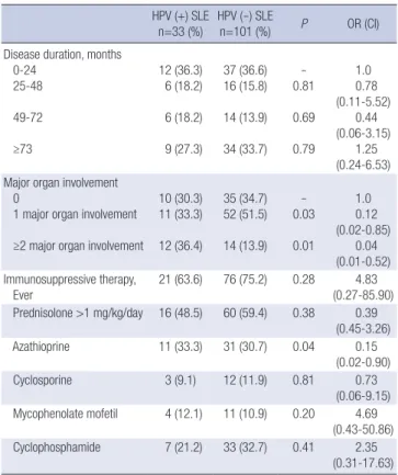
High risk HPV positive and negative SLE patients were compar-ed to determine the possible disease relatcompar-ed risk factors for high risk HPV infection in SLE patients (Table 4). Odds ratios adjust-ed for age, adjust-education, smoking, parity, number of sexual part-ners, age at first intercourse, and history of sexually transmitted diseases were determined by multiple logistic regression analy-sis. Significant increases in the number of patients with major organ involvement and the use of azathioprine were found in
the high risk HPV positive SLE patients compared to the high risk SLE negative patients. However, adjusted odds ratios for these factors were not increased, thus not identified as a disease related risk factors for high risk HPV infection. No significant differences were found in the use of immunosuppressive agents ever, and the disease duration between patients with high risk HPV infection and those without (Table 4).
DISCUSSION
Our study suggests that Korean patients with SLE have a signifi-cantly increased prevalence of high-risk HPV infection and ab-normal cervical cytology compared to controls. SLE itself ap-pears to be the major risk factor for HPV infection.
Increased prevalence of HPV infection has been reported for small groups of SLE patients. In a study of 30 SLE patients by Nath et al. (8) from the United Kingdom, tests for high risk gen-otype HPV-16 was positive in 57% of SLE patients compared to 1% of controls. A study from of 85 SLE patients from China re-vealed intermediate- or high-risk HPV genotype infection in 10.6% of SLE patients versus 4.2% in controls (9). In a French study consisted of 11 women with SLE, HPV-positivity was ob-served in 37.5% of the patients compared to 14.7% of the con-trols (13). We observed that 24.6% of the 134 Korean SLE pa-tients were infected with high-risk HPV. This represents 3.8 fold
Table 2. Unadjusted odds ratio for potential risk factors associated with high risk HPV infection
No. of subjects
HPV positivity
Prevalence (%) Unadjusted OR (95% CI) History of SLE Yes No 4,595134 24.6 (33/134)7.9 (362/4,595) 3.8 (2.5-5.7)1 Smoking Yes No 4,488238 12.6 (30/238)8.1 (365/4,488) 0.6 (0.4-0.9)1 Parity 0 1 2 ≥3 278 638 2,477 1,329 33.8 (94/278) 19.6 (125/638) 14.3 (353/2,477) 13.9 (185/1,329) 1 0.3 (0.2-0.5) 0.7 (0.5-0.9) 1.0 (0.8-1.3) No. of sexual partners
1 2 ≥3 3,992 467 263 14.1 (561/3,992) 27.6 (129/467) 52.1 (124/238) 0.6 (0.1-9.1) 7.2 (5.4-9.5) 3.4 (2.4-5.0) Abnormal PAP smear
Yes
No 4,580149 45.6 (68/149)7.1 (327/4,580) 18.9 (11.0-32.6)1 Table 3. Odds ratios (95% confidence interval) determined by multiple logistic reg-ression model using high risk HPV infection status as a variable
P Adjusted OR (95% CI)
History of SLE 0.001 6.9 (2.5-18.9)
Abnormal PAP smear 0.001 97.3 (6.5-1456.7) ≥2 sexual partners 0.035 8.5 (1.2-61.6)
Table 4. Disease related risk factors for high risk HPV infection in SLE patients* HPV (+) SLE
n=33 (%) HPV (-) SLE n=101 (%) P OR (CI) Disease duration, months
0-24 25-48 49-72 ≥73 12 (36.3) 6 (18.2) 6 (18.2) 9 (27.3) 37 (36.6) 16 (15.8) 14 (13.9) 34 (33.7) -0.81 0.69 0.79 1.0 0.78 (0.11-5.52) 0.44 (0.06-3.15) 1.25 (0.24-6.53) Major organ involvement
0
1 major organ involvement ≥2 major organ involvement
10 (30.3) 11 (33.3) 12 (36.4) 35 (34.7) 52 (51.5) 14 (13.9) -0.03 0.01 1.0 0.12 (0.02-0.85) 0.04 (0.01-0.52) Immunosuppressive therapy, Ever 21 (63.6) 76 (75.2) 0.28 4.83 (0.27-85.90) Prednisolone >1 mg/kg/day 16 (48.5) 60 (59.4) 0.38 0.39 (0.45-3.26) Azathioprine 11 (33.3) 31 (30.7) 0.04 0.15 (0.02-0.90) Cyclosporine 3 (9.1) 12 (11.9) 0.81 0.73 (0.06-9.15) Mycophenolate mofetil 4 (12.1) 11 (10.9) 0.20 4.69 (0.43-50.86) Cyclophosphamide 7 (21.2) 33 (32.7) 0.41 2.35 (0.31-17.63) *Values are the number (%) of patients.
increases in the prevalence of high-risk HPV in Korean SLE pa-tients compared to the general population. Our data confirmed the previous observation in other ethnic group that SLE patients were more prone to HPV infection.
The major risk factor for acquisition of HPV infection is high-risk sexual behavior (14). In this regard, the highest prevalence of HPV infection in the general population was noted in sexual-ly active women of 20-24 yr of age, and those with multiple life-time sexual partners (15). We also found that women with 2 or more sexual partners were 8.5 folds more at risk of acquiring high risk HPV infection. In turn, infection with high risk HPV increased the risk of developing abnormal cytology approxi-mately 100 folds. However, other risk factor previously shown to be associated with HPV infection, cigarette smoking was not associated in our study. This may be due to much lower rate of cigarette smoking in our study population compared to other studies (9, 14). In addition to the external factors such as sexual behavior and cigarette smoking, host factor such as immuno-compromised status as in SLE patients, can play a role in the perpetuation of HPV infection.
The possible mechanism for progression of HPV infection, from acquisition to development of cervical dysplasia, is incom-pletely understood in immunocompromised host such as pa-tients with SLE. However, it can be speculated that multiple de-rangements of the immune system found in SLE can alter the natural course of the cervicovaginal infection to a more persis-tent and recurrent infection. Immune derangements in SLE pa-tients include activated humoral immunity, ineffective cell-me-diated immunity, and a shift in balance from Th1 to Th2 cyto-kines (16-18). In healthy individuals, T helper type 1 cells pro-duce immunoregulatory cytokines that increase tumor immu-nity (19). Therefore, CD4+ cells infiltrating the cervical epitheli-um can induce regression of low-grade cervical lesions caused by low-risk HPV infection. In the general population, 70% of HPV infections are short-lived and spontaneously resolve in less than 1 yr due to effective viral clearance, especially in indi-viduals under 30 yr of age (20). In cases of ineffective cytotoxici-ty performed by CD8+ T cells (17) or a shift in the balance of Th1 to Th2 cytokines (16) as seen in SLE, inefficient clearance of HPV occurs, resulting in the persistence of infection. Recurrent and persistent HPV infection has been shown to occur in patients with SLE (8, 9). Our observation that significantly increased num-ber of patients with major organ involvement was found in the high risk HPV positive SLE patients compared to the HPV nega-tive SLE patients suggests that the severity of the immune dys-regulation of the host contributes to the increased susceptibility of SLE patients to HPV infection. However, due to small num-ber of patients analyzed, risk for HPV infection was not increased in SLE patients with major organ involvement.
Immunosuppressive drugs used to treat the disease can be another risk factor for HPV infection in SLE patients.
Immuno-suppressive drugs can directly induce oncogenic cellular muta-tion or result in ineffective immune protecmuta-tion against oncogen-ic infection (7). Almost all studies to date reported increases in cervical dysplasia related to immunosuppressive drug use (21-23). In the one study reporting no differences, there was a fail-ure to control concurrently for important clinical variables (9). We did not find a difference in HPV positivity between patients treated with or without immunosuppressive therapy, although use of azathioprine was more common in the high risk HPV pos-itive SLE patients. We did not find increased risk for high risk HPV infection related to the immunosuppressive drug use in the SLE patients. This may be due to the small number of the patients analyzed, or may be related to low sexual activity in ac-tive severe SLE patients, which may decrease the chance of HPV infection by sexual intercourse.
The importance of high risk HPV infection is that it is the most important, and a requisite factor for development of cervical cancer, although other factors such as smoking, diet, cervical trauma, HIV co-infection and sexually transmitted infections may also contribute (24). Cervical cancer is still one of the lead-ing causes of death worldwide and half a million of cervical can-cers are attributable to HPV infection (25). The significance of HPV infection in SLE patients is greater since most SLE patients are women (10), and they are highly susceptible to become in-fected with high risk, oncogenic HPV types due to the immuno-compromised state of the host, which in turn, lead to cervical cancer (8, 9). The prevalence of abnormal cervical cytology in SLE patients was reported to be much greater than that in the general population (9, 26). We found that cervical cytological abnormalities were increased to 16.4% in SLE patients compar-ed to 2.8% in the general population.
The incidence of cervical cancer has decreased significantly because of the availability of screening tests for cervical cytolo-gy combined with HPV tests. The United States recommenda-tions state that high-risk HPV test should be done every 3 yr for immuno-competent women over 30 yr of age with no past his-tory of dysplasia (27). In Korea, current screening for cervical cancer in the general population only includes yearly Pap smears in females with a coitus history (28). However, development of guidelines including HPV testing in cervical cancer screening is currently under discussion. In addition, recent availability of HPV vaccines marked an important milestone in efforts to pre-vent cervical cancer (29, 30). With these efforts, cervical cancer has become one of the preventable cancers. Since result of our study in accordance with the others alerts the medical commu-nity of increased risk of cervical cancer in SLE patients, more stringent criteria should be applied to cervical cancer screening in SLE patients. To achieve this goal, studies examining cost effec-tiveness of the screening procedure, and efficacy of HPV vacci-nation in SLE patients should be conducted.
compari-son sample comes from a different baseline general population in regards to regional residence, sexual activity and volunteer bias for low socioeconomic group, these differences might have introduced somewhat biased results. Cross-sectional studies possess limitations compared to prospective studies in evaluat-ing the incidence and the time course of development of HPV infections. However, the strength of this study includes the de-tailed evaluations of HPV status in a relatively large number of SLE patients. Age and region-matched prospective case-control study will provide more detailed information about risk factors for HPV infection in Korean SLE patients.
High-risk HPV infection and abnormal cervical cytology are more prevalent in Korean female patients with SLE compared with the controls. SLE itself appears to be a risk factor for HPV infection in Korean women. Based on our observations, close monitoring of HPV infections and abnormal PAP smears are recommended for immune-compromised individuals with SLE. ACKNOWLEDGMENTS
The authors would like to thank the gynecologists who partici-pated in this study. Without their dedication, the project would not have been possible.
REFERENCES
1. Walboomers JM, Jacobs MV, Manos MM, Bosch FX, Kummer JA, Shah KV, Snijders PJ, Peto J, Meijer CJ, Munoz N. Human papillomavirus is a necessary cause of invasive cervical cancer worldwide. J Pathol 1999; 189: 12-9.
2. Clifford GM, Smith JS, Plummer M, Munoz N, Franceschi S. Human papillomavirus types in invasive cervical cancer worldwide: a meta-analysis. Br J Cancer 2003; 88: 63-73.
3. Roka S, Rasoul-Rockenschaub S, Roka J, Kirnbauer R, Muhlbacher F, Salat A. Prevalence of anal HPV infection in solid-organ transplant pa-tients prior to immunosuppression. Transpl Int 2004; 17: 366-9. 4. Heard I, Tassie JM, Schmitz V, Mandelbrot L, Kazatchkine MD, Orth G.
Increased risk of cervical disease among human immunodeficiency vi-rus-infected women with severe immunosuppression and high human papillomavirus load(1). Obstet Gynecol 2000; 96: 403-9.
5. Sun XW, Kuhn L, Ellerbrock TV, Chiasson MA, Bush TJ, Wright TC Jr. Human papillomavirus infection in women infected with the human immunodeficiency virus. N Engl J Med 1997; 337: 1343-9.
6. Stricker H, Colucci G, Godio M, Mossi G, Mombelli G. The influence of a prolonged sitting position on the biochemical markers of coagulation activation in healthy subjects: evidence of reduced thrombin generation. J Thromb Haemost 2003; 1: 380-1.
7. Buell JF, Gross TG, Woodle ES. Malignancy after transplantation. Trans-plantation 2005; 80(2 Suppl): S254-64.
8. Nath R, Mant C, Luxton J, Hughes G, Raju KS, Shepherd P, Cason J. High risk of human papillomavirus type 16 infections and of development of cervical squamous intraepithelial lesions in systemic lupus erythemato-sus patients. Arthritis Rheum 2007; 57: 619-25.
9. Tam LS, Chan AY, Chan PK, Chang AR, Li EK. Increased prevalence of squamous intraepithelial lesions in systemic lupus erythematosus: asso-ciation with human papillomavirus infection. Arthritis Rheum 2004; 50: 3619-25.
10. Rus V ME, Hochberg MC. The epidemiology of systemic lupus erythe-matosus. In: Wallace DJ, Hahn BH, editors,. Dubois’ lupus erythemato-sus. Philadelphia: Lippincott Williams & Wilkins, 2007; 65-83.
11. Oh JK, Franceschi S, Kim BK, Kim JY, Ju YH, Hong EK, Cahang YC, Rha SH, Kim HH, Kim JH, Kim CY, Shin HR. Prevalence of human papillo-mavirus and Chlamydia trachomatis infection among women attend-ing cervical cancer screenattend-ing in the Republic of Korea. Eur J Cancer Prev 2009; 18: 56-61.
12. Kim CJ, Jeong JK, Park M, Park TS, Park TC, Namkoong SE, Park JS. HPV oligonucleotide microarray-based detection of HPV genotypes in cervical neoplastic lesions. Gynecol Oncol 2003; 89: 210-7.
13. Berthier S, Mougin C, Vercherin P, Desmurs H, Gil H, de Wazieres B, Dupond JL. Does a particular risk associated with papillomavirus infec-tions exist in women with lupus? Rev Med Interne 1999; 20: 128-32. 14. Moscicki AB, Hills N, Shiboski S, Powell K, Jay N, Hanson E, Miller S,
Clayton L, Forhat S, Broering J, Darragh T, Palefsky J. Risks for incident human papillomavirus infection and low-grade squamous intraepithe-lial lesion development in young females. JAMA 2001; 285: 2995-3002. 15. Datta SD, Koutsky LA, Ratelle S, Unger ER, Shlay J, McClain T, Weaver B,
Kerndt P, Zenilman J, Hagensece M, Suhr CJ, Weinstock H, Helmerhorst TJ. Human papillomavirus infection and cervical cytology in women screened for cervical cancer in the United States, 2003-2005. Ann Intern Med 2008; 148: 493-500.
16. Bais AG, Beckmann I, Lindemans J, Ewing PC, Meijer CJ, Snijders PJ, Helmerhorst TJ. A shift to a peripheral Th2-type cytokine pattern during the carcinogenesis of cervical cancer becomes manifest in CIN III lesions. J Clin Pathol 2005; 58: 1096-100.
17. Padula SJ, Clark RB, Korn JH. Cell-mediated immunity in rheumatic disease. Hum Pathol 1986; 17: 254-63.
18. Pisetsky DS. The role of innate immunity in the induction of autoimmu-nity. Autoimmun Rev 2008; 8: 69-72.
19. Alcocer-Gonzalez JM, Berumen J, Tamez-Guerra R, Bermudez-Morales V, Peralta-Zaragoza O, Hernandez-Pando R, Moreno J, Gariglio P, Ma-drid-Marina V. In vivo expression of immunosuppressive cytokines in human papillomavirus-transformed cervical cancer cells. Viral Immu-nol 2006; 19: 481-91.
20. Ho GY, Bierman R, Beardsley L, Chang CJ, Burk RD. Natural history of cervicovaginal papillomavirus infection in young women. N Engl J Med 1998; 338: 423-8.
21. Nyberg G, Eriksson O, Westberg NG. Increased incidence of cervical atyp-ia in women with systemic lupus erythematosus treated with chemo-therapy. Arthritis Rheum 1981; 24: 648-50.
22. Bateman H, Yazici Y, Leff L, Peterson M, Paget SA. Increased cervical dysplasia in intravenous cyclophosphamide-treated patients with SLE: a preliminary study. Lupus 2000; 9: 542-4.
23. Ognenovski VM, Marder W, Somers EC, Johnston CM, Farrehi JG, Sel-vaggi SM, McCune WJ. Increased incidence of cervical intraepithelial neoplasia in women with systemic lupus erythematosus treated with in-travenous cyclophosphamide. J Rheumatol 2004; 31: 1763-7.
24. Castellsague X, Munoz N. Chapter 3: Cofactors in human papillomavi-rus carcinogenesis--role of parity, oral contraceptives, and tobacco
smok-ing. J Natl Cancer Inst Monogr 2003; 31: 20-8.
25. World Health Organization.int [website on the internet]: World Health Organization, c2008 [updated 2007]. Available from http://www.who. int/hpvcentre/en. [accessed on Sep 5, 2008].
26. Bernatsky S, Boivin JF, Joseph L, Rajan R, Zoma A, Manzi S, Ginzler E, Urowitz M, Gladman D, Fortin PR, Petri M, Edworthy S, Barr S, Gordon C, Bae SC, Sibley J, Isenberg D, Rahman A, Aranow C, Dooley MA, Steins-son K, Nived O, Sturfelt G, Alarcon G, Senecal JL, Zummer M, Hanly J, Ensworth S, Pope J, El-Gabalawy H, McCarthy T, St Pierre Y, Ramsey-Goldman R, Clarke A. An international cohort study of cancer in system-ic lupus erythematosus. Arthritis Rheum 2005; 52: 1481-90.
27. Wright TC Jr, Schiffman M, Solomon D, Cox JT, Garcia F, Goldie S, Hatch K, Noller KL, Roach N, Runowicz C, Saslow D. Interim guidance for the use of human papillomavirus DNA testing as an adjunct to cervical cy-tology for screening. Obstet Gynecol 2004; 103: 304-9.
28. Cancer.go.kr [website on the internet]: National Cancer Information Center. C2008. Available from http://www.cancer.go.kr. [accessed on Aug 28, 2008].
29. Harper DM, Franco EL, Wheeler CM, Moscicki AB, Romanowski B, Roteli-Martins CM, Jenkins D, Schuind A, Costa Clemens SA, Dubin G. Sustained efficacy up to 4.5 yr of a bivalent L1 virus-like particle vaccine against human papillomavirus types 16 and 18: follow-up from a ran-domised control trial. Lancet 2006; 367: 1247-55.
30. Villa LL, Ault KA, Giuliano AR, Costa RL, Petta CA, Andrade RP, Brown DR, Ferenczy A, Harper DM, Koutsky LA, Kurman RJ, Lehtinen M, Malm C, Olsson SE, Ronnett BM, Skjeldestad FE, Steinwall M, Stoler MH, Wheeler CM, Taddeo FJ, Yu J, Lupinacci L, Railkar R, Marchese R, Esser MT, Bryan J, Jansen KU, Sings HL, Tamms GM, Saah AJ, Barr E. Immunologic responses following administration of a vaccine targeting human papillomavirus Types 6, 11, 16, and 18. Vaccine 2006; 24: 5571-83.