Osteogenic differentiation of
human mesenchymal stem cells
by type I BMP receptor, ALK-2
Hyang Kim
Department of Medical Science
The Graduate School, Yonsei University
Osteogenic differentiation of
human mesenchymal stem cells
by type I BMP receptor, ALK-2
Hyang Kim
Department of Medical Science
The Graduate School, Yonsei University
Osteogenic differentiation of
human mesenchymal stem cells
by type I BMP receptor, ALK-2
Directed by Professor Seong-Hwan Moon
Doctoral Dissertation
submitted to the Department of Medical Science,
the Graduate School of Yonsei University
in partial fulfillment of the requirements for the degree of
Doctor of Philosophy of Medical Science
Hyang Kim
June 2010
This certifies that the Doctoral Dissertation of
Hyang Kim is approved.
_______________________________________________
Thesis Supervisor: Seong-Hwan Moon
_______________________________________________
Thesis Committee Member #1:
Hwan-Mo Lee
_______________________________________________
Thesis Committee Member #2: Hyun-Woo Kim
_______________________________________________
Thesis Committee Member #3:
Dong-Wook Kim
_______________________________________________
Thesis Committee Member #4: Chae-Ok Yun
The Graduate School
Yonsei University
CONTENTS
ABSTRACT……….1
Ⅰ
. INTRODUCTION……….4
Ⅱ
. MATERIALS AND METHODS………..8
1. Cell culture of human mesenchymal stem cells (hMSCs) ……..………...8
A. Primary culture of hMSCs………...8
B. Three-dimensional (3D) culture of hMSCs………9
2. Construction and preparation of adenoviral vectors………...11
3. Transduction efficiency of adenovirus vector to hMSCs………...………..…12
A.
5-bromo-4-chloro-3-indolyl- β-D-galactopyranoside (X-gal) staining………. 12B.
The expression of ALK-2 and BMP-2 in hMSCs………134. Assay of osteogenic differentiation of hMSCs...14
A. Histochemical staining of osteogenic phenotype……….14
B. Reverse-transcription-polymerase chain reaction (RT-PCR) of osteogenic marker genes………..15
C.
SEM and SEM-EDX of Ad/ALK-2-transduced hMSCs-seeded bDBM sponge……….165. Assay of MAPK signaling in ALK-2-overexpressed hMSCs………...17
6. Histological assay to the in vivo implantation of Ad/ALK-2-transduced
hMSCs-seeded bDBMs . ………..………..19
A. Implantation of genetic modified hMSCs-seeded bDBM scaffold...19
B. Hematoxylin & eosin (H&E) staining of the implants……….20
Ⅲ
. RESULTS……….20
1. Transduction efficiency of the prepared adenovirus vectors to hMSCs…….21
2. Overexpression of ALK-2 in hMSCs induces expression of osteocalcin mRNA
………..22
3. Overexpression of ALK-2 in hMSCs stimulates mRNA expression of
osteogenic transcription factor………..23
4. Overexpression of ALK-2 differentiates hMSCs to osteoblastic cells……….24
5. 3D culture of Ad/ALK-2-transduced hMSCs used by bDBM sponges……...25
6. ALK-2 activates Smad1/5/8 signaling and the phosphorylation of p38 protein
………..32
7. Implantation of Ad/ALK-2-transduced hMSCs to immunodeficient mice…34
Ⅳ
. DISCUSSION………..38
Ⅴ
. CONCLUSION………. ………..43
REFERENCES………..44
LIST OF FIGURES
Figure 1. Scheme of 3D culture with adenovirus-transduced hMSCs on bDBM
scaffolds……….10
Figure 2. Implantation of genetic modified hMSCs-adhered bDBM sponges to nude
mice. ………..19
Figure 3. X-gal staining of Ad/LacZ-transduced hMSCs………...21
Figure 4. Expression of ALK-2 protein and osteocalcin mRNA on Ad/ALK-2-
transduced
hMSCs………..………...22
Figure 5. RT-PCR of runx2, dlx5, and osterix mRNA as the osteogenic
transcriptional factor……….23
Figure 6. Ostegenic phenotype staining of Ad/ALK-2-transduced hMSCs……….24
Figure 7. SEM photograph of only bDBM sponge and hMSCs-adhered bDBM
sponge……….27
Figure 8. SEM photograph of Ad/LacZ-transduced hMSCs-adhered bDBM sponge
………..28
Figure 9. SEM photograph of Ad/ALK-2-transduced hMSCs-adhered bDBM
sponge………..…29
Figure 10. SEM photograph of Ad/ALK-2-transduced hMSCs-adhered bDBM
sponge………..30
Figure 11. SEM-EDX analysis of the minerals surface on Ad/ALK-2-transduced
hMSCs………...31
Figure 12. Smad1/5/8 and MAPK expression in Ad/ALK-2-transduced and
Figure 13. H&E staining of the hMSCs-adhered bDBM sponges from nude mice
at 2 weeks after the implantation………35
Figure 14. H&E staining of the hMSCs-adhered bDBM sponges from nude mice
at 4 weeks after the implantation………36
Figure 15. H&E staining of the hMSCs-adhered bDBM sponges from nude mice
at 4 weeks after the implantation………37
Figure 16. Expression of human ALK-2 protein in the implanted site of nude
LIST OF TABLES
Table 1. Sequence of the oligonucleotide primers for PCR analysis……….18
Table 2. Amplification condition of osteogenic marker genes………...18
ABSTRACT
Osteogenic differentiation of human mesenchymal stem cells
by type I BMP receptor, ALK-2
Hyang Kim
Department of Medical Science
The Graduate School, Yonsei University
(Directed by Professor Seong-Hwan Moon)
Human mesenchymal stem cells (hMSCs) render a potential interest to investigators in the fields of tissue engineering, gene therapy and cellular transplantation because hMSCs can differentiate to multifunctional mesenchyme-origin cells. Specially, with respect bone regeneration, many scientists have researched the osteogenic functions of bone morphogenetic proteins (BMPs) with hMSCs. The overexpression of bone morphogenetic protein-2 (BMP-2) gene can stimulate osteogenic differentiation of hMSCs and also induce bone formation in animal model have been reported. As well as, osteogenesis can be induced by the overexpression of type I BMP receptor gene in chondrocytes and myoblasts as similar as BMP-2 gene has been reported. In the present study, we investigated the osteogenic differentiation of hMSCs by the overexpression of activin receptor-like kinase-2 (ALK-2) gene as one of type I BMP receptors by in vitro experiments and verified bone formation in immunodeficiency mice by the implantation of genetic modified hMSCs.
hMSCs were isolated from bone marrow of patients with spinal stenosis. Adenoviral vector containing with ALK-2 gene (Ad/ALK-2) was produced from 293A cell. After hMSCs were transduced with Ad/ALK-2, osteogenic staining and RT-PCR to osteogenic marker genes were performed to observe osteogenic differentiation of hMSCs. To verify the overexpression of ALK-2 gene in hMSCs trigger osteogenic cell signaling, westem blot to Smad1/5/8 and MAPK was performed. For 3-dimensional (3D) culture of Ad/ALK-2-transduced hMSCs, cells were seeded to bovine demineralized bone matrix (bDBM) as scaffold and the cell morphology was observed by scanning electron microscopy (SEM). Also, for analyzing newly synthesized surface material of ALK-2-overexpressed hMSCs, SEM-energy-dispersive X-ray spectroscopy (SEM-EDX) was performed. Finally, Ad/ALK-2-transduced hMSCs-adhered bDBMs were implanted to immunodeficiency mice. At 2 and 4 weeks, each group was sacrificed and the implants were harvested. For histological analysis, hematoxylin and eosin (H&E) staining and immunofluorescent (IF) staining were performed.
We observed Ad/ALK-2-transduced hMSCs were stained by osteogenic staining and mRNA expression of osteocalcin in hMSCs was increased with depending on virus titer of Ad/ALK-2. Also, mRNA expression of osteogenic transcription factors such as runx2, osterix, dlx5 was activated by ALK-2. Western blot analysis of osteogenic cell signaling protein showed that the overexpression of ALK-2 activated Smad1/5/8 signaling pathway and increased phosphorylation of p38.On 3D culture, a deposit of calcium phosphate at surface on these cells was observed. On the in vivo experiment, the overexpression of ALK-2 could not stimulated enough to bone regeneration compared to the overexpression of BMP-2 as positive control at 2 weeks, but the ALK-2-overexpressed groups stimulated osteogenesis at 4 weeks were observed by histological staining. And ALK-2 protein in Ad/ALK-2-transduced hMSCs-adhered implants was overexpressed for 4 weeks was verified by immunofluorescent (IF) staining.
Overexpression of type I BMP receptor, ALK-2 induced osteogenic phenotype in hMSC was verified by in vitro and in vivo studies. Therefore, ALK-2 is a potential therapeutic candidate for osteogenic gene therapy was confirmed by this study.
Key words : human mesenchymal stem cells, type I BMP receptor, activin receptor-like kinase-2, gene therapy
Bone formation of human mesenchymal stem cell induced by
adenovirus-based gene transfer of ALK-2
Hyang Kim
Department of Medical Science
The Graduate School, Yonsei University
(Directed by Professor Seong-Hwan Moon)
Ⅰ
. INTRODUCTION
Bone grafting is commonly used in orthopedic reconstruction surgeries such as spinal fusion, revision of total joint arthroplasty, or repair of skeletal defects following trauma or the removal of tumor 1. In the past, many experimental and clinical studies have shown that fresh autogenous bone grafts are vastly superior to allogenous bone graft in skeletal repair and remodeling. However, due to the size limitation of autogenous bone grafts, problems with chronic pain at the donor site, and also complications of the procedures, processed allograft remains an attractive substitute for bone grafting 1-3. Extensive research has shown that the critical difference between autograft and allograft bone healing is the role of the grafted cells 2. These previous studies demonstrated that there are no significant differences between the healing of an allograft and an isolated and processed graft from a genetically identical animal, using a murine model of femoral graft healing 4,5.
The repair and incorporation of bone graft is regulated process that is very similar to fracture-healing. The initial phase is characterized by inflammation and vascular invasion from the host bed, which facilitates recruitment of MSC that will differentiate into the bone-forming cells. In the case of autograft, both graft and host bones contribute these osteogenic cells. In contrast, since allograft does not contain any live cells, healing relies upon invasion of the graft by host cells and tissues. While the later phases of graft healing are characterized by remodeling, allografts remodel very slowly, and in the case of large structural allografts, remodeling along the allograft is very limited 6-8.
There are two conceivable approaches by which osteoinductive and remodeling properties can be conferred on processed allograft. The first is to engraft MSC that will act as an artificial periosteum to promote bone formation from the graft and subsequent vascular ingrowth and remodeling. While several groups have demonstrated the efficacy of this approach, many issues remain regarding its clinical potential, including the source of the cells, reproducible engraftment of cells onto the graft, and added cost and complexity 6,7,9-15. The other approach is to introduce the critical biological factor onto the allograft directly. This approach has come to fruition in the form of Food and Drug Administration (FDA)-approved BMP 1,2,16-18. However, requirement of high dose and short protein half-life of BMP limit this strategy for large structural grafts. So then, the most orthopedic researchers believe gene therapy offers a cost-effective solution to these problems.
BMPs play a particularly important role in skeletal formation as a previous descried 19. Although BMPs have been shown to have a broad spectrum of action on proliferation, differentiation, and apoptosis in numerous cellular systems, they are the only members of the TGF-β superfamily that have the ability to stimulate ectopic bone formation by recapitulating all the events occurring during endochondral ossification, and to potentiate chondrocyte and
osteoblast differentiation in vitro 19,20. Because of these reasons, many studies of BMPs and MSC were reported on the orthopeadic parts, especially BMP-2 and also bmp-2 gene have been used as a candidate gene of gene therapy for bone regeneration 21-28.
BMPs signal cells through two types of serine/threonine kinase receptors, which phosphate and activate the type II and type I receptors upon ligand binding 19,29,30. This activated signaling pathway finally induces the osteogenesis of MSCs. ALK-2 as the major target of the present study, is one of these BMP type I receptors, originally named activin receptor-like kinase (ALK) have been cloned in mammals 19. ALK-2 has been shown to bind activin, BMP-2, and BMP-7, and also to mediate Műllerian inhibiting substance signaling. The other receptors of BMP-2 and BMP-7 were reported ALK-3 (termed also BMPR-IA) and its close structural and functional homologue ALK-6 (BMPR-IB). The type I receptors act downstream from type II receptors and are the effectors of the signal transduction. When activated, the type I receptors phosphorylate intracellular mediators, the Smad proteins. Eight Smad proteins have been identified in mammals, and these Smads are classified into three groups according to their function: the receptor-regulated Smad (R-Smad), the common mediator Smad (Co-Smad) and the inhibitory Smads (I-Smad). The R-Smads are phosphorylated by the type I receptors upon ligand binding. Specially, Smads 1, 5, 8 are involved in the osteogenic cell signaling pathway of BMP. The phosphorylated R-Smads interact with the single Co-Smad identified in mammals, Smad4. The heteromeric complex formed then translocates into the nucleus, and is responsible for the transcriptional regulation of target genes such as osteogenic transcriptional factors 20,31-34.
Some researchers have reported that overexpression of active ALK-1, ALK-2, ALK-3, and ALK-6 induces alkaline phosphatase activity in myoblastic C2C12 cells and then ALK-2 induces highly osteogenic differentiation compared to other ALKs 29,35. In addition, other
research group showed that ALK-2 allows chondrocytes to undergo further differentiation into osteoblastic cells, as attested by osteocalcin mRNA expression and by the highest alkaline phosphatase activity observed in mouse chondrocyte cell line, MC615 cells and embryonic chondrocyte producing active ALK-2 32,36. They proved that ALK-2 is the only receptor to induce osteocalcin gene expression and Smads are not sufficient to promote osteocalcin gene expression. It was hypothesized that overexpression of ALK-2 in hMSCs could be similar to osteogenic property in murine cell line at the previous reports and also genetic modification of BMP type I receptor in hMSCs could represent to bone formation in vivo system.
The purposes of the current study are to verify the overexpression of BMP type I receptor, ALK-2 affect to osteogenic differentiation of hMSCs and to prove ALK-2 protein mediates a same or a different MAPK signaling pathway compared to BMP-2 at in vitro experiment. Final goals are to estimate the osteogenic properties of alk-2 gene-transduced hMSCs-seeded scaffolds induce new bone formation on in vivo murine model by comparing to bmp-2 gene-transduced hMSC groups.
Ⅱ
. MATERIALS AND METHODS
All of the experimental protocols were approved by the human subjects Institutional
Review Board of the institution.
1. Cell culture of human mesenchymal stem cells (hMSCs)
A. Primary culture of hMSCs
Bone marrow samples were obtained from iliac crest aspirates from ten patients (average age: 65 years) during surgical procedures of spinal stenosis.
Briefly, 10ml of bone marrow was taken from different donors over heparin (BD lifescience, Franklin Lakes, NJ, USA). Low-density mononuclear cells were separated on Ficoll-Hypaque (Pharmacia Diagnostic, Freiburg, Germany) density gradient. Mononuclear cells were plated at a concentration of 106cells/ml of DMEM-LG medium supplemented with 10% fetal bovine serum (FBS), 1% penicillin-streptomysin solution (all from Invitrogen, Rockville, MD, USA) in T-25 cm2 tissue culture flasks (Nalgene Nunc International, Rocklide, Denmark) and incubated at 37℃ in humidified, 5% CO2 atmosphere. After 1 week, non-adherent cells were removed. hMSCs were expanded to fourth passage and experimented at fifth passage through sixth passage.
B. Three-dimensional (3D) culture of hMSCs
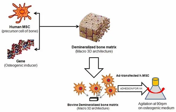
hMSCs and genetic modified hMSCs were trypsinized and resuspended in 2 x 10⁵ cells/ 20µl culture medium supplemented with 50µg/ml of ascorbic acid (Sigma-Aldrich, St. Louis, MO, USA) and 5mM of β-glycerol phosphate (Sigma-Aldrich, St. Louis, MO, USA) as osteogenic media. The concentrated cell suspensions were seeded onto the prepared scaffolds. The used scaffold was bDBM sponge. Briefly, bDBM sponge was prepared to 0.5 x 0.5 x 0.1 cm3 size and demineralized used by standard HCl/Triton X-100 treatment. The demineralized bDBM sponges were sterilized by the γ-irradiation at 25kGy. Cells were allowed to adhere to bDBM for 1hour and transferred to 125ml Erlenmeyer flask (Corning, Steuben County, NY, USA). Each flask was contained 5 each cells-seeded scaffolds. 50ml of osteogenic media was added to flask (Figure 1). For animal study, a part of scaffold was collected from flask at 24 hour and implanted to subcutaneous on nude mouse. The rest of scaffolds were cultured for 14 days and analyzed cell morphology by a scanning electron microscopy (SEM) and SEM-energy-dispersive X-ray spectroscopy (SEM-EDX). Culture media were changed every 3 days.
Figure 1. Scheme of 3D culture with adenovirus-transduced hMSCs on bDBM scaffolds bDBM sponges were prepared to 0.5 x 0.5 x 0.1 cm3 size and adhered adenovirus-transduced hMSCs for 1 hour. Cells-adhered sponges were transferred to 125ml Erlenmeyer flask. Every group was cultured with agitating at 90 rpm.
2. Construction and preparation of adenoviral vectors
The human alk-2(halk-2) cDNA was generated by the standard PCR protocol using set consisting of BamHI restriction site contained upstream primer and HindIII restriction site contained downstream primer. The former had a Kosak sequence at position -3 in the original sequence. The latter had a stop codon (encodes 509 amino acids). The PCR product of human
alk-2 cDNA contains 1 GS domain and 1 protein kinase domain. The human bmp-2 (hbmp-2)
cDNA as the positive control to osteogenesis was also generated by a similar method, and it encoded 396 amino acids. These receptor and ligand cDNAs were primarily subcloned into TOPO-TA cloning vector (Invitrogen, San Diego, CA, USA). The nucleotide sequence of the amplified cDNA was verified using an ABI PRISM377 automatic DNA sequencer. The sequence confirmed cDNA were digested by BamHI (NEB, Beverly, MA, USA) and HindIII (NEB, Beverly, MA, USA) at 37℃ for 1hour and eluted by Gel extraction kit (QIAGEN Inc., Hilden, Germany). The pCA14 shuttle vector was also excised by BamHI and HindIII at same condition and prepared by cleanup method for ligation with cDNAs. Restriction enzyme digested pCA14 vector and halk-2 cDNA or hbmp-2 cDNA were ligated at room temperature for 2 hours and then transformed to Escherichia coli DH5α competent cell. Positive transformants (named each vector, pCA14/halk-2 and pCA14/hbmp-2) were collected and verified used by the standard PCR method. To finally clone each cDNA to adenoviral vector, type 5 adenoviral vector, vmRL-H5dl324Bst was linearized by BstBI (BMS, IN, USA) digestion and pCA14/halk2 was linearized by NdeI/ScaI (BMS, IN, USA) digestion. Also, pCA14/hbmp2 was linearized by XmnI (NEB, Beverly, MA, USA) digestion. Type 5 adenoviral vector was kindly provided by Dr. Yoon at Cancer Research Center, Yonsei university. The linearized pCA14/halk2 or pCA14/hbmp2 was co-tranformed into E. coli
BJ5183 competent cell together with BstBI-digested vmRL-H5dl324Bst for homologous recombination. To verify the respective homologous recombinants, the plasmid DNA purified from overnight E.coli culture was digested with HindIII, and the digestion pattern was analysis. Also, the plasmid DNA (vmRL-H5dl324Bst/halk-2 and vmRL-H5dl324Bst/hbmp-2) was checked PCR to each cDNA to verify finally. The proper homologous recombinant adenoviral plasmid DNA was digested with PacI (NEB, Beverly, MA, USA) and transfected into 293A cells (Invitrogen, Rockville, MD, USA) to generate each cDNA-contained adenovirus, Ad/ALK-2 and Ad/BMP-2.
To generate LacZ-expressing adenovirus, a shuttle vector was created in which the lacZ gene was excised from plasmid pcDNA/hygro/LacZ (Invitrogen, Rockville, MD, USA) and cloned into pCA14 shuttle vector. Ad/lacZ preparation method was same as the previous Ad/ALK-2.
The titer (multiplicity of infection, MOI) used in this study was determined by absorbency of the dissociated virus at A260nm, where 1 absorbency unit is equivalent to 1012 viral particles per milliliter. The particle-to-infection unit (PFU) ratio was 100:1.
3. Transduction efficiency of adenovirus vector to hMSCs
A. 5-bromo-4-chloro-3-indolyl- β-D-galactopyranoside (X-gal) staining
hMSCs were plated onto 24-well plates at 2 x 10⁴ cells per well. At 24 hour, cells were transduced with Ad/lacZ adenovirus at various MOIs. β-Galactosidase (β-gal) activity was visualized 48hours later by 5-bromo-4-chloro-3-indolyl- β-D-galactopyranoside (X-gal;
Sigma-Aldrich, Steinheim, Germany) staining according to standard technique. Individual experiments were carried out with three sets of cells and all experiments were repeated at least three times.
B. The expression of ALK-2 and BMP-2 in hMSCs
To verify the expression of ALK-2 on Ad/ALK-2-transduced hMSCs, hMSCs were plated onto 6-well plates at 2 x 10⁵ cells per well and maintained for 24 hours in DMEM-LG medium supplemented with 10% FBS. The medium was replaced with fresh Hank’s balanced salt solution (HBSS; Invitrogen, Rockville, MD, USA) containing with 1% FBS at 1 hour before the transduction of adenovirus. The cells were transduced with Ad/ALK-2 adenovirus at 150 MOI and were added at 1 hour with DMEM-LG containing with 10% FBS. Negative control and mock (Ad/lacZ) group were treated at the same condition. After overnight incubation, the medium was replaced with fresh complete culture medium and transduced cells were cultured for another 2 days at 37℃, 5% CO2 atmosphere.
Cells transduced with Ad/ALK-2 and control were washed twice with PBS and solubilized in a cell lysis buffer containing with 20mM Tris-HCl, pH7.4, 150mM NaCl, 1% (w/v) Triton X-100, and protease inhibitor cocktail (Roche Applied Science, Branford, CT, USA). Lysates were briefly sonicated and cleared by centrifugation. Protein concentration was determined by the Bradford method (Bio-Rad Laboratories Inc., Hercules, CA, USA). 10µg of each sample was separated by SDS-PAGE. The proteins were transferred to nitrocellulose membranes. The membranes were then blocked for 1hour at room temperature in 50mM Tris-HCl, pH 7.6, 150mM NaCl, 0.1% Tween 20 containing with 5% nonfat dry milk (BD Biosciences, Franklin Lakes, NJ, USA). After blocking, the membranes were incubated with
anti-human ACVR antibody (R&D Systems, Minneapolis, MN, USA). The secondary antibody was used by a goat anti-mouse IgG conjugated to horseradish peroxidase (R&D systems, Minneapolis, MN, USA). Finally, the blots were visualized using enhanced chemiluminescence plus (ECL-PLUS; GE, Buckinghamshire, UK).
4. Assay to osteogenic differentiation of hMSCs
A. Histochemical staining of osteogenic phenotype
Histochemical analysis of alkaline phosphatase activity was carried out as describes. hMSCs were plated onto 24-well plates at 2 x 10⁴ cells per well and transduced with Ad/ALK-2 adenovirus at a various MOI. The transduced cells were maintained for 7 days and the medium was changed twice during the course of the experiment. The cells were washed with PBS and fixed with 60% citrate buffered acetone at room temperature for 30 seconds. After washing with distilled water, cells were incubated for 30 minutes with a mixture of 0.1mg/ml naphthol AS-MX phosphate (Sigma-Aldrich, St. Louis, MO, USA), 0.5% N,N-dimethylformamide, 2mM MgCl2, and 0.6mg/ml fast blue BB salt (Sigma-Aldrich, St. Louis, MO, USA) in 0.1M Tris-HCl, pH8.5, at room temperature, and rinsed with distilled water. Counter staining was used by Mayer’s hematoxylin solution (Sigma-Aldrich, St. Louis, MO, USA). Finally, the stained cells were air-dried and photographed.
For the determination of mineralized nodules, hMSCs were plated and transduced as same as the previous describes. Cells were maintained for 7 days and the medium was changed twice during the course of the experiment. The cultured medium was supplemented with 50µg/ml
ascorbic acid and 5mM β-glycerol phosphate. At the end of the treatment, the cells were washed twice with PBS, and the formation of in vitro mineralized nodules was determined by alizarin red-S staining and von Kossa staining. Alizarin red-S is a dye that binds selectively to calcium salts and binds about 2 mol of calcium/mol of dye.
For alizarin red-S staining, cells were fixed at 4℃ for 1 hour with 70% ethanol and rinsed with distilled water. The fixed cells were stained for 10 minutes at room temperature with 40mM alizarin red-S (Sigma-Aldrich, St. Louis, MO, USA) in 0.1M borate buffer, pH4.0 and washed with PBS for 5 minutes. Finally, the stained cells were air-dried and photographed.
For von Kossa staining, cells were fixed with 10% neutral buffered formalin for 1hour and rinsed with distilled water. The fixed cells were stained with 3% fresh made AgNO3 (Sigma-Aldrich, St. Louis, MO, USA) and developed in front of a 60W lamp until the calcium turn to black. And then stained cells were counterstained with nuclear fast red (Sigma-Aldrich, St. Louis, MO, USA). Finally, the stained cells were air-dried and photographed.
B. Reverse-transcription-polymerase chain reaction (RT-PCR) of osteogenic marker genes
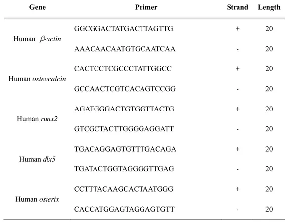
Total cellular RNA was eluted by selective binding to a silica gel-based membrane using an RNeasy mini kit (QIAGEN Inc., Hilden, Germany). For reverse transcription, a 50µl RT premix reaction (Bioneer, Deajun, Korea) contained 1µg total RNA and 12.5ng/µl oligo (dT)12-18 primers (Invitrogen, Rockville, MD, USA). Reactions were carried out at 42℃ for 1 hour, followed by inactivation of the enzyme at 95℃ for 5 minutes. For PCR amplification, each 20µl PCR premix reaction (Bioneer, Deajun, Korea) contained 1µl of RT aliquot and 10nmol each primer set (Table 1; Bioneer, Deajun, Korea). Following an initial denaturation step of 2
minutes at 94℃, amplification consisted of 25~35cycles of 5 seconds at 94℃, 5~30 seconds at optimal temperature (Table 2), and 30 seconds at 72℃, followed by final extension step for 5 minutes at 72℃. Amplification was performed in a ThermoHybrid Px2 system (ThemoHybaid, Franklin, MA, USA). Specific primers were designed from sequence available in the data banks (Table 2). The RT-PCR products were separated by electrophoresis on 2% agarose gels and stained with ethidium bromide. The intensity of the products was quantified using the BioImage Visage 110 system (Bio-Rad Laboratories Inc., Hercules, CA, USA).
No PCR product was observed when the RT reaction volume was replaced with water in PCR reaction (data not shown). β-actin gene was used to ascertain that an equivalent amount
of cDNA was synthesized from the different samples.
C. SEM and SEM-EDX of Ad/ALK-2-transduced hMSCs-seeded bDBM sponge
For the biological sample preparation of SEM, hMSCs-seeded bDBM sponges were fixed with 2.5% glutaraldehyde (Sigma-Aldrich, St. Louis, MO, USA), and subjected to a serial graded ethanol for dehydration (50, 70, 95, and 100%). After dehydration, the specimens were immersed in hexamethyldisilazane (HMDS; EM sciences, Hatfield, AR, USA) and air-dried. Upon drying, they were mounted on the SEM stubs. The specimens were coated with gold to improve the conductivity. The secondary electron mode was applied during SEM observation. EDX was also obtained during SEM observation, which was coupled with EDX detector.
5. Assay of MAPK signaling in ALK-2-overexpressed hMSCs
To determine the correlation of ALK-2 or BMP-2 to MAPK inhibition, MAPK inhibitors and ALK inhibitor were treated to hMSCs at 72 hours after adenovirus transduction. Cells were incubated for 16 hours with 50µΜ PD98059, 5 µΜ SB203580, 10 µΜ SP600125 (all reagent from Sigma-Aldrich, St. Louis, MO, USA), or 5 µΜ dorsomorphin dihydrochloride (Tocris Bioscience, Ellisville, MO, USA).
MAPK or ALK inhibitor treated hMSCs were washed with PBS and solubilized in a cell lysis buffer containing containing 20mM Tris-HCl, pH7.4, 150mM NaCl, 1%(w/v) Triton X-100, and protease inhibitor cocktail. Lysates were briefly sonicated and cleared by centrifugation. Protein concentration was determined by the Bradford method. 10µg of each sample was separated by SDS-PAGE. The proteins were transferred to nitrocellulose membranes and immunoblotted with anti-Smad1 antibody, anti-phospho-Smad1/5/8 antibody, anti-p44/42 antibody, anti-phsopho-p44/42 antibody, anti-p38 antibody, anti-phospho-p38 antibody, anti-SAPK/JNK antibody, or anti-phospho-SAPK/JNK antibody (all from Cell Signaling, Beverly, MA, USA). The blots were visualized using enhanced chemiluminascence detection system (GE, Buckinghamshire, UK).
TABLE 1. Sequence of the oligonucleotide primers for PCR analysis
Gene Primer Strand Length
Human β-actin GGCGGACTATGACTTAGTTG AAACAACAATGTGCAATCAA + - 20 20 Human osteocalcin CACTCCTCGCCCTATTGGCC GCCAACTCGTCACAGTCCGG + - 20 20 Human runx2 AGATGGGACTGTGGTTACTG GTCGCTACTTGGGGAGGATT + - 20 20 Human dlx5 TGACAGGAGTGTTTGACAGA TGATACTGGTAGGGGTTGAG + - 20 20 Human osterix CCTTTACAAGCACTAATGGG CACCATGGAGTAGGAGTGTT + - 20 20
TABLE 2. Amplification condition of osteogenic marker genes
Gene Temperature (℃) Cycle Product size(bp)
Human β-actin 53 26 238 Human osteocalcin 62 28 299 Human runx2 58 30 189 Human dlx5 58 30 225 Human osterix 55 30 299 - 18 -
6. Histological assay to the in vivo implantation of Ad/ALK-2-transduced hMSCs-seeded bDBMs
A. Implantation of genetic modified hMSCs-seeded bDBM scaffold
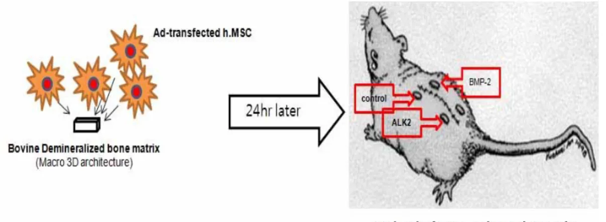
All animal studies were conducted in accordance with principles and procedures approval by the University of Yonsei Committee for Animal Resources. Implantation of Ad/ALK-2 or Ad/BMP-2-transduced hMSCs-seeded bDBM scaffolds was performed at specific pathogen free (SPF) zone. Briefly, 8-week-old Balb/c-nu/nu mice were anesthetized, and 5mm-long incision was made on the back skin. hMSCs-adhered scaffolds were implanted to subcutaneous part and the incision was closed with absorbable sutures. The mice were maintained for 2 weeks or 4 weeks (Figure 2). At the times, each group was sacrificed by CO2 inhalation and the implanted scaffolds were harvested. The harvested scaffolds were performed to histological analysis.
Figure 2. Implantation of genetic modified hMSCs-adhered bDBM sponges to nude mice Sponges were implanted to subcutaneous part of mice and maintained for 2 or 4 weeks.
B. Hematoxylin & eosin (H&E) staining of the implants
The harvested scaffolds were fixed in 10% neutral buffered formalin (NBF) and decalcified with 5% formic acid in 10% NBF for 3 day with gently shaking. During the decalcification, the decalcified solution was changed every day. And then 3µm paraffin-embedded sections were prepared and stained by standard H&E staining method. The H&E stained slides were observed by bright-field microscopy (Olympus, Tokyo, Japan).
C. Immunofluorescent (IF) staining of ALK-2 protein
To verify the expression of ALK-2 on the harvested implants, IF staining was performed with anti-human ACVR antibody (R&D Systems, Minneapolis, MN, USA). For antigen retrieval of formalin-fixed paraffin embedded specimens, paraffin sections were immersed in 10mM Tris, 1mM EDTA, and 0.05% Tween-20, pH9.0 for 30 minutes and allowed to cool for 20 minutes at room temperature. Antigen retrieval sections were rinsed with 0.05% Tween-20 and blocked for 30 minutes. The sections were followed by incubation with primary antibody to human ALK-2 and then incubated with Alexa488-conjugated antibody against mouse immunoglobulin (Molecular Probes Inc., Eugene, OR, USA). After a final wash, the sections were covered with the mounting media contained DAPI (Vector Laboratories Inc., Burlingame, Canada) and analyzed by confocal laser-scanning microscopy (Carl Zeiss Microimaging GmbH, Göttingen, Germany).
Ⅲ
. RESULTS
1. Transduction efficiency of the prepared adenovirus vectors to hMSCs
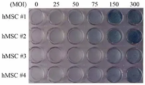
For the decision of optimal virus titer, Ad/LacZ was transduced to hMSCs and stained by X-gal staining (Figure 3). The difference of transduction efficiency of hMSCs from different patients was observed. However, the average transduction efficiency was over 80% at 150MOI of Ad/LacZ. These experiments were repeated at three times.
Figure 3. X-gal staining of Ad/LacZ-transduced hMSCs Cells were plated onto 24-well plate at 2 x 10⁴ cells per well. Ad/LacZ was diluted with HBSS and transduced for 1 hour. X-gal staining was performed at 48 hours after adenovirus transduction. Negative control (0 MOI) was applied with the same volume of HBSS solution.
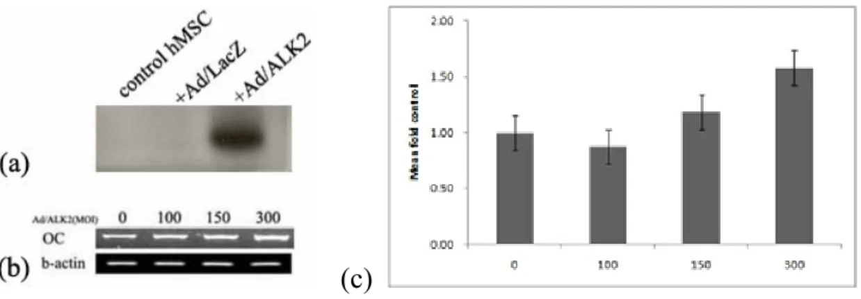
2. Overexpression of ALK-2 in hMSCs induces expression of osteocalcin mRNA
ALK-2 proteins were over-expressed in Ad/ALK-2-transduced hMSCs compared to control and Ad/LacZ group (Figure 4-a). These increased expression of ALK-2 stimulated expression of osteocalcin mRNA, osteogenic marker gene in hMSCs (Figure 4-b). Expression level of osteocalcin mRNA at 300 MOI of Ad/ALK-2 increased 1.5-fold compared to control group (Figure 4-c). These results suggested ALK-2 protein activates osteogenic cell signaling and then stimulates the expression of terminal differentiation marker gene.
(c)
Figure 4. Expression of ALK-2 protein and osteocalcin mRNA in Ad/ALK-2-transduced hMSCs Cell extract and total RNA were extracted at 72 hours after virus transduction. (a) ALK-2 protein expression in adenovirus-transduced hMSCs was detected by western blot analysis. Cells were transduced with 150MOI of adenoviral vector and control group was treated with HBSS as same as virus volume. (b) Result of RT-PCR to human osteocalcin mRNA. β-actin mRNA was used as internal control. (c) Densitometric analysis of osteocalcin
mRNA expression according to increase of Ad/ALK-2-transduction titer to hMSCs (p < 0.001).
3. Overexpression of ALK-2 in hMSCs stimulates mRNA expression of osteogenic
transcription factor
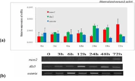
To verify the overexpression of ALK-2 stimulates another osteogenic marker gene expression at transcriptional level, RT-PCR of a typical osteogenic transcription factors such as
runx2, dlx5, and osterix was performed (Figure 5). Overexpression of ALK-2 stimulated
expression of these mRNA in hMSCs. Expression of dlx5 mRNAs was upregulated at 3 hours after transduction, and osterix and runx2 mRNA was upregulated at 24 hours and 48 hours after transduction. These results demonstrated the overexpression of ALK-2 in hMSCs activates expression of runx2, dlx5, and osterix mRNA.
(a)
(b)
Figure 5. RT-PCR of runx2, dlx5, and osterix mRNA as the osteogenic transcription factor Total RNA was extracted from Ad/ALK-2-transduced hMSCs and virus titer was 150MOI. Every total RNA was extracted according to time period after virus transduction. (a) Densitometric analysis to RT-PCR of runx2, dlx5, and osterix mRNA. (b) Electrophoresis of RT-PCR products. Every end product was loaded 5μl and visualized by EtBr staining.
4. Overexpression of ALK-2 differentiates hMSCs to osteoblastic cells
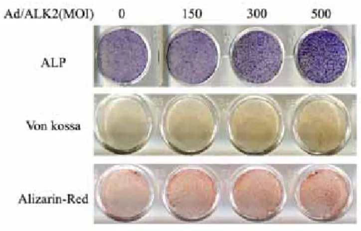
To verify the osteogenic phenotype of Ad/ALK-2-transduced hMSCs, alkaline phosphatase staining, Von kossa staining, and alizarin-red staining were performed (Figure 6). Alkaline phosphatase staining is a very common staining to osteoblastic differentiation. Von Kossa staining and alizarin-red staining show a mineral deposit of the surface on osteoblasts. At 7 day after transfection of Ad/ALK-2 to hMSCs, all osteogenic stainings showed the titer-dependent increased intensity of pigments. Specially, a mineral deposit of hMSCs induced by the transduction of Ad/ALK-2 was observed at very early time by von Kossa and alizarin-red staining. Most studies related to osteogenesis have demonstrated the mineral deposit was the terminal osteogenic differentiation marker and this mineral deposit was observed at over 2 weeks with stimulating continuously. However, the mineral deposit of Ad/ALK-2-transduced hMSCs was observed at 7 days. These results showed the accumulation of osteogenic stimulation by the overexpression of ALK-2 accelerated the mineral deposit surface on hMSCs.
Figure 6. Ostegenic phenotype staining of Ad/ALK-2-transduced hMSCs All stainings were performed at 7 days after adenovirus transduction. Culture medium was supplemented with 50µg/ml ascorbic acid and 5mM β-glycerol phosphate.
5. 3D culture of Ad/ALK-2-transduced hMSCs used by bDBM sponges
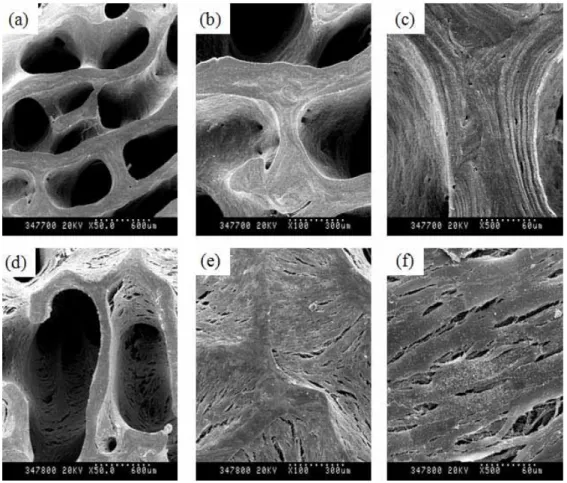
To prove the feasibility of Ad/ALK-2-transduced hMSCs for developing the optional tools of ex vivo bone regeneration therapy, Ad/ALK-2-transduced hMSCs was adhered on bDBM sponge and cultured with agitating. And to observe cell morphology with hMSCs-adhered scaffolds, SEM and SEM-EDX were performed.
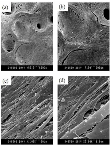
The prepared bDBMs were observed to a collagenous matrix, which maintained a bony structure and had not any other cells (Figure 7-a~c). Average diameter of inner pore was 300µm over. To check cell adhesion surface on bDBM sponge, only hMSCs were adhered for 1hour on bDBM sponge and immediately transferred to Erlenmeyer culture flask for the agitation culture. hMSCs were well adhered to bDBM sponge and spread out in spite of harsh agitation at 90rpm (Figure 7-d~f). However, any other morphological change without well adhesion was not observed.
To use mock control, Ad/LacZ was transduced to hMSCs and adhered to bDBM as same method as only MSC group (Figure 8). Unexpectedly, proliferation of Ad/LacZ-transduced hMSCs increased and spread out widely surface on bDBM. In this group, the inner pore of bDBM sponge was covered in Ad/LacZ-transduced hMSCs. β-Galactosidase has been well known as a control marker gene in vector system. But in animal, β-galactosidase also has been known which plays functional roles in the formation of extracellular elastic fibers (elastogenesis) and in the development of connective tissue. It seems to be identical to the elastin-binding protein (EBP), a major component of the non-integrin cell surface receptor expressed on fibroblasts, smooth muscle cells, chondroblasts, leukocytes, and certain cancer cell types 37. Accordingly, we thought this function of β-galactosidase could demonstrate the increase proliferation of Ad/LacZ-transduced hMSCs on DBM sponge with agitating culture.
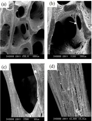
Newly formation of porous channel on Ad/ALK-2-transduced hMSCs at 150 MOI was observed (Figure 9), which was not observed at only hMSC group or Ad/LacZ-transduced group. Also, mineral deposits on cell surface were observed when hMSCs were treated with 1,000 MOI of Ad/ALK-2 (Figure 10), which was very similar to hydroxyapatite crystal.
To analyze these materials, SEM-EDX was performed (Figure 12). Because the specimen of SEM-EDX was used with the same specimen of SEM (Figure 12-a), the gold (Au) peak in the result of SEM-EDX was withdrawn from final analysis. Hydroxyapatite is a major component of bone mineral, which consist of calcium (Ca) and phosphate (PO4). Figure 12-b showed three peaks of calcium and this portion was over 10% versus total composite. Finally, we demonstrated that 3D culture of high titer of Ad/ALK-2-transduced hMSCs with agitating induces deposit of calcium phosphate surface on hMSCs within 14 days.
Figure 7. SEM photograph of only bDBM sponge (a~c) and hMSCs-adhered bDBM sponge (d~f) All groups were cultured with the osteogenic media for 14 days at 90rpm. About 2 x 10⁵ cells of hMSCs were adhered on bDBM (d~f). (a, d) magnification x 50, (b, e) magnification x 100, (c, f) magnification x 500.
Figure 8. SEM photograph of Ad/LacZ-transduced hMSCs-adhered bDBM sponge Cells-adhered bDBM sponges were cultured with the osteogenic media for 14 days at 90rpm. Virus titer of Ad/LacZ was 150MOI. (a) magnification x 50, (b) magnification x 100, (c) magnification x 1,000, (d) magnification x 5,000.
Figure 9. SEM photograph of Ad/ALK-2-transduced hMSCs-adhered bDBM sponge Cell-adhered bDBM sponges were cultured with the osteogenic media for 14 days at 90rpm. Virus titer of Ad/ALK-2 was 150MOI. (a) magnification x 50, (b) magnification x 100, (c) magnification x 500, (d) magnification x 2,000. Arrow mark; newly cell made porous channel.
Figure 10. SEM photograph of Ad/ALK-2-transduced hMSCs-adhered bDBM sponge Cells-adhered bDBM sponges were cultured with the osteogenic media for 14 days at 90rpm. Virus titer of Ad/ALK-2 was 1,000 MOI. (a) magnification x 50, (b) magnification x 100, (c) magnification x 5,000, (d) magnification x 50,000.
(a)
(b)
Figure 11. SEM-EDX analysis of the minerals surface on Ad/ALK-2-transduced hMSCs (a) Pre-scanning photograph of Ad/ALK-2 (1,000MOI)-transduced hMSCs-adhered bDBM sponge before EDX analysis. Red square is a target of EDX analysis, Spectrum 9. (b) Analysis graph of spectrum 9. Au is gold and Ca is calcium.
6. ALK-2 activates Smad1/5/8 signaling and the phosphorylation of p38 protein
ALK-2 protein is a membrane receptor to TGF-β family. To verify the overexpression of ALK-2 in hMSCs activates type I TGF-β receptor-mediated cell signaling, western blot to Smad proteins was performed. Smad1/5/8 proteins are known to BMP-mediated osteogenic cell signaling molecule. These proteins are phosphorylated by type I TGF-β receptor such as ALK-2. Phosphorylation of smad1/5/8 proteins was activated by increasing virus titer of Ad/ALK-2 to hMSCs (Figure12-a). This demonstrated the overexpression of ALK-2 only without a ligand stimulated downstream cell signaling over membrane receptor.
BMP-2 protein was known also as MAPK stimulator because BMP-2 poses a various function for cell differentiation and proliferation. To verify the cell signaling by overexpression of ALK-2 represent to the same pattern of BMP-2 cell signaling, western blot to Smad1/5/8 and MAPK proteins was performed after adenovirus transduction and MAPK or ALK inhibition (Figure12-b,c). Phosphorylation of Smad1/5/8 protein on Ad/ALK-2-transduced hMSCs showed as same as Ad/BMP-2-transduced group, which was verified by western blot after the treatment of ALK inhibitor, dorsomorphin. This result demonstrated that phosphorylation of Smad1/5/8 depend on the activation of type I receptor protein. Expression pattern of Erk2 (p44/42) and SAPK/JNK proteins showed as same as both group. However, phosphorylation of p38 on Ad/ALK-2-transduced group increased over 20-fold compared to Ad/BMP-2-transduced group. This over-phosphorylation of p38 in Ad/ALK-2-transduced group was reported in Fibrodysplasia ossificans progressive (FOP), which disease is a rare autosomal dominant disorder characterized by congenital malformation of the great toes and by progressive heterotopic bone formation in muscle tissue. This disorder was reported by the mutation of BMP type I receptor, ALK-2.
(a)
(b)
(c)
Figure 12. Smad1/5/8 and MAPK expression in Ad/ALK-2-transduced and Ad/BMP-2-transduced hMSCs (a) Western blot to ALK-2 and phospho-Smad1/5/8 in Ad/ALK-2-transduced hMSCs. Virus titer of Ad/ALK-2 was 0, 100, 150 and 300 MOI. (b) Western blot of Smad1/5/8 and MAPK in Ad/ALK-2-transduced hMSCs and (c) Ad/BMP-2-transduced hMSCs. (b) and (c) were treated with 150MOI virus titer. 10μg protein sample was loaded for electrophoresis. ; C, control; L, Ad/LacZ-transduced group; (-), transduction of only Ad/ALK-2 or Ad/BMP-Ad/ALK-2 ; the following groups was treated with MAPK inhibitor or ALK inhibitor after adenovirus-transduced hMSCs, PD, treatment of 50µΜ PD98059; SB, treatment of 5 µΜ SB203580; SP, 10 µΜ of SP600125, or DO, 5 µΜ of dorsomorphin dihydrochloride.
7. Implantation of Ad/ALK-2-transduced hMSCs to immunodeficient mice
To prove the osteogenic activity of ALK-2 in vivo model, 3D cultured adenovirus-transduced hMSCs-adhered bDBM sponges were implanted to subcutaneous in nude mice (Figure2). Each group was divided 2 weeks and 4 weeks and maintained. At 2 weeks, only Ad/BMP-2-transduced group as the positive control showed the histological phenotype of osteogenic differentiation, which group was observed osteoblastic lining cells and fat marrow around hMSCs-adhered implants. All groups were observed the implanted cell and sponge well took its place, and the boundary between the implanted cell and mouse cell was observed clearly (Figure 13). At 4 weeks, control groups were not observed osteogenic differentiation, but Ad/BMP-2-transduced groups were observed newly synthesized bone matrix and bone marrow formation. Most of newly bone in Ad/BMP-2 groups was generated from the outside of implanted sponge. This demonstrated a common phenotype of BMP-2 action related to new bone formation because a fresh nutrient and oxygen for new tissue generation was existed abundant around the outside of implantation. Ad/ALK-2 groups were observed the newly synthesized the collagenous matrix, but did not show osteogenic differentiation phenotype at 300 MOI of Ad/ALK-2 (Figure 14). However, 1,000MOI-applied Ad/ALK-2 groups showed osteogenic differentiation phenotype in some part of the implants compared to control. This phenotype was observed at the outside of implants (Figure 15).
IF staining to ALK-2 in the implants showed ALK-2 protein was overexpressed in the implanted hMSCs at 4 weeks (Figure 16). However, these results demonstrated the overexpression of ALK-2 protein could not stimulate enough to induce osteogenesis at in vivo model compared to BMP-2 overexpressed groups, and on the case of Ad/BMP-2 groups, a paracrine effect of BMP-2 induced a robust osteogenic differentiation at in vivo. Ad/ALK-2
groups showed a delayed new bone formation at in vivo, which demonstrated that because ALK-2 protein is a membrane intercalating receptor, which could not induced a powerful bone formation even though they had the stimulating potential of osteogenic cell signaling and
osteocalcin mRNA expression at in vitro.
Figure 13. H&E staining of the hMSCs-adhered bDBM sponges from nude mice at 2 weeks after the implantation The used virus titer was 150MOI. (a, b) only hMSC-DBM group, (c, d) Ad/BMP-2-transduced hMSCs-bDBM group, (e, f) Ad/ALK-2-transduced group. (Upper) magnification x 40, (Bottom) magnification x 100.
Figure 14. H&E staining of the hMSCs-adhered bDBM sponges from nude mice at 4 weeks after the implantation The used virus titer was 150MOI. (a, b) only hMSCs-bDBM group, (c, d) Ad/BMP-2-transduced hMSCs-bDBM group, (e, f) Ad/ALK-2-transduced group. (Upper) magnification x 40, (Bottom) magnification x 100.
Figure 15. H&E staining of the hMSCs-adhered bDBM sponges from nude mice at 4 weeks after the implantation The used virus titer was 1,000MOI. (a) Ad/BMP-2-transduced hMSCs-bDBM group, (b) Ad/ALK-2-transduced group. Magnification x 100.
Figure 16. Expression of human ALK-2 protein in the implanted site of nude mouse The implants were Ad/ALK-2-transduced hMSCs-bDBM group at 2 weeks (a) and at 4 weeks (b). Primary antibody to ALK-2 protein was conjugated with Alexa 488 and rodamine F-actin as counterstain was used.
Ⅳ
. DISCUSSION
Musculoskeletal conditions are increasingly becoming one of the major health concerns worldwide because of an aging population and increased occurrence of sports-related injuries according to be improve life style 38. Entering 2005, there is increase approximately to 240 billion dollars worldwide to musculoskeletal disease care, especially fractures healing market, and between 5% and 10% of these result in nonunion or delayed union 39. Collectively, this represents a substantial cause of morbidity, missed work, and medical cost. Biologics that promote bone-healing are needed in the treatment of established nonunions as well as in the acute treatment of certain fractures associated with many clinical options for stimulating bone formation, but each has substantial limitations 40-43. To date, autologous bone remains the gold-standard graft material 44,45. However, its harvest can cause substantial morbidity, including hematoma formation, infection, numbness at the incision site, and persistent pain. In addition, the limited quantity of autologous bone available for harvest may not be sufficient for the treatment of large defects 46.
Recombinant human BMPs (rhBMPs) have recently emerged as a bone-graft substitute. RhBMP-2 has been approved by the U.S. FDA for use in anterior lumbar interbody fusions and the treatment of open tibial fractures 47,48. RhBMP-7 (osteogenic protein-1) has been approved under a “Humanitarian Device Exemption (HDE)” for the treatment of recalcitrant long-bone nonunions and for use in revision posterolateral spinal arthrodeses 49. There is evidence that BMPs are more effective than autograft for promoting fracture-healing and spinal fusion; consequently, the introduction of BMPs has been met with a great deal of enthusiasm by orthopaedic community. However, the use of BMP has not been optimized. High doses of growth factor are needed to produce an adequate bone formation response. Presently, rhBMP is
being administrated at doses that a million times greater than its normal concentration in bone, and there are concerns about both safety and the cost of such supraphysiologic doses. Clinical trials and preclinical studies have both shown a potential for ectopic bone formation as well as edema 50-53. These observations might partly be attributed to the collagen carriers used to deliver BMP, which have been hypothesized to be inefficient protein delivery systems.
These concerns have led to investigations of alternative protein delivery mechanisms to promote bone repair 23,26. Regional gene therapy offers a novel approach to difficult clinical problem. Genetic sequences encoding for growth factors can be transferred to cells at the fracture site, resulting in the production of osteogenic proteins in a localized, sustained, and physiologic manner. Preclinical animal models have demonstrated the tremendous potential of these techniques. In tissue-engineering strategy that will include a spectrum of treatment options such as autologous bone-marrow injection, and stem cell therapies. It is envisioned that gene therapy options will initially be available for the most severe clinical situations such as massive bone loss and recalcitrant nonunions 21. Despite its tremendous promise, the clinical application of gene therapy must be approached with caution. Thus far, clinical trials for gene therapy for inflammatory arthritis and metabolic disease have led two deaths. Any substantial morbidity will not be accepted in the treatment of nonfatal musculoskeletal conditions 24,25,54.
Gene therapy is a tool that can be used to deliver osteoinductive proteins at a desired location. It may be a more efficient growth factor delivery system than are the current methods of rhBMP delivery, which have substantial limitations, such as a short duration of action. The commercially available products deliver BMPs with a type I collagen carrier. There is an initial burst of BMP release with a half-life of less than ten minutes, followed by a second phase of gradual release with a half-life of between three and five days. A more prolonged expression of BMP might enhance the fracture healing process. Gene therapy may offer a solution. Cells
present within the body can be genetically manipulated to produce osteogenic proteins in the area of interest. This would provide a more physiologic delivery system, with continuous in vivo production of protein at a relative constant level for sustained period 23,49,55.
Stem-cell-based therapies are another emerging option for promoting bone regeneration as the previous describes. Stem cells are defined by their distinct ability to self-renew and to differentiate into multiple cell types. The cell that has been most extensively studied for orthopaedic applications is the mesenchymal stem cell. This is an adult stem cell that is found in tissues of mesoderm origin such as bone marrow, adipose tissue, muscle, and skin 56-58. When exposed to the appropriate growth factors, these multipotent cells can differentiate into chondrocytes or osteocytes and may contribute to bone formation 27,56,59. Mesenchymal stem cells seeded onto scaffolds such as hydroxyapatite have induced healing of critical-sized bone defects in severe animal models 60,61. Moreover, percutaneous injection of bone marrow aspirates has been used to treat tibial nonunions with moderate success 25,62-65.
Stem-cell-based gene therapy may be another way to increase the power of these techniques. Mesenchymal stem cells can be genetically modified to overexpress osteogenic protein such as BMPs. These growth factors stimulate the mesenchymal stem cells to differentiate into bone forming cells through autocrine signaling, and they recruit host osteogenic progenitors through paracrine signaling. Then, almost all scientist possessed of the previous concepts have been questioned how choice the combination of these therapeutic tools.
The purpose of the present study was to find a new candidate gene for bone specific regeneration and a new method for combining stem-cell based tissue engineering. BMP-2 has been chosen and researched by most orthopeadic researchers because which have the robust osteogenic and paracrine effect. However, BMP-2 participates to the many biological processes from embryonic development to adult and from osteogenesis or chondrogenesis to
adipogenesis, which protein is indeed a multiplayer in the human body. There has been thought BMP-2 is a protein very hard to regulate at in vivo environment because of their paracrine effect.
In the past studies, some researchers on orthopeadic therapy have demonstrated the ALK-2 protein as BMP-ALK-2 and BMP-7 type I receptor has the osteogenic potential at in vitro. ALK-ALK-2 is a protein kinase itself, which possess a GS domain and a protein kinase domain. Using this kinase activity, Ulrich and colleagues also have proved the continuous active ALK-2 (caALK-2) simulates continuously osteogenesis 36,66. However, the previous study used by caALK-2 was doubted that the continuous stimulation of caALK-2 cannot be safe to bone regeneration therapy in human body because some researchers demonstrated wild type ALK-2 has a potential enough to stimulate osteogenic differentiation of MSCs. Also. the past studies have reported that osteocalcin expression was not upregulated by overexpression of the receptor Smad protein only or Co-Smad. These observations have been raised to the possibility that additional pathway, such as ALK-2-dependent, might be required for osteocalcin expression, a unique trait specific of the osteoblast phenotype.
In this present study, the overexpression of ALK-2 protein used by adenoviral vector system stimulated enough to differentiate hMSCs to osteoblasts in vitro and induced Smad1/5/8 activation and MAPK stimulation in hMSCs was observed. Specially, the up-regulation of p38 MAPK by the overexpression of ALK-2 have recently shown to control BMP-2-induced osteocalcin expression in myoblastic C2C12 cells 29,35,67. At the present in vivo experiments using by Ad/ALK-2 and 3D scaffolds, bone formation by the overexpression of ALK-2 on the implanted site was failed. These results might be explained that ALK-2 expression did not recruit the precursor cell enough to bone regeneration from nearby environments because ALK-2 protein is not a secreted protein.
However, we thought the present studies demonstrated that on the case of bone union in elderly patients, the restoration of this BMP receptor in hMSCs can increase the sensitivity to growth factors, such as BMP-2, and also if the restoration of this BMP receptor in hMSCs combine with rhBMPs at in vivo therapy, this approach can increase bone regeneration and decrease the using dosage of rhBMPs for bone union. Also, in ex vivo therapy, if the combination of BMP receptor and BMP gene with the appropriate scaffold join to bioreactor culture system, hand-made bone formation could be realized on the extra-corporeal environment.
Based on these results of the present study, we are undergoing studies to verify the potentialities of the combination therapy using by Ad/ALK-2 and Ad/BMP-2 or rhBMP-2 and also to develop the simulated bioreactor system to bone regeneration, which bioreactor can scale-up to 3D culture with Erlenmeyer flask and agitation stress on the previous before.
Ⅴ
. CONCLUSION
Overexpression of BMP type I receptor ALK-2 induces osteogenic phenotype in hMSCs. Therefore, ALK-2 gene can be a new therapeutic candidate for osteogenic gene therapy and for restoration of receptor to growth factor in hMSCs.
REFERENCES
1. Damien, C.J. & Parsons, J.R. Bone graft and bone graft substitutes: a review of current technology and applications. J Appl Biomater 2, 187-208 (1991).
2. Awad, H.A., et al. Recent advances in gene delivery for structural bone allografts. Tissue
Eng 13, 1973-1985 (2007).
3. Lee, K.J., Roper, J.G. & Wang, J.C. Demineralized bone matrix and spinal arthrodesis.
Spine J 5, 217S-223S (2005).
4. Tiyapatanaputi, P., et al. A novel murine segmental femoral graft model. J Orthop Res 22, 1254-1260 (2004).
5. Wheeler, D.L. & Enneking, W.F. Allograft bone decreases in strength in vivo over time.
Clin Orthop Relat Res, 36-42 (2005).
6. Anderegg, C.R., Martin, S.J., Gray, J.L., Mellonig, J.T. & Gher, M.E. Clinical evaluation of the use of decalcified freeze-dried bone allograft with guided tissue regeneration in the treatment of molar furcation invasions. J Periodontol 62, 264-8 (1991).
7. Kakiuchi, M. [Bone allograft and its clinical application--defatted, gas-sterilized bone allograft]. Nippon Seikeigeka Gakkai Zasshi 68, 26-35 (1994).
8. Mattout, P., Nowzari, H. & Mattout, C. Clinical evaluation of guided bone regeneration at exposed parts of Branemark dental implants with and without bone allograft. Clin Oral
Implants Res 6, 189-195 (1995).
9. Skoff, H.D. Bone marrow/allograft component therapy. A clinical trial. Am J Orthop (Belle
Mead NJ) 24, 40-7 (1995).
10. Masters, L.B., Mellonig, J.T., Brunsvold, M.A. & Nummikoski, P.V. A clinical evaluation of demineralized freeze-dried bone allograft in combination with tetracycline in the treatment of periodontal osseous defects. J Periodontol 67, 770-781 (1996).
11. Gurinsky, B.S., Mills, M.P. & Mellonig, J.T. Clinical evaluation of demineralized freeze-dried bone allograft and enamel matrix derivative versus enamel matrix derivative alone for the treatment of periodontal osseous defects in humans. J Periodontol 75, 1309-1318 (2004).
12. Aichelmann-Reidy, M.E., Heath, C.D. & Reynolds, M.A. Clinical evaluation of calcium sulfate in combination with demineralized freeze-dried bone allograft for the treatment of human intraosseous defects. J Periodontol 75, 340-7 (2004).
13. Schwartz, Z., et al. Clinical evaluation of demineralized bone allograft in a hyaluronic acid carrier for sinus lift augmentation in humans: a computed tomography and histomorphometric study. Clin Oral Implants Res 18, 204-211 (2007).
14. Ilgenli, T., Dundar, N. & Kal, B.I. Demineralized freeze-dried bone allograft and platelet-rich plasma vs platelet-platelet-rich plasma alone in infrabony defects: a clinical and radiographic evaluation. Clin Oral Investig 11, 51-9 (2007).
15. Bianchini, M.A., et al. The use of freeze-dried bone allograft as an alternative to autogenous bone graft in the atrophic maxilla: a 3-year clinical follow-up. Int J
Periodontics Restorative Dent 29, 643-7 (2009).
16. Slosar, P.J., Josey, R. & Reynolds, J. Accelerating lumbar fusions by combining rhBMP-2 with allograft bone: a prospective analysis of interbody fusion rates and clinical outcomes.
Spine J 7, 301-7 (2007).
17. Nevins, M., Hanratty, J. & Lynch, S.E. Clinical results using recombinant human platelet-derived growth factor and mineralized freeze-dried bone allograft in periodontal defects.
Int J Periodontics Restorative Dent 27, 421-7 (2007).
18. Omura, S., et al. A carrier for clinical use of recombinant human BMP-2: dehydrothermally cross-linked composite of fibrillar and denatured atelocollagen sponge.
Int J Oral Maxillofac Surg 27, 129-134 (1998).
19. Chen, D., Zhao, M. & Mundy, G.R. Bone morphogenetic proteins. Growth Factors 22, 233-241 (2004).
20. Miyazono, K. & Miyazawa, K. Id: a target of BMP signaling. Sci STKE 2002, pe40 (2002). 21. Franceschi, R.T. Biological approaches to bone regeneration by gene therapy. J Dent Res
84, 1093-1103 (2005).
22. Yu, P.B., et al. BMP type I receptor inhibition reduces heterotopic [corrected] ossification.
Nat Med 14, 1363-9 (2008).
23. Keskin, D.S., Tezcaner, A., Korkusuz, P., Korkusuz, F. & Hasirci, V. Collagen-chondroitin
sulfate-based PLLA-SAIB-coated rhBMP-2 delivery system for bone repair. Biomaterials 26, 4023-4034 (2005).
24. Carofino, B.C. & Lieberman, J.R. Gene therapy applications for fracture-healing. J Bone
Joint Surg Am 90 Suppl 1, 99-110 (2008).
25. Peterson, B., et al. Healing of critically sized femoral defects, using genetically modified mesenchymal stem cells from human adipose tissue. Tissue Eng 11, 120-9 (2005).
26. Kamakura, S., Nakajo, S., Suzuki, O. & Sasano, Y. New scaffold for recombinant human bone morphogenetic protein-2. J Biomed Mater Res A 71, 299-307 (2004).
27. Hennig, T., et al. Reduced chondrogenic potential of adipose tissue derived stromal cells correlates with an altered TGFbeta receptor and BMP profile and is overcome by BMP-6. J
Cell Physiol 211, 682-691 (2007).
28. Keilhoff, G., Stang, F., Goihl, A., Wolf, G. & Fansa, H. Transdifferentiated mesenchymal stem cells as alternative therapy in supporting nerve regeneration and myelination. Cell
Mol Neurobiol 26, 1235-1252 (2006).
29. Fujii, M., et al. Roles of bone morphogenetic protein type I receptors and Smad proteins in osteoblast and chondroblast differentiation. Mol Biol Cell 10, 3801-3813 (1999).
30. Goumans, M.J. & Mummery, C. Functional analysis of the TGFbeta receptor/Smad pathway through gene ablation in mice. Int J Dev Biol 44, 253-265 (2000).
31. Bondestam, J., et al. Engagement of activin and bone morphogenetic protein signaling pathway Smad proteins in the induction of inhibin B production in ovarian granulosa cells.
Mol Cell Endocrinol 195, 79-88 (2002).
32. Valcourt, U., Kowanetz, M., Niimi, H., Heldin, C.H. & Moustakas, A. TGF-beta and the Smad signaling pathway support transcriptomic reprogramming during epithelial-mesenchymal cell transition. Mol Biol Cell 16, 1987-2002 (2005).
33. Lebrin, F., Deckers, M., Bertolino, P. & Ten Dijke, P. TGF-beta receptor function in the endothelium. Cardiovasc Res 65, 599-608 (2005).
34. Bonafoux, D. & Lee, W.C. Strategies for TGF-beta modulation: a review of recent patents.
Expert Opin Ther Pat 19, 1759-1769 (2009).
35. Aoki, H., et al. Synergistic effects of different bone morphogenetic protein type I receptors
on alkaline phosphatase induction. J Cell Sci 114, 1483-9 (2001).
36. Valcourt, U., Gouttenoire, J., Moustakas, A., Herbage, D. & Mallein-Gerin, F. Functions of transforming growth factor-beta family type I receptors and Smad proteins in the hypertrophic maturation and osteoblastic differentiation of chondrocytes. J Biol Chem 277, 33545-33558 (2002).
37. Hinek, A. Biological roles of the non-integrin elastin/laminin receptor. Biol Chem 377, 471-480 (1996).
38. Laurencin, C.T., Ambrosio, A.M., Borden, M.D. & Cooper, J.A., Jr. Tissue engineering: orthopedic applications. Annu Rev Biomed Eng 1, 19-46 (1999).
39. Vargas, B., Lutz, N., Dutoit, M. & Zambelli, P.Y. Nonunion after fracture of the anterior tibial spine: case report and review of the literature. J Pediatr Orthop B 18, 90-2 (2009). 40. Fuji, T., et al. Interspinous wiring without bone grafting for nonunion or delayed union
following anterior spinal fusion of the cervical spine. Spine (Phila Pa 1976) 11, 982-7 (1986).
41. Nagel, D.A., Kramers, P.C., Rahn, B.A., Cordey, J. & Perren, S.M. A paradigm of delayed union and nonunion in the lumbosacral joint. A study of motion and bone grafting of the lumbosacral spine in sheep. Spine (Phila Pa 1976) 16, 553-9 (1991).
42. Lee, C., Dorcil, J. & Radomisli, T.E. Nonunion of the spine: a review. Clin Orthop Relat
Res, 71-5 (2004).
43. As-Sultany, M., Tambe, A. & Clark, D.I. Nonunion of a scapular spine fracture: Case report and management with open reduction, internal fixation, and bone graft. Int J
Shoulder Surg 2, 64-7 (2008).
44. Endres, S., et al. [Biocompatibility testing of different sterilised or disinfected allogenous bone grafts in comparison to the gold standard of autologous bone grafts--an "in vitro" analysis of immunomodulation]. Z Orthop Ihre Grenzgeb 143, 660-8 (2005).
45. Sen, M.K. & Miclau, T. Autologous iliac crest bone graft: should it still be the gold standard for treating nonunions? Injury 38 Suppl 1, S75-80 (2007).
46. Pacaccio, D.J. & Stern, S.F. Demineralized bone matrix: basic science and clinical applications. Clin Podiatr Med Surg North Am 22, 599-606, vii (2005).