www.impactjournals.com/oncotarget/ Oncotarget, 2017, Vol. 8, (No. 23), pp: 37807-37816
Role of systematic lymphadenectomy as part of primary
debulking surgery for optimally cytoreduced advanced ovarian
cancer: Reappraisal in the era of radical surgery
Kyung Jin Eoh1,*, Jung-Yun Lee1,*, Jung Won Yoon1, Eun Ji Nam1, Sunghoon Kim1,
Sang-Wun Kim1 and Young Tae Kim1
1Department of Obstetrics and Gynecology, Institute of Women’s Life Medical Science, Yonsei University College of Medicine,
Seoul, Korea
*These authors have contributed equally to this work
Correspondence to: Young Tae Kim, email: [email protected]
Keywords: lymphadenectomy, advanced epithelial ovarian cancer, primary debulking surgery, optimal cytoreductive surgery,
lymph node dissection
Received: July 07, 2016 Accepted: November 14, 2016 Published: November 29, 2016
Copyright: Eoh et al. This is an open-access article distributed under the terms of the Creative Commons Attribution License 3.0 (CC BY 3.0), which permits unrestricted use, distribution, and reproduction in any medium, provided the original author and source are credited.
ABSTRACT
primary debulking surgery for advanced-stage ovarian cancer remains unclear. This study aimed to evaluate the survival impact of lymph node dissection (LND) in patients treated with optimal cytoreduction for advanced ovarian cancer. Data from 158 consecutive patients with stage IIIC–IV disease who underwent optimal cytoreduction
Progression-free (PFS) and overall survival (OS) were analyzed using the Kaplan–Meier method. Among the included patients, 96 and 62 patients underwent LND and LNS as primary debulking surgery, respectively. There were no differences in the extent of debulking surgical procedures, including extensive upper abdominal surgery, between
underwent LNS. In a subgroup with negative lymphadenopathy on preoperative computed tomography scans, revealed LND correlated with a better PFS and OS (P = 0.042, 0.001, respectively). Follow-ups of subsequent recurrences observed
CI, 0.137–0.456). In conclusion, systematic LND might have therapeutic value and improve prognosis for patients with optimally cytoreduced advanced ovarian cancer.
INTRODUCTION
More than two-thirds of patients with epithelial ovarian cancer (EOC) have advanced disease at the time of diagnosis; hence, EOC remains a major cause of gynecologic cancer-related mortality [1]. Currently, the primary standard treatment for advanced-stage EOC comprises debulking surgery and adjuvant taxane- and platinum-based chemotherapies [2].
Radical debulking surgery is a critical treatment strategy for advanced ovarian cancer, and several studies have supported the importance of maximal cytoreductive surgical efforts to minimize residual disease [3, 4].
Notably, the role of systematic lymph node dissection (LND) in the treatment of stage IIIC–IV ovarian cancer remains controversial because this procedure does not is uncertain [5–7]. Consensus has not yet been reached Clinical Research Paper
of retroperitoneal lymph node metastasis was reported as a major risk factor for poor prognosis [8–10]. Current National Comprehensive Cancer Network (NCCN) guidelines do not recommend systematic LND other than the removal of suspicious and/or enlarged nodes in patients with advanced disease. In addition, 2 previous randomized controlled trials LND for overall survival (OS) [5, 11], whereas retrospective studies have demonstrated the potential favorable impact of this procedure on OS [9, 10, 12, 13]].
Previous studies were, however, performed before radical surgery was generally accepted as a standard therapy for advanced ovarian cancer [14, 15]. Therefore, the role of systematic LND merits further investigation in the era of radical surgery. The present study aimed to evaluate the survival impact of systematic LND as part of optimal primary debulking surgery for the treatment of advanced ovarian cancer.
RESULTS
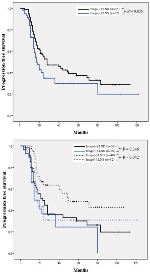
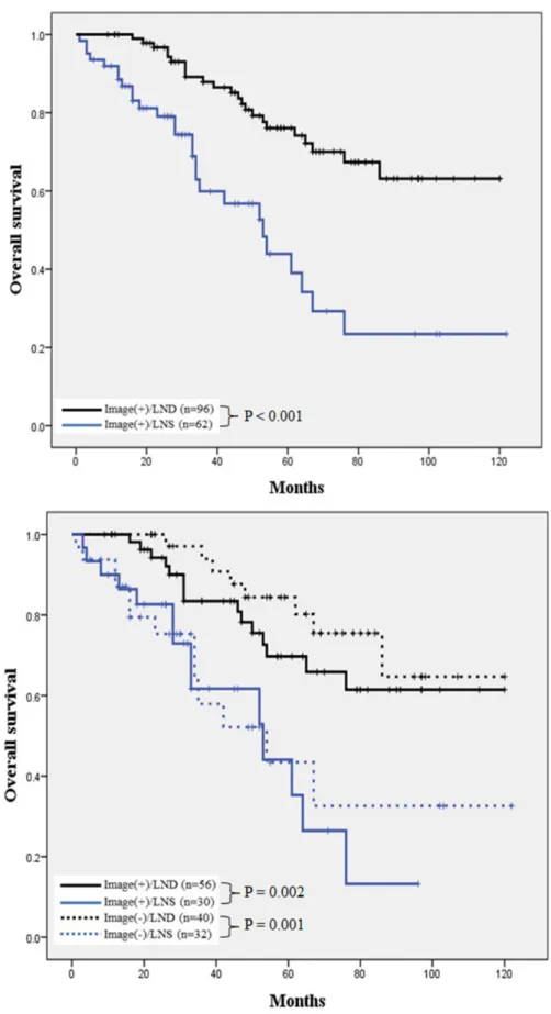
Among 274 patients who were diagnosed with advanced epithelial ovarian cancer at our institution from 2006 to 2015, optimal cytoreduction was achieved in 175 (63.9%); of these, the lymphadenectomy status could characteristics of the patients included in this study are listed in Table 1. A total of 158 consecutive patients with optimally cytoreduced primary International Federation of Gynecology and Obstetrics (FIGO) stage IIIC and IV disease who had received adjuvant platinum-based chemotherapy during the study period were analyzed. Of these patients, 62 (39.2%) underwent lymph node sampling (LNS) and 96 (60.8%) underwent LND (including both pelvic and para-aortic lymphadenectomy) as part of primary debulking surgery. The groups were similar with respect to age, preoperative cancer antigen (CA) 125 level, suspected lymph node metastasis on preoperative computed tomography (CT) scan, histology, FIGO stage, and grade. In addition, the groups did not differ with regard to the radicality of surgical procedures, including bowel resection, diaphragm resection, peritonectomy, and video-assisted thoracoscopic surgery (VATS), but did differ nodes (P < 0.001; Table 2). Furthermore, the rates of no gross residual disease after debulking surgery did not differ A Kaplan–Meier survival analysis indicated an apparently favorable progression-free survival (PFS) in the LND group, compared to the LNS group, and this better PFS when compared with LNS among patients with negative lymphadenopathy on a preoperative CT scan
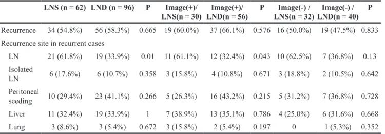
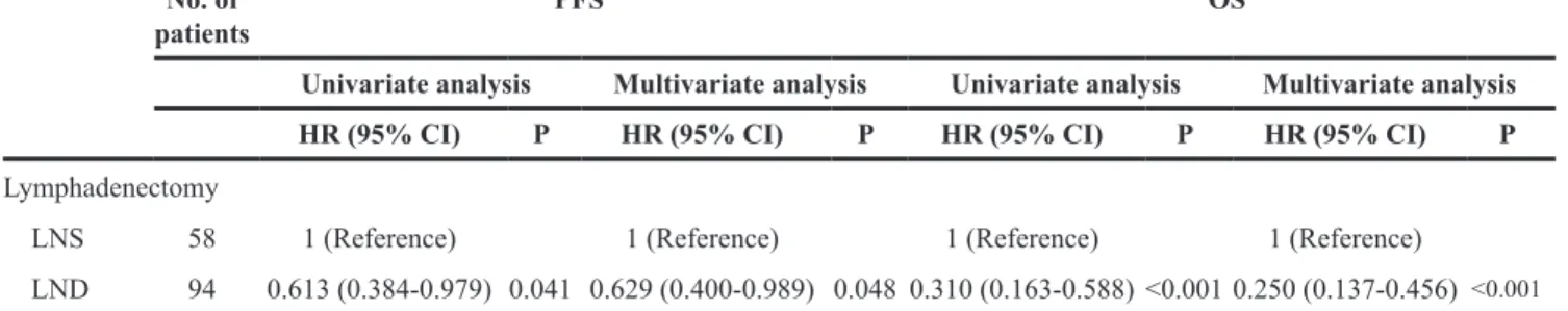
those who underwent LNS (P < 0.001), and a subgroup longer OS, regardless of the suspected lymphadenopathy A comparison of the characteristics of patients who experienced subsequent recurrence is shown in Table 3. between the LNS and LND groups (54.8% vs. 58.3%, P underwent LNS, compared to those who underwent LND correlated with a lower nodal recurrence rate, regardless of the suspected lymphadenopathy status determined by preoperative CT scan (LNS vs. LND: 61.1% vs. 32.4% in Univariate and multivariate Cox regression analyses of PFS and OS in all patients are shown in Table 4. The CA 125 level >500 U/ml, histologically proven nodal 0.03, 0.003, and 0.045, respectively). The hazard ratio (HR)
for OS was 0.250 (95% CI, 0.137–0.456).
DISCUSSION
improvement in PFS in patients who underwent pelvic and para-aortic systematic LND during primary optimal debulking surgery for advanced-stage ovarian cancer. Moreover, in a subgroup analysis according to the gross lymphadenopathy status as assessed by preoperative LND on both PFS and OS were observed in patients with negative lymphadenopathy on a preoperative CT be attributed to the contribution of this procedure to the detection and removal of occult and chemoresistant lymph node metastases, as inferred from our data regarding the characteristics of subsequent nodal recurrence, the incidence of which might be decreased by LND.
Multidisciplinary treatment, which includes cytoreductive surgery and platinum-based chemotherapy, is the mainstay of management for women with advanced-stage ovarian cancer [16]. Although medical treatment is nearly homogenous, surgical treatment is individualized according to the disease extent and patient characteristics and therefore remains heterogeneous. Moreover, the role of complete lymphadenectomy in a primary staging operation with the intent to gain information regarding
prognostic relevance remains under debate by many authors [5, 6, 10, 11, 13, 17–22], and therefore a consensus regarding the therapeutic role of this procedure has not been established, particularly after radical surgery was accepted as standard EOC management.
Previous studies have demonstrated the potential importance of systematic LND for the detection of occult lymph node metastases [5, 11, 18]. In two previous RCTs, patients with EOC who were treated with systematic LND
had a higher rate of histologically proven lymph node metastasis when compared with those who underwent macroscopic lymph node removal (22% vs. 9% for early-stage disease; 70% vs. 42% for advanced-early-stage disease) [5, 11], suggesting a potential increase in the opportunity to detect occult lymph node metastasis via systematic LND in patients with advanced ovarian cancer. This the reported detection rates of lymph node metastasis in Table 1: Characteristics of patients in the study
Variables LNS (n = 62) LND (n = 96) P-value
Age, median (range) 53 (27-81) 55 (32-78) 0.359
BMI (kg/m2) 23.1 (11.6-35.3) 22.5 (17.5-31.4) 0.192
Preoperative serum CA 125 (U/
mL) 1643.6 (16.3–17223.1) 1637.1 (13.5–11582.7) 0.987
Suspected LN metastasis on CT scan
Yes 30 (48.4%) 56 (58.3%) 0.22 No 32 (51.6%) 40 (41.7%) Histology Serous 45 (72.6%) 85 (88.5%) 0.182 Mucinous 0 1 (1.0%) Endometrioid 7 (11.3%) 4 (4.2%) Clear cell 3 (4.8%) 3 (3.1%) Mixed 3 (4.8%) 2 (2.1%) Undifferentiated 1 (1.6%) 0 Unknown 3 (4.8%) 1 (1.0%) Stage IIIC 47 (75.8%) 74 (77.1%) 0.853 IV 15 (24.2%) 22 (22.9%) Tumor grade Grade 1 1 (1.6%) 6 (6.3%) 0.209 Grade 2 21(33.9%) 39 (40.6%) Grade 3 40 (64.5%) 51 (53.1%) LN metastasis Yes 33 (53.2%) 70 (72.9%) 0.011 No 29 (46.8%) 26 (27.1%) Residual disease NGR 11 (17.7%) 28 (29.2%) 0.141 R < 0.5 cm 30 (48.4%) 33 (34.4%) R < 1 cm 21 (33.9%) 35 (36.5%)
LNS, lymph node sampling; LND, lymph node dissection; SD, standard deviation; N/A, not available; LN, lymph node, NGR, no gross residual disease; R, residual disease
patients with peritoneally advanced ovarian cancer ranged from 48% to 75% [18]. Of note, the data from our study of a preoperative CT scan for the detection of lymph node metastasis were 65.0% and 65.5%, respectively. Among 86 patients with suspected lymphadenopathy on preoperative CT scans, nodal metastasis was histologically proven in 67 patients (77.9%). In addition, the false negative rate, or detection rate of occult lymph node metastasis among patients with preoperative CT scans indicating negative lymphadenopathy, was 50% (36 of 72 patients). This observation suggests that a negative preoperative imaging result on lymph node metastasis could not justify the omission of systematic LND during debulking surgery for advanced ovarian cancer.
Incompletely resected occult lymph node metastases may give rise to chemoresistance, a possibility that is supported by the “pharmacologic sanctuary hypothesis” of poor prognosis in patients with EOC with lymph that the diminished blood supply of lymph node metastases might promote resistance to chemotherapy [23], and further implies that systematic LND might be a favorable prognostic factor in patients with advanced ovarian cancer who have an increased risk of occult lymph node metastasis [24]. Because ovarian cancer is known to spread simultaneously both intraperitoneally and retroperitoneally, the lymphatic spread of a tumor might persist despite achieving optimal cytoreduction of
intraperitoneal dissemination, thus contributing to a poor prognosis [25].
LND for patients with optimally cytoreduced advanced ovarian cancer, these data should be interpreted with caution. We attempted to minimize potential bias by accounting for all known prognostic variables associated with both the tumors and patients; however, selection bias regarding patient recruitment cannot be ruled out because of the retrospective nature of this study. Moreover, decisions regarding whether to perform systematic lymphadenectomy were made according to each surgeon’s discretion, rather Notably, the prognostic relevance of lymphadenectomy is currently under investigation in the prospective phase III trial “Randomized, Multicentre Trial for Lymphadenectomy in Ovarian Neoplasms” (https://clinicaltrials.gov/ct2/show/ NCT00712218). This ongoing study compares the prognostic outcomes associated with systematic lymphadenectomy vs. no lymphadenectomy in patients without macroscopic systematic pelvic and para-aortic lymphadenectomy. This trial is expected to clarify the status of this important issue.
In conclusion, the present study has demonstrated the potential therapeutic value of systematic LND with respect to improved prognosis following the optimal removal of intra-abdominal peritoneal metastases, regardless of the preoperatively suspected lymphadenopathy status on CT lower nodal recurrence rate with systematic LND vs. LNS. Table 2: Characteristics of surgical procedures applied to patients in this study
LNS (n = 62) LND (n = 96) P Number of resected LNs 10 (0–19) 37 (20–97) <0.001 Radical surgery Bowel resection 15 (24.2%) 19 (19.8%) 0.511 Diaphragm resection 14 (22.6%) 22 (22.9%) 1 Peritonectomy 24 (38.7%) 32 (33.3%) 0.49 VATS 4 (6.5%) 11 (11.5%) 0.294 Ureter resection 3 (4.8%) 2 (2.1%) 0.381 Liver resection 4 (6.5%) 3 (3.1%) 0.434 Splenectomy 2 (3.2%) 8 (8.3%) 0.318 Cholecystectomy 4 (6.5%) 2 (2.1%) 0.212 Residual disease NGR 11 (17.7%) 28 (29.2%) 0.141 R < 0.5 cm 30 (48.4%) 33 (34.4%) R < 1 cm 21 (33.9%) 35 (36.5%)
LNS, lymph node sampling; LND, lymph node dissection; LN, lymph node; VATS, video-assisted thoracoscopic surgery; NGR, no gross residual disease; R, residual disease
Figure 1: Comparison of PFS in patients who underwent LNS and LND. A. Overall analysis of PFS. Patients who underwent B. Subgroup analysis of PFS according to lymphadenopathy status
Figure 2: Comparison of OS in patients who underwent LNS and LND. A. Overall analysis of OS. Patients who underwent B. Subgroup analysis of OS according to
Table 3: Characteristics of recurrence LNS (n = 62) LND (n = 96) P Image(+)/ LNS(n = 30) Image(+)/ LND(n = 56) P Image(-) / LNS(n = 32) Image(-) / LND(n = 40) P Recurrence 34 (54.8%) 56 (58.3%) 0.665 19 (60.0%) 37 (66.1%) 0.576 16 (50.0%) 19 (47.5%) 0.833 Recurrence site in recurrent cases
LN 21 (61.8%) 19 (33.9%) 0.01 11 (61.1%) 12 (32.4%) 0.043 10 (62.5%) 7 (36.8%) 0.13 Isolated LN 6 (17.6%) 6 (10.7%) 0.358 3 (15.8%) 4 (10.8%) 0.671 3 (18.8%) 2 (10.5%) 0.642 Peritoneal seeding 10 (29.4%) 23 (41.1%) 0.266 5 (26.3%) 16 (43.2%) 0.215 5 (31.2%) 7 (36.8%) 0.728 Liver 11 (32.4%) 19 (33.9%) 1 7 (38.9%) 13 (35.1%) 0.786 4 (25.0%) 6 (31.6%) 0.668 Lung 3 (8.6%) 3 (5.4%) 0.672 3 (15.8%) 2 (5.4%) 0.197 0 1 (5.3%) 0.352
LNS, lymph node sampling; LND, lymph node dissection; Image(+), positive lymphadenopathy on preoperative CT scan; Image(-), negative lymphadenopathy on preoperative CT scan; LN, lymph node
Table 4: Univariate and multivariate analyses of various factors for PFS and OS No. of
patients
PFS OS
Univariate analysis Multivariate analysis Univariate analysis Multivariate analysis
HR (95% CI) P HR (95% CI) P HR (95% CI) P HR (95% CI) P
Age, years
(continuous) 158 1.016 (0.994-1.039) 0.154 1.043 (1.013-1.075) 0.005 1.044 (1.014-1.075) 0.004 Histology
Non-serous 28 1 (Reference) 1 (Reference)
Serous 124 1.226 (0.611-2.462) 0.567 1.072 (0.454-2.530) 0.874
Tumor grade
Grade 1 7 1 (Reference) 1 (Reference)
Grade 2-3 151 1.364 (0.576-3.186) 0.227 1.140 (0.345-3.765) 0.83 Preoperative CA 125, U/ml
58 1 (Reference) 1 (Reference) 1 (Reference)
>500 94 1.722 (1.030-2.881) 0.038 1.713 (1.053-2.789) 0.033 1.540 (0.782-3.033) 0.212 Suspected LN metastasis on preoperative imaging studies
No 69 1 (Reference) 1 (Reference)
Yes 83 1.326 (0.809-2.171) 0.263 1.461 (0.746-2.860) 0.269
LN metastasis
No 55 1 (Reference) 1 (Reference) 1 (Reference)
Yes 103 2.060 (1.162-3.651) 0.013 2.238 (1.304-3.841) 0.004 1.331 (0.628-2.820) 0.455 Residual disease
NGR 39 1 (Reference) 1 (Reference)
R < 1 cm 113 1.102 (0.604-2.011) 0.752 1.251 (0.498-3.142) 0.634
MATERIALS AND METHODS
Study designis presented in Figure 3. Patients who were diagnosed with advanced-stage (FIGO IIIC and IV) EOC from January 2006 to December 2015 and underwent optimal cytoreduction, or had residual disease of <1 cm were included in this study. The retrospective study protocol of this study was approved by our Institutional Review Board.
A retrospective chart review was performed to identify all patients who underwent primary debulking surgery, including hysterectomy, bilateral oophorectomy, omentectomy, and retroperitoneal lymph node excision with or without various radical surgeries (e.g., bowel resection, diaphragm resection, peritonectomy) and received adjuvant standard platinum-based chemotherapy. A gynecologic oncology team comprising 5 surgeons at a single institute conducted all procedures, and 2 dedicated radiologists at the same institute reviewed preoperative computed tomography (CT) scan data.
No. of patients
PFS OS
Univariate analysis Multivariate analysis Univariate analysis Multivariate analysis
HR (95% CI) P HR (95% CI) P HR (95% CI) P HR (95% CI) P
Lymphadenectomy
LNS 58 1 (Reference) 1 (Reference) 1 (Reference) 1 (Reference)
LND 94 0.613 (0.384-0.979) 0.041 0.629 (0.400-0.989) 0.048 0.310 (0.163-0.588) <0.001 0.250 (0.137-0.456) <0.001
gross residual disease; R, residual disease; LNS, lymph node sampling; LND, lymph node dissection
To determine the therapeutic value of systematic [11, 26, 27]. Decisions regarding whether to perform LND or LNS were made according to each surgeon’s discretion. as systematic lymphadenectomy (total resected node count: node dissection. Systemic pelvic LND included the resection of all lymph nodes and fatty tissue between the external and internal iliac arteries from the bifurcation of the common nerve. Systemic para-aortic LND included the resection of all lymph nodes and fatty tissue overlying the common iliac artery, vena cava, and aorta anteriorly up to the renal vessels and laterally to the edge of the psoas major muscle. The surgical procedures and characteristics of recurrence and prognosis were compared between the groups.
Additionally, a subgroup analysis was conducted to investigate the role of LND with respect to apparent nodal involvement on a preoperative CT scan. Both the suspected lymphadenopathy status on preoperative CT scans. Image(+) indicated positive preoperatively
lymphadenopathy on a preoperative CT scan.
Statistical analysis
IBM SPSS version 20 for Windows (SPSS Inc., Chicago, IL, USA) was used for the statistical analysis. The Kolmogorov–Smirnov test was used to verify standard normal distributional assumptions. Pearson’s chi square test, Fisher’s exact test, and the Mann–Whitney U test were used in the univariate analysis. Survival outcomes were determined through a Kaplan–Meier survival analysis. Univariate and multivariate analyses of the effects of various prognostic factors on survival were performed using the Cox proportional hazards model. Multivariate analysis was performed with variables that
CONFLICTS OF INTEREST
None.
REFERENCES
1. Siegel RL, Miller KD, Jemal A. Cancer statistics, 2015. Cancer J Clin. 2015; 65:5-29.
2. Bookman MA, Greer BE, Ozols RF. Optimal therapy of advanced ovarian cancer: carboplatin and paclitaxel vs.
cisplatin and paclitaxel (GOG 158) and an update on GOG0 182-ICON5. Int J Gynecol Cancer. 2003; 13:735-740. 3. Peiretti M, Zanagnolo V, Aletti GD, Bocciolone L, Colombo
Role of maximal primary cytoreductive surgery in patients with advanced epithelial ovarian and tubal cancer: Surgical and oncological outcomes. Single institution experience. Gynecol Oncol. 2010; 119:259-264.
4. Chang SJ, Hodeib M, Chang J, Bristow RE. Survival impact of complete cytoreduction to no gross residual disease for advanced-stage ovarian cancer: a meta-analysis. Gynecol Oncol. 2013; 130:493-498.
5. Panici PB, Maggioni A, Hacker N, Landoni F, Ackermann S, Campagnutta E, Tamussino K, Winter R, Pellegrino A, Greggi S, Angioli R, Manci N, Scambia G, et al. Systematic aortic and pelvic lymphadenectomy versus resection of bulky nodes only in optimally debulked advanced ovarian cancer: a randomized clinical trial. J Natl Cancer Inst. 2005; 97:560-566.
6. Pereira A, Perez-Medina T, Magrina JF, Magtibay PM, Millan I, Iglesias E. The role of lymphadenectomy in node-positive epithelial ovarian cancer. Int J Gynecol Cancer. 2012; 22:987-992.
7. Scarabelli C, Gallo A, Visentin MC, Canzonieri V, Carbone A, Zarrelli A. Systematic pelvic and para-aortic lymphadenectomy in advanced ovarian cancer patients with no residual intraperitoneal disease. Int J Gynecol Cancer. 1997; 7:18-26.
8. Takeshima N, Hirai Y, Umayahara K, Fujiwara K, Takizawa K, Hasumi K. Lymph node metastasis in ovarian cancer: difference between serous and non-serous primary tumors. Gynecol Oncol. 2005; 99:427-431.
9. Chan JK, Munro EG, Cheung MK, Husain A, Teng NN, Berek JS, Osann K. Association of lymphadenectomy and survival in stage I ovarian cancer patients. Obstet Gynecol. 2007; 109:12-19.
10. Chan JK, Urban R, Hu JM, Shin JY, Husain A, Teng NN, Berek JS, Osann K, Kapp DS. The potential therapeutic role of lymph node resection in epithelial ovarian cancer: a study of 13918 patients. Br J Cancer. 2007; 96:1817-1822.
11. Maggioni A, Benedetti Panici P, Dell’Anna T, Landoni F, Lissoni A, Pellegrino A, Rossi RS, Chiari S, Campagnutta E, Greggi S, Angioli R, Manci N, Calcagno M, et al. Randomised study of systematic lymphadenectomy in patients with epithelial ovarian cancer macroscopically
12. Isonishi S, Niimi S, Sasaki H, Ochiai K, Yasuda M, lymphadenectomy during cytoreductive surgery in optimally debulked stages IIIc and IV ovarian cancer. Gynecol Oncol. 2004; 93:647-652.
13.
surgery in patients with advanced ovarian cancer. Gynecol Oncol. 2012; 126:381-386.
14. Heitz F, Harter P, Alesina PF, Walz MK, Lorenz D, Groeben H, Heikaus S, Fisseler-Eckhoff A, Schneider S, Ataseven B, Kurzeder C, Prader S, Beutel B, et al. Pattern of and reason for postoperative residual disease in patients with advanced ovarian cancer following upfront radical debulking surgery. Gynecol Oncol. 2016; 141:264-270.
15. Elattar A, Bryant A, Winter-Roach BA, Hatem M, Naik R. Optimal primary surgical treatment for advanced epithelial ovarian cancer. Cochrane Database Syst Rev. 2011. doi:10.1002/14651858.CD007565. pub2:Cd007565.
16. Bookman MA. Optimal primary therapy of ovarian cancer. Ann Oncol. 2016; 27:i58-i62.
17. Aletti GD, Dowdy S, Podratz KC, Cliby WA. Role of lymphadenectomy in the management of grossly apparent advanced stage epithelial ovarian cancer. Am J Obstet Gynecol. 2006; 195:1862-1868.
18. Pereira A, Magrina JF, Rey V, Cortes M, Magtibay PM. Pelvic and aortic lymph node metastasis in epithelial ovarian cancer. Gynecol Oncol. 2007; 105:604-608. 19. Hacker NF, Valmadre S, Robertson G. Management of
retroperitoneal lymph nodes in advanced ovarian cancer. Int J Gynecol Cancer. 2008; 18:7-10.
20. du Bois A, Reuss A, Harter P, Pujade-Lauraine E, Ray-advanced ovarian cancer: a combined exploratory analysis
of three prospectively randomized phase III multicenter trials. J Clin Oncol. 2010; 28:1733-1739.
21. Bachmann C, Bachmann R, Brucker SY, Staebler A, Fend F, Grischke EM, Wallwiener D. Role of Pelvic and Para-aortic Lymph Node Metastases in Optimally Cytoreduced Advanced Ovarian Cancer. Anticancer Res. 2015; 35:3479-3484.
22. Bachmann C, Brucker SY, Kraemer B, Rothmund R, Staebler A, Fend F, Wallwiener D, Grischke EM. The prognostic relevance of node metastases in optimally cytoreduced advanced ovarian cancer. J Cancer Res Clin Oncol. 2015; 141:1475-1480.
23.
occult macroscopically positive retroperitoneal nodes in patients with epithelial ovarian cancer. Gynecol Oncol. 2001; 82:143-149.
24. Berek JS. Lymph node-positive stage IIIC ovarian cancer: a separate entity? Int J Gynecol Cancer. 2009; 19:S18-20. 25. Paik ES, Lee YY, Lee EJ, Choi CH, Kim TJ, Lee JW, Bae
DS, Kim BG. Survival analysis of revised 2013 FIGO
Obstet Gynecol Sci. 2015; 58:124-134.
26. Chan JK, Kapp DS. Role of complete lymphadenectomy in endometrioid uterine cancer. Lancet Oncol. 2007; 8:831-841.
27. Hacker NF, Wain GV, Nicklin JL. Resection of bulky positive lymph nodes in patients with cervical carcinoma. Int J Gynecol Cancer. 1995; 5:250-256.