Hepatocellular carcinoma in Budd-Chiari syndrome: A single
center experience with long-term follow-up in South Korea
Hana Park, Jin Young Yoon, Kyeong Hye Park, Do Young Kim, Sang Hoon Ahn, Kwang-Hyub Han, Chae Yoon Chon, Jun Yong Park
Hana Park, Jin Young Yoon, Kyeong Hye Park, Do Young Kim, Sang Hoon Ahn, Kwang-Hyub Han, Chae Yoon Chon, Jun Yong Park, Department of Internal Medicine, Yonsei University College of Medicine, Seoul 120-752, South Korea
Do Young Kim, Sang Hoon Ahn, Kwang-Hyub Han, Chae Yoon Chon, Jun Yong Park, Institute of Gastroenterology, Yonsei University College of Medicine, Seoul 120-752, South Korea Do Young Kim, Sang Hoon Ahn, Kwang-Hyub Han, Chae Yoon Chon, Jun Yong Park, Liver Cirrhosis Clinical Research Center, Seoul 120-752, South Korea
Sang Hoon Ahn, Kwang-Hyub Han, Brain South Korea 21 Pro-ject for Medical Science, Seoul 120-752, South Korea
Author contributions: Park H designed the research/study, ana-lyzed the data, performed the study and wrote the paper; Yoon JY designed the research/study and reviewed the data of study popu-lation; Park KH performed the study and wrote the paper; Kim DY collected the data; Ahn SH collected the data; Han KH col-lected the data; Chon CY colcol-lected the data; and Park JY designed the research/study, collected the data and performed the study. Supported by A grant of the South Korea Healthcare Technol-ogy R and D Project, Ministry of Health and Welfare, South Ko-rea, No. A102065
Correspondence to: Jun Yong Park, MD, Department of In-ternal Medicine, Yonsei University College of Medicine, Seoul 120-752, South Korea. [email protected]
Telephone: +82-2-22281994 Fax: +82-2-3936884 Received: September 26, 2011 Revised: October 27, 2011 Accepted: February 16, 2012
Published online: April 28, 2012
Abstract
AIM: To evaluate long-term clinical course of Budd-Chiari syndrome (BCS) and predictive factors associated with the development of hepatocellular carcinoma (HCC) and survival.
METHODS: We analyzed 67 patients with BCS be-tween June 1988 and May 2008. The diagnosis of BCS was confirmed by hepatic venous outflow obstruction shown on abdominal ultrasound sonography, computed tomography, magnetic resonance imaging, or
venogra-phy. The median follow-up period was 103 ± 156 [in-terquartile range (IQR)] mo.
RESULTS: The median age of the patients was 47 ± 16 (IQR) years. At diagnosis, 54 patients had cirrhosis, 25 (37.3%) Child-Pugh class A, 23 (34.3%) Child-Pugh class B, and six (9.0%) patients Child-Pugh class C. During the follow-up period, HCC was developed in 17 patients, and the annual incidence of HCC in patients with BCS was 2.8%. Patients in HCC group (n = 17) had higher hepatic venous pressure gradient (HVPG) than those in non-HCC group (n = 50) (21 ± 12 mmHg vs 14 ± 7 mmHg, P = 0.019). The survival rate of BCS patients was 86.2% for 5 years, 73.8% for 10 years, and 61.2% for 15 years. In patients with BCS and HCC, survival was 79% for 5 years, 43.1% for 10 years, and 21.5% for 15 years.
CONCLUSION: The incidence of HCC in patients with BCS was similar to that in patients with other etiologic cirrhosis in South Korea. The HVPG is expected to pro-vide additional information for predicting HCC develop-ment in BCS patients.
© 2012 Baishideng. All rights reserved.
Key words: Budd-Chiari syndrome; Hepatocellular carci-noma; Prognosis
Peer reviewer: Guang-Wen Cao, MD, PhD, Professor and Chairman, Department of Epidemiology, The Second Military Medical University, 800 Xiangyin Road, Shanghai 200433, China Park H, Yoon JY, Park KH, Kim DY, Ahn SH, Han KH, Chon CY, Park JY. Hepatocellular carcinoma in Budd-Chiari syn-drome: A single center experience with long-term follow-up in South Korea. World J Gastroenterol 2012; 18(16): 1946-1952 Available from: URL: http://www.wjgnet.com/1007-9327/full/ v18/i16/1946.htm DOI: http://dx.doi.org/10.3748/wjg.v18. i16.1946
BRIEF ARTICLE
INTRODUCTION
Budd-Chiari syndrome (BCS) is a rare hepatic disease caused by occlusion of the hepatic venous outflow. Thrombogenic conditions among the known causes of BCS are well documented, such as coagulopathy, chronic intake of contraceptives, myeloproliferative diseases, au-toimmune diseases, and others[1]. Hepatic venous outflow
block can also be caused by malignant tumors[2]. BCS
was initially defined as a symptomatic occlusion of the hepatic veins, but with increasing reports on various ob-structive cases in the hepatic portion of the inferior vena cava (IVC), it became to include obstructive IVC lesions as well as the major hepatic veins. BCS induces chronic liver congestion so that it causes hepatomegaly, ascites, leg edema, collateral venous dilatation in the body trunk and portal hypertension[3]. Several studies have suggested
that hepatic congestion caused by obstruction of hepatic venous outflow can lead to cirrhosis and hepatocellular carcinoma (HCC)[4,5]. The incidence of HCC in patients
with BCS has varied according to regions and
investiga-tors[3,6-9]. Japan and South Africa showed relatively higher
incidences compared to those of United States and France: 6.4%-47.5% vs 4%-20%[5,7,9,10]. Prognosis of HCC
in BCS has varied as well[5,6,11].
We followed BCS patients treated at our hospital for more than 20 years, and our long term follow-up data can be helpful to understand the prognosis of BCS patients and natural course of the disease. Thus, we (1) evaluated the incidence and cumulative annual risk of HCC in BCS patients; (2) analyzed the characteristics associated with the development of HCC in BCS patients; and (3) inves-tigated the prognosis of BCS and HCC in BCS patients.
MATERIALS AND METHODS
Patients
From June 1988 to May 2008, 95 consecutive patients who were diagnosed with BCS at Severance Hospital were studied retrospectively. Among them, 28 patients were excluded based on the criteria as follows: patients with secondary BCS [occlusion of the hepatic venous outflow by an outside structure (HCC, a klatskin tumor, and renal cell carcinoma)], hematologic diseases which could result venous obstructive disease, hepatic septic emboli of colon cancer, or post-operative complication of HCC. Finally 67 patients who were diagnosed with primary BCS were investigated. The study protocol con-formed to the ethical guidelines of the 1975 Helsinki Declaration and was approved by the institutional review board of our institute.
Methods
We retrospectively reviewed patients’ age, gender, and presenting symptoms of decompensate liver cirrhosis such as ascites, encephalopathy and variceal bleeding. We also investigated their history of medication which can induce thrombosis such as oral contraceptive, herbal
medication, and steroid. Patients were assessed for other risk factors of cirrhosis and HCC such as alcohol, hepati-tis B virus and hepatihepati-tis C virus (HCV) infections.
Laboratory tests included complete blood count, pro-thrombin time, alanine aminotransferase (ALT), bilirubin, albumin, creatinine, viral markers such as hepatitis B virus surface antigen (HBsAg) and anti-HCV antibody. Based on laboratory and physical examination results, Model for End-Stage Liver Disease (MELD) score and Child Pugh score were calculated to evaluate liver function. Every 3-6 mo, imaging studies such as computed tomography (CT), ultrasonography or magnetic resonance imaging (MRI) were performed, and alpha-fetoprotein (AFP) level was checked for HCC surveillance.
Diagnosis of BCS and HCC
The diagnosis of BCS was confirmed by hepatic venous outflow obstruction shown on ultrasound sonography (US), contrast-enhanced CT, MRI, or venography. Patients were confirmed to have HCC according to the American Association for the Study of Liver Disease practice guide-lines for the HCC diagnosis[12]. Briefly, patients were
di-agnosed with HCC if they had a tumor with a maximum diameter of > 2 cm and the typical features of HCC on dynamic CT (defined as enhancement in the arterial phase and early washout in the portal phase), and an AFP > 200 ng/mL[12]. If the maximum diameter of the tumor was 1
cm to 2 cm, dynamic CT and MRI were performed. HCC was diagnosed if coincidental typical features of HCC were noted. If the tumor did not satisfy above criteria, a biopsy was performed. During hospital stay, abdominal ultrasonography was carried out to evaluate the presence of cirrhosis and hepatocellular carcinoma.
Measurement of hepatic venous pressure gradient
The hepatic venous pressure gradient (HVPG) was mea-sured through catheterization during venography or an-gioplasty. Forty-five patients underwent venography or angioplasty when diagnosed with BCS or during follow up period. Patients were diagnosed with clinical liver cirrhosis if they satisfied one or more of the following 3 condi-tions: (1) a platelet count < 100 000/μL and ultrasono-graphic findings suggestive of cirrhosis including a blunt-ed, nodular liver edge accompanied by splenomegaly (> 12 cm)[13,14]; (2) the presence of esophageal or gastric varices;
and (3) overt complications of liver cirrhosis, including ascites, variceal bleeding, and hepatic encephalopathy.
Statistical analysis
Statistical analysis was performed using the Statistical Package for the Social Sciences (SPSS, version 17.0). De-scriptive statistics were computed for all variables, includ-ing median ± interquartile range (IQR) and percentiles for continuous variables and frequencies for categorical factors. Mann-Whitney U test and χ2-test were used to
compare HCC and non-HCC groups. Kaplan-Meier anal-ysis was performed to estimate the cumulative incidence
of HCC from the time of BCS diagnosis and survival of all BCS patients and those who developed HCC. To eval-uate the factors associated with the development of HCC in patients with BCS, multivariate Cox regression analysis was used. A two-sided P value < 0.05 was considered to
indicate a significant difference.
RESULTS
Baseline clinical characteristics of the patients
The baseline characteristics of the 67 patients are shown in Table 1. The site of obstruction was IVC in 56 patients (83.6%), hepatic vein in five patients (7.4%), and both IVC and hepatic vein sites in six patients who were clas-sified as “combined”. There was no patient with history of chronic use of medication such as oral contraceptive, herbal medication, and steroid. Patients with heavy alco-hol consumption defined as over 80 g/d were 20 (29.9%). HBsAg was positive in 3 patients (4.5%) and there was no patient with positive for anti-HCV antibody.
Fifty-four patients had underlying liver cirrhosis at the time of BCS diagnosis. Twenty-five of them (37.3%) were classi-fied as Child-Pugh class A, 23 (34.3%) as class B, and six (9.0%) as class C. The median MELD score was 11 ± 6 (IQR). Twenty three patients (34.3%) had decompensated liver cirrhosis symptoms such as variceal bleeding, un-controlled ascites or hepatic encephalopathy higher than grade 3. Twenty-seven (40.3%) patients were treated with percutaneous angioplasty, four (5.9%) with shunt opera-tion, three (4.5%) with transjugular intrahepatic portosys-temic shunt (TIPS), and one (1.5%) with thrombolysis. Thirty-two (47.7%) patients received only symptomatic medical treatment. The median follow up period was 103 ± 156 (IQR) mo.
Development of HCC in the patients with BCS
During follow-up periods, HCC was occurred in 17 pa-tients. At the time of diagnosis of HCC, the median age of the patients was 53 ± 12 (IQR) years, and time pe-riod between diagnoses of BCS and HCC was 51 ± 115 (IQR) mo (Table 2). HCC was histologically confirmed in 2 patients with hepatic resection. According to the Kaplan-Meier analysis, as shown in Figure 1, the cumula-tive probability was 18.5% at 5 years, 30.3% at 10 years, and 42.6% at 15 years. The annual occurrence of HCC in BCS patients was 2.8%.
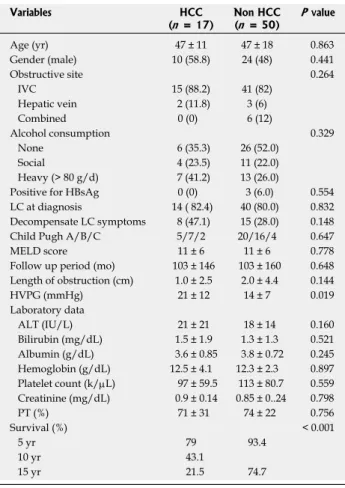
We compared the baseline characteristics (at the time of diagnosis with BCS) of HCC group (n = 17) with
non-HCC group (n = 50). The differences between the two
groups are shown in Table 3. There were no significant differences in the comparison of age, gender, obstruction site, alcohol consumption and presence of viral markers between two groups. HVPG was significantly higher in HCC group [21 ± 12 (IQR) mmHg] than in non-HCC Table 1 Baseline clinical characteristics of patients with
Budd-Chiari syndrome (n = 67)
Characteristics Budd-Chiari syndrome
Age (yr) 47 ± 16 Gender (male) 34 (50.7) Obstruction site IVC 56 (83.6) Hepatic vein 5 (7.5) Combined 6 (9.0) Alcohol consumption None/social/heavy (> 80 g/d) 32 (47.8)/15 (22.3)/20 (29.9) Positive viral marker
HBsAg/anti-HCV 3 (4.5)/0 (0) Liver cirrhosis at diagnosis 54 (80.6) Child-Pugh A/B/C 25/23/6 (37.3/34.3/9.0) MELD score 11 ± 6 Decompensate LC symptoms 23 (34.3) Length of obstruction (cm) 2.0 ± 4.0 HVPG (mmHg) 15 ± 10 Laboratory data ALT (IU/L) 19 ± 17 Bilirubin (mg/dL) 1.4 ± 1.3 Albumin (g/dL) 3.8 ± 0.7 Hemoglobin (g/dL) 12.3 ± 2.6 Platelet count (k/μL) 109 ± 78 Creatinine (mg/dL) 0.9 ± 0.2 PT (%) 73 ± 27 Treatment modality Angioplasty 27 (40.3) Shunt operation 4 (5.9) TIPS 3 (4.5) Thrombolysis 1 (1.5) Symptomatic medical treatment 32 (47.7) Median follow up period (mo) 103 ± 156
Data were expressed as median ± IQR or n (%). IVC: Inferior vena cava; HCV: Hepatitis C virus; HbsAg: Hepatitis B surface antigen; MELD: Model for End-Stage Liver Disease; LC: Liver cirrhosis; HVPG: Hepatic venous pressure gradient; ALT: Alanine aminotransferase; PT: Prothrombin time; TIPS: Transjugular intrahepatic portosystemic shunt; IQR: Interquartile range.
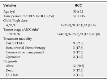
Table 2 Characteristics of the patients with hepatocellular carcinoma at the time point of diagnosis (n = 17)
Variables HCC
Age (yr) 53 ± 12
Time period from BCS to HCC (mo) 51 ± 115 Child-Pugh class
A/B/C 6 (35.3)/8 (47.1)/3 (17.6) Tumor stage (AJCC 6th)1
Ⅰ/Ⅱ/Ⅲ/Ⅳ 8 (47.1)/6 (35.3)/3 (17.6)/0 (0) Treatment modality TACE/TACI 9 (52.9) Intra-arterial chemotherapy 3 (17.6) Conservative management 3 (17.6) Operation 2 (11.9) Prognosis Alive 12 (70.5) Death 3 (17.6) F/U loss 2 (11.9)
Data were expressed as median ± IQR or n (%). BCS: Budd-Chiari syn-drome; HCC: Hepatocellular carcinoma; TACE: Transarterial chemo-embolization; TACI: Transarterial chemo-infusion; F/U: Follow up; IQR: Interquartile range. 1AJCC 6th, American Joint Committee on Cancer stag-ing system, 6th edition.
Prognosis and survival of patients with BCS and patients who were diagnosed with HCC
We estimated the overall survival rates of all 67 BCS patients using the Kaplan-Mayer method. The overall survival rate was 86.2% at 5 years, 73.8% at 10 years, and 61.2% at 15 years (Figure 2). During the follow-up peri-ods, three patients among 17 who were diagnosed with HCC died, and two of them died from hepatic failure and the other from massive variceal bleeding. In HCC group, the 5-year survival rate was 79%, 10-year rate 43.1%, and 15-year rate 21.5% (Figure 3). Meanwhile, non-HCC group (n = 50) showed significantly higher
sur-vival rates than HCC group: 93.4% for 5 years and 74.7% for 15 years (P < 0.001).
DISCUSSION
BCS is caused by obstruction of hepatic venous outflow at any level from the small hepatic veins to the junction of the IVC with the right atrium. There are two forms of BCS according to the obstruction site: primary hepatic vein obstruction (classical BCS) and obstruction of the group [14 ± 7 (IQR) mmHg] (P = 0.019). However, in
the multivariate Cox regression analysis, HVPG showed no significant difference between in HCC group and in non-HCC group (P = 0.452).
Annual probability: 2.8%/yr
15 yr: 42.6% 10 yr: 30.3% 5 yr: 18.5% 1.0 0.8 0.6 0.4 0.2 0.0 0 50 100 150 200 250 300 t/mo Cumulativ e pr obabili ty
Figure 1 Cumulative probability of hepatocellular carcinoma in patients with Budd-Chiari syndrome (n = 17).
Table 3 Comparison of baseline clinical characteristics be-tween hepatocellular carcinoma group and non hepatocellular carcinoma group Variables HCC (n = 17) Non HCC (n = 50) P value Age (yr) 47 ± 11 47 ± 18 0.863 Gender (male) 10 (58.8) 24 (48) 0.441 Obstructive site 0.264 IVC 15 (88.2) 41 (82) Hepatic vein 2 (11.8) 3 (6) Combined 0 (0) 6 (12) Alcohol consumption 0.329 None 6 (35.3) 26 (52.0) Social 4 (23.5) 11 (22.0) Heavy (> 80 g/d) 7 (41.2) 13 (26.0)
Positive for HBsAg 0 (0) 3 (6.0) 0.554 LC at diagnosis 14 ( 82.4) 40 (80.0) 0.832 Decompensate LC symptoms 8 (47.1) 15 (28.0) 0.148 Child Pugh A/B/C 5/7/2 20/16/4 0.647 MELD score 11 ± 6 11 ± 6 0.778 Follow up period (mo) 103 ± 146 103 ± 160 0.648 Length of obstruction (cm) 1.0 ± 2.5 2.0 ± 4.4 0.144 HVPG (mmHg) 21 ± 12 14 ± 7 0.019 Laboratory data ALT (IU/L) 21 ± 21 18 ± 14 0.160 Bilirubin (mg/dL) 1.5 ± 1.9 1.3 ± 1.3 0.521 Albumin (g/dL) 3.6 ± 0.85 3.8 ± 0.72 0.245 Hemoglobin (g/dL) 12.5 ± 4.1 12.3 ± 2.3 0.897 Platelet count (k/μL) 97 ± 59.5 113 ± 80.7 0.559 Creatinine (mg/dL) 0.9 ± 0.14 0.85 ± 0..24 0.798 PT (%) 71 ± 31 74 ± 22 0.756 Survival (%) < 0.001 5 yr 79 93.4 10 yr 43.1 15 yr 21.5 74.7
Data were expressed as median ± IQR or n (%). HCC: Hepatocellular carcinoma; IVC: Inferior vena cava; LC: Liver cirrhosis; MELD: Model for End-Stage Liver Disease; HVPG: Hepatic venous pressure gradient; ALT: Alanine aminotransferase; PT: Prothrombin time; IQR: Interquartile range. 1.0 0.8 0.6 0.4 0.2 0.0 0 50 100 150 200 250 300 t/mo Ov er al l surviv al 5 yr: 86.2% 10 yr: 73.8% 15 yr: 61.2%
Figure 2 Overall survival of patients with Budd-Chiari syndrome. 1.0 0.8 0.6 0.4 0.2 0.0 0 50 100 150 200 250 300 t/mo Ov er al l surviv al HCC group
Figure 3 Comparison of survival between hepatocellular carcinoma Group (n = 17) and non-hepatocellular carcinoma group (n = 50). HCC:
Hepatocellular carcinoma.
Non HCC group
hepatic portion of the inferior vena cava (IVCO). The IVCO form is common in Asia and Africa but rarely reported in Western countries[9,15]. Most of our patients
(83.6%) had the IVCO form, which is similar to previous studies which reported the predominance of IVCO form in Asia. The major difference between classical BCS and IVCO is that the former is rarely associated with HCC, while the latter is frequently complicated by HCC[3,8]. In
our study, HCC was developed in 17 of 67 patients, and the annual incidence was 2.8%, similar to the incidence in patients with other etiologic cirrhosis in South Korea[16,17].
Until now, the accurate pathogenesis of HCC in BCS has not been elucidated yet. Gwon et al[6] suggested that
chronic liver injuries and congestion caused by obstruc-tion of hepatic venous outflow might contribute to a fi-brotic process and development of nodular type of HCC. Prolonged congestion can lead to hepatocyte necrosis, and its replacement with fibrous tissue results in fibrosis, which is assumed to be the mechanism of cirrhosis and HCC development[18-20]. This hypothesis is supported by
frequent findings of liver parenchymal cirrhotic change adjacent to HCC in BCS context[7].
The HVPG was significantly higher in HCC group than non-HCC group in our study. HVPG has been ac-cepted as the gold standard for assessing the severity of portal hypertension[21]. With the pathogenesis proposed
above, the higher pressure gradient means a greater degree of portal hypertension and hepatic congestion; this might have contributed to a higher pressure gradient in HCC group in our study. Our study that showed significantly
high HVPG in HCC group is worthy, which supports the hypothesis of development of HCC in patients with BCS. Until now, there were several published reports to analyze the risk factors for HCC in patients with BCS. Although Moucari et al[7] showed that BCS patients with
HCC compared with those without HCC presented with IVC obstruction more frequently, there was no report that showed direct differences of pressure gradient as our data presented. For comparison, other published data concern-ing the prevalence and characteristics of HCC in patients with BCS were reviewed in Table 4.
Varying survival results of BCS have been reported, and the 5-year survival rate ranges from 69% to 87%[22,23].
In our data, the overall survival rate was 86.2% for 5 years, 73.8% for 10 years, and 61.2% for 15 years. In pa-tients with BCS and HCC, the survival rate was 79% for 5 years, 43.1% for 10 years, and 21.5% for 15 years in our study. This is comparable with the results of other pub-lished reports[6,24]. Improvement in availability and
tech-niques of diagnostic tools and development of treatment modalities may allow earlier diagnosis of BCS patients and better prognosis[18-20].
Although our study has an advantage of long-term follow up data of patients with BCS, there are still some limitations. First, this was retrospective and therefore has the limitations of such an investigational design. Another limitation was that the values of HPVG obtained during venography were not checked in all patients. Because of invasiveness of venography, it could not be performed on all patients for HVPG measurement. These days, Table 4 Prevalence and characteristics of hepatocellular carcinoma in patients with Budd-Chiari syndrome from the literature
Ref. Matsui et al[3] Shin et al[24] Moucari et al[7] Gwon et al[6]
Year, country 2000, Japan 2004, South Korea 2008, France 2010, South Korea Study design Retrospective Retrospective Retrospective Retrospective Study period Apr 1968-Feb 1999 Mar 1989-Aug 2001 1987-2005 Mar 1990-Nov 2008
No. of patient with BCS 12 73 97 98
HCC (%) 3 (25) 15 (20.5) 11 (11.3) 23 (23)
Cumulative incidence of HCC - - 4 yr 3% 1 yr 7.3%
7 yr 6% 5 yr 13.5% 14 yr 12% 10 yr 31.8% Tx. for HCC Resection (1) TACE (11) TACE (7) TACE (20) TAE (1) Resection (2) LT (3) TACE + LT (3) iA chemotherapy (1) Conservative tx. (2) Conservative tx. (1)
Survival rate of HCC in patients with BCS
Median survival period (mo) - 58 (range, 3-59) -
Cumulative survival - 1 yr 93% - 1 yr 90%
2 yr 84% 2 yr 85%
3 yr 72% 3 yr 61%
4 yr 61% 5 yr 46% Risk factors for HCC in patients with BCS
Risk factors Chronic congestion in the liver, caused by an outflow block of hepatic veins
- Male gender, Female gender2 Coagulopathy1,
IVC obstruction
Analysis method No statistical analysis (mere presumption) - Univariate analysis Multivariate analysis HCC: Hepatocellular carcinoma; BCS: Budd-Chiari syndrome; Tx.: Treatment; TAE: Transarterial embolization; TACE: Transarterial chemoembolization; iA: Intra-arterial; LT: Liver transplantation. 1Coagulopathy harbored factor V Leiden; 2Female gender showed an odds ratio of 6.02 with P < 0.001 in Cox regression analysis; IVC: Inferior vena cava.
Doppler US is used to non-invasively assess the HPVG with portal vein velocity. Considering the low incidence of BCS, a multicenter study should be performed to overcome the limitations of patient number and insuf-ficient information. Despite these limitations, this study is worthy to analyze the incidence and prognosis of HCC in BCS with long-term follow-up periods in South Korea.
In conclusion, the annual incidence of HCC in pa-tients with BCS was similar to the incidence in papa-tients with other etiologic cirrhosis in South Korea. Further-more, the HVPG can be a possible predictive factor of BCS-associated HCC development. Thus, BCS patients who are expected to have high pressure gradient should be actively managed as a high risk group for HCC de-velopment. An intervention to decrease the pressure gradient in BCS patients may be helpful to reduce the in-cidence of HCC. A large-scale study will be necessary to further investigate whether the treatment of congestion decreases the incidence of HCC.
ACKNOWLEDGMENTS
The authors are grateful to Dong-Su Jang (Medical Illus-trator, Medical Research Support Section, Yonsei Univer-sity College of Medicine, Seoul, South Korea) for his help with the figures.
COMMENTS
Background
Budd-Chiari syndrome (BCS) is a rare disease caused by obstruction of the hepatic venous outflow. BCS induces chronic liver congestion so that it causes hepatomegaly, ascites, leg edema, collateral venous dilatation in the body trunk, and portal hypertension. Several studies have suggested that hepatic congestion caused by obstruction of hepatic venous outflow can lead to cir-rhosis and hepatocellular carcinoma (HCC). However, the incidence of HCC in patients with BCS has varied according to regions and investigators and there has been a lack of reports for long-term prognosis of HCC in patients with BCS.
Research frontiers
Although BCS is a relatively rare disease as contrasted with other viral liver disease that can lead to advanced liver disease such as liver cirrhosis or HCC, several studies have reported the association between BCS and HCC. Long-term follow-up data of BCS may help to understand not only the prognosis of BCS but also the process of HCC in patients with BCS.
Innovations and breakthroughs
This study showed that the hepatic venous pressure gradient (HVPG) was significantly higher in HCC group than non-HCC group. Although the accurate pathogenesis of HCC in BCS has not been elucidated, there were several suggestions that chronic liver injuries and congestion caused by obstruction of hepatic venous outflow might contribute to a fibrotic process and development of HCC. The study is worthy because it supports this hypothesis about the de-velopment of HCC in patients with BCS.
Applications
From the study, it was suggested that the HVPG can be a possible predictive factor of BCS-associated HCC development. Thus, BCS patients who are expected to have a high pressure gradient should be actively managed as a high risk group for HCC development. An intervention to decrease the pressure gradient in BCS patients may be helpful to reduce the incidence of HCC.
Peer review
This study provides basic and essential data for the clinical care of the patients with BCS.
REFERENCES
1 Dhiman RK, Saraswat VA, Radhakrishnan S, Parashar A,
Agarwal DK, Naik SR. Multiple venous thromboses and membranous obstruction of inferior vena cava in association with hereditary protein C deficiency: a case report. J Gastro-enterol Hepatol 1992; 7: 434-438
2 Nakashima T, Okuda K, Kojiro M, Jimi A, Yamaguchi R,
Sakamoto K, Ikari T. Pathology of hepatocellular carcinoma in Japan. 232 Consecutive cases autopsied in ten years. Can-cer 1983; 51: 863-877
3 Matsui S, Ichida T, Watanabe M, Sugitani S, Suda T,
Taka-hashi T, Asakura H. Clinical features and etiology of hepa-tocellular carcinoma arising in patients with membranous obstruction of the inferior vena cava: in reference to hepatitis viral infection. J Gastroenterol Hepatol 2000; 15: 1205-1211 4 Tanaka M, Wanless IR. Pathology of the liver in
Budd-Chi-ari syndrome: portal vein thrombosis and the histogenesis of veno-centric cirrhosis, veno-portal cirrhosis, and large regen-erative nodules. Hepatology 1998; 27: 488-496
5 Okuda K. Inferior vena cava thrombosis at its hepatic
por-tion (obliterative hepatocavopathy). Semin Liver Dis 2002; 22: 15-26
6 Gwon D, Ko GY, Yoon HK, Sung KB, Kim JH, Lee SS, Lee
JM, Ohm JY, Shin JH, Song HY. Hepatocellular carcinoma associated with membranous obstruction of the inferior vena cava: incidence, characteristics, and risk factors and clinical efficacy of TACE. Radiology 2010; 254: 617-626
7 Moucari R, Rautou PE, Cazals-Hatem D, Geara A, Bureau C,
Consigny Y, Francoz C, Denninger MH, Vilgrain V, Belghiti J, Durand F, Valla D, Plessier A. Hepatocellular carcinoma in Budd-Chiari syndrome: characteristics and risk factors. Gut 2008; 57: 828-835
8 Eapen CE, Mammen T, Moses V, Shyamkumar NK.
Chang-ing profile of Budd Chiari syndrome in India. Indian J Gastro-enterol 2007; 26: 77-81
9 Kew MC, McKnight A, Hodkinson J, Bukofzer S, Esser JD.
The role of membranous obstruction of the inferior vena cava in the etiology of hepatocellular carcinoma in Southern African blacks. Hepatology 1989; 9: 121-125
10 Rector WG, Xu YH, Goldstein L, Peters RL, Reynolds TB. Membranous obstruction of the inferior vena cava in the United States. Medicine (Baltimore) 1985; 64: 134-143
11 Okuda H, Yamagata H, Obata H, Iwata H, Sasaki R, Imai F, Okudaira M, Ohbu M, Okuda K. Epidemiological and clinical features of Budd-Chiari syndrome in Japan. J Hepatol 1995; 22: 1-9
12 Bruix J, Sherman M. Management of hepatocellular carci-noma. Hepatology 2005; 42: 1208-1236
13 Jung KS, Kim SU, Ahn SH, Park YN, Kim do Y, Park JY, Chon CY, Choi EH, Han KH. Risk assessment of hepatitis B virus-related hepatocellular carcinoma development using liver stiffness measurement (FibroScan). Hepatology 2011; 53: 885-894
14 Di Lelio A, Cestari C, Lomazzi A, Beretta L. Cirrhosis: diag-nosis with sonographic study of the liver surface. Radiology 1989; 172: 389-392
15 Parker RG. Occlusion of the hepatic veins in man. Medicine (Baltimore) 1959; 38: 369-402
16 Jang JW. Current status of liver diseases in Korea: liver cir-rhosis. Korean J Hepatol 2009; 15 Suppl 6: S40-S49
17 Song IH, Kim KS. Current status of liver diseases in Korea: hepatocellular carcinoma. Korean J Hepatol 2009; 15 Suppl 6: S50-S59
18 Lee BB, Villavicencio L, Kim YW, Do YS, Koh KC, Lim HK, Lim JH, Ahn KW. Primary Budd-Chiari syndrome: outcome of endovascular management for suprahepatic venous
COMMENTS
struction. J Vasc Surg 2006; 43: 101-108
19 Valla D. Hepatic venous outflow tract obstruction etiopatho-genesis: Asia versus the West. J Gastroenterol Hepatol 2004; 19: S204-S211
20 Ghaferi AA, Hutchins GM. Progression of liver pathology in patients undergoing the Fontan procedure: Chronic pas-sive congestion, cardiac cirrhosis, hepatic adenoma, and hepatocellular carcinoma. J Thorac Cardiovasc Surg 2005; 129: 1348-1352
21 Schepke M, Raab P, Hoppe A, Schiedermaier P, Brensing KA, Sauerbruch T. Comparison of portal vein velocity and the hepatic venous pressure gradient in assessing the acute portal hemodynamic response to propranolol in patients with cirrhosis. Am J Gastroenterol 2000; 95: 2905-2909
22 Darwish Murad S, Valla DC, de Groen PC, Zeitoun G, Hop-mans JA, Haagsma EB, van Hoek B, Hansen BE, Rosendaal FR, Janssen HL. Determinants of survival and the effect of portosystemic shunting in patients with Budd-Chiari syn-drome. Hepatology 2004; 39: 500-508
23 Slakey DP, Klein AS, Venbrux AC, Cameron JL. Budd-Chi-ari syndrome: current management options. Ann Surg 2001;
233: 522-527
24 Shin SH, Chung YH, Suh DD, Shin JW, Jang MK, Ryu SH, Park NH, Lee HC, Lee YS, Suh DJ. Characteristic clinical features of hepatocellular carcinoma associated with Budd-Chiari syndrome: evidence of different carcinogenic process from hepatitis B virus-associated hepatocellular carcinoma. Eur J Gastroenterol Hepatol 2004; 16: 319-324