Locking Plate in Proximal Tibial Fracture: A Correlation between
the Coronal Alignment of Tibia and Joint Screw Angle
Jong-Keon Oh,
1Jin-Ho Hwang,
2Lalrinliana Varte,
2Jae-Han Ko,
2Chang-Wug Oh,
3Duk-Young Jung,
4Hyonggin An,
5and Jae-Woo Cho
11Department of Orthopaedic Surgery, Korea University School of Medicine, Guro Hospital, Seoul; 2Department of Orthopaedic Surgery, Yonsei University College of Medicine, Seoul; 3Department of Orthopaedic Surgery, School of Medicine, Kyungpook National University, Daegu;
4Technical Support Team, Techno-Park, Senior Products Industrial Center, Busan; 5Department of Biostatistics, College of Medicine, Korea University, Seoul, Korea.
Received: May 2, 2012 Revised: July 11, 2012 Accepted: July 12, 2012
Corresponding author: Dr. Jin-Ho Hwang, Department of Orthopaedic Surgery, Yonsei University College of Medicine, 50 Yonsei-ro, Seodaemun-gu, Seoul 120-752, Korea.
Tel: 82-2-2228-2185, Fax: 82-2-363-1139 E-mail: [email protected]
∙ The authors have no financial conflicts of interest.
© Copyright:
Yonsei University College of Medicine 2013
This is an Open Access article distributed under the terms of the Creative Commons Attribution Non-Commercial License (http://creativecommons.org/ licenses/by-nc/3.0) which permits unrestricted non-commercial use, distribution, and reproduction in any medium, provided the original work is properly cited.
Purpose: The purpose of this study is to evaluate the relationship between the an-gle formed between the proximal most screw through the locking compression plate-proximal lateral tibia (LCP PLT) and the joint line, and to evaluate if this an-gle can be used intraoperatively as an assessment tool to determine normal align-ment of the tibia in the coronal plane. Materials and Methods: There are two parts to this study: in the first part, LCP PLT was applied to 30 cadaveric adult tib-ia. The angle between the joint line and the proximal most screw was measured and termed as the ‘joint screw angle’ (JSA). In the second part, 56 proximal tibial fractures treated with LCP PLT were retrospectively studied. Two angles were measured on the radiographs, the medial proximal tibial angle (MPTA) and the JSA. Their relationship was analyzed statistically. Results: The average JSA was 1.16 degrees in the anatomical study. Statistical analysis of the clinical study showed that the normal MPTA had a direct correlation with an acceptable JSA. Conclusion: We therefore conclude that the JSA can be used intraoperatively to assess the achievement of a normal coronal axis.
Key Words: Proximal tibial fracture, locking plate, tibial coronal alignment
INTRODUCTION
Unilateral plating for proximal tibial fractures with conventional plating system has often been blamed for secondary loss of reduction due to the lack of angular stability.1-3 Dual plating, however, is commonly associated with serious
complica-tions involving soft tissue breakdown.4,5 The introduction of anatomically
pre-con-toured locking plate such as less invasive stabilization system (LISS), Locking compression plates-proximal lateral tibia (LCP PLT, Synthes®, West Chester, PA,
USA) and biological plating concept, has made unilateral plating for complex proximal tibial fractures very popular in recent years.6-19 With this new technique,
line in normal adult tibia of cadaver limbs (this angle was termed as the ‘JSA’) and to establish a normal JSA range. Subsequently, we evaluated our results in a clinical setting to assess if the normal JSA has any constant relationship with the coronal alignment of tibia. This was done in two parts. In the first part, we conducted a cadaveric study using 30 adult tibial bones. In the second part, in a clinical set up, we retro-spectively analyzed the relationship between the postopera-tive alignment and the radiological guideline (JSA). The study was approved by the Konkuk University Hospital In-stitutional review board.
Anatomical study
We retrieved 30 tibial shafts from adult cadavers of 24 males and 16 females. The mean age was 62 years (range 31-90 years). An 11-hole LCP PLT was applied to the lateral as-pect of the proximal tibia. The LCP PLT was applied such that the plate nestled up with the lateral aspect of proximal metaphysis and was parallel to the axis of the tibial shaft. The two proximal most locking screws were then placed. One locking screw was placed on the shaft according to the manufacturer’s recommendations. We grossly observed the plate and bone construct to see if there was any part where the plate did not fit to the bone surface well. The anteropos-terior (AP) and lateral views of the proximal tibia were tak-en. On the AP image, we measured the angle between prox-imal tibial joint line and the locking screw, and termed the reported. The main problem with this technique, however,
is malalignment.8,12 Although many technical tips have
been described to assess the alignment intraoperatively,20
there is still a need for more methods which could be used to assess the alignment intraoperatively.
The purpose of this study is to evaluate whether the angle formed between the proximal most screw through the LCP PLT and the joint line has any relationship with the coronal plane alignment of the tibia.
With this objective in mind, we measured the angle be-tween the proximal most locking screw through LCP PLT and the joint line in normal adult tibia of cadaver limbs [this angle was termed as the ‘joint screw angle’ (JSA)] and es-tablished a normal JSA range. Then, we evaluated our re-sults in a clinical setting to assess if the JSA can be used as a radiological guideline intraoperatively, for the assessment of final coronal alignment.
Our hypothesis is that the position of the proximal most screw through LCP PLT in relation to the joint line can be used as a rough guideline to assess the alignment in the cor-onal plane.
MATERIALS AND METHODS
Our basic methodology was to measure the angle between the proximal most locking screw through LCP PLT and the joint
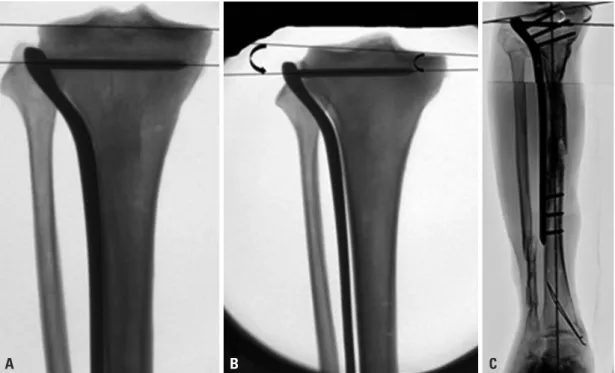
Fig. 1. (A) JSA=0: proximal most screw (PMS) is parallel to the joint line. (B) JSA (-): PMS tilted in varus with tip pointing towards joint line.
(C) JSA (+): PMS tilted in valgus with tip pointing away from joint line. JSA, joint screw angle.
sured on the medial side formed between the tibial plateau line and the line along the tibial axis) was measured in both the tibiae on the postoperative orthoradiogram and the ference was calculated. Malalignment was defined as a dif-ference of more than 5° in the MPTA. In three cases with bilateral proximal tibial fractures, the average population MPTA (87°) was used as a reference angle.
(2) The JSA was measured simultaneously on both intra-operative and postintra-operative images in 29 cases in whom the C-arm images were also saved in addition to the post-operative X-rays. For the remaining 27 patients, the JSA was measured only on the postoperative orthoradiogram. The screw was considered acceptable if it is approximately parallel to the joint line and the JSA was found to be in the range of 0±5°.
In the 29 patients, we did not find a difference of more than 1 degree between the JSA on the intraoperative image and the JSA on the postoperative image. On the basis of this interim analysis, we decided to use the postoperative mea-surement of the JSA as an alternative to the JSA on the in-traoperative image in the remaining 27 patients. We then statistically analyzed the relationship between the JSA and the postoperative malalignment (MPTA) in all 56 cases. All the radiographic measurements were made by two indepen-dent blinded observers (two orthopedic surgeons: first and corresponding authors).
Statistical analysis
Inter-observer variance was assessed using the paired ‘t’ test. No difference was found between the observations made by the two observers (p<0.05).
Patients were categorized into two groups based on their postoperative coronal plane alignment: one group with mal alignment (MPTA within ±5° of the contralateral nor-mal limb) and the other group with nor-malalignment. Patients were also divided into two groups based on the degree of the JSA: one group with acceptable JSA (within ±5°) and the other group with unacceptable JSA. The kappa statistics was calculated to assess the agreement of alignment status between the MPTA and the JSA. We also conducted an ex-act test version of McNemar test to evaluate the agreement between MPTA and JSA. In this test, the null hypothesis was agreement between two variables while the alternative hypothesis was difference between two variables. All analy-ses were conducted using the SAS version 9.1.3 (SAS In-stitute Inc., Cary, NC, USA). The level of significance was set at 0.05.
angle as ‘JSA’ (Fig. 1A). We designated a negative value to the JSA when the screw was tilted in a varus position to the joint line, such that the tip of the screw pointed towards the joint line, resulting in a lateral opening angle (Fig. 1B). If the screw was tilted in a valgus position, such that the tip pointed away from the joint line, resulting in a medial opening angle, we described it as a positive JSA value (Fig. 1C).
Clinical study
We retrospectively reviewed 56 cases of proximal tibial frac-tures that were treated with LCP PLT. They comprised 44 males and 12 females. The mean age was 52 years (range, 30-88 years). Forty-nine fractures were treated in an acute setting within 2 weeks after the injury. There were six non-union and one malnon-union of the proximal metaphysis. The fresh fractures were classified according to the AO-OTA classification. They were of the following types: 41A1(2), 41A2(1), 41A3(7), 41B2(1), 41C2(1), 41C3(18), 42A1(1), 42A2(3), 42A3(1), 42B2(5), 42C1(1), 42C2(3), 42C3(5). All operations were performed by two experienced trauma sur-geons or they were performed under their guidance. In all the fractures, the reduction was achieved first and then LCP PLT was applied via a lateral approach. The plate was ap-plied over the lateral surface of the tibia, anterior to fibular articular surface of the tibia. The first screw was placed in the mid diaphysis and then the proximal screws were in-serted. All efforts were made to align the plate well with the tibial surface proximally and with the shaft.
X-ray analysis
The radiologic analysis formed the backbone of our study. This was done based on the radiograms from 56 patients. In 29 of these patients, we could retrieve the intraoperative imag-es as well as the postoperative radiographs. For the remaining 27 patients, we had the postoperative radiographs only.
1) Postoperative radiograms were taken at approximately 4-20 weeks postoperatively or as soon as patient was able to stand and bear weight on the operated leg. Postoperative radiograms were standing orthoradiograms of the lower ex-tremity centering at the knee joint with the patient facing the radiographic tube and the patella pointing forward.
2) Intraoperative images were taken with the patient posi-tioned supine, knee in full extension, patella pointing to-wards the ceiling, and the image intensifier aligned perpen-dicular to the knee joint. Following measurements were made on the radiograms. Two angles were measured:
mea-were found to have a normal alignment. Out of the 6 pa-tients with an abnormal JSA, 4 papa-tients (67%) showed ma-lalignment. The kappa value for the measure of agreement between JSA and MPTA was 0.565 (95% confidence inter-val: 0.225, 0.905) (Table 1).
The p-value from the exact test version of McNemar’s test was 1.000, indicating a good agreement between JSA and MPTA and clearly stating that the association between the MPTA and the JSA was very relevant and not just coin-cidental.
DISCUSSION
Although the minimal invasive Locking plate fixation has shown promising results in reducing infections (4%) and achieving high rates of union, it has given rise to more trou-blesome, alignment related issues. The published data in lit-erature show varied complication rates of this technique. According to Ricci, et al.,14 obtaining proper alignment
with the use of LISS is technically demanding. Malalign-ment in the coronal plane after the use of LISS in treatMalalign-ment of various types of proximal tibial fractures has been re-ported in many papers.8,12,15,21
Our anatomical study was carried out to identify the rela-tionship between the coronal plane alignment and the JSA. We found, the proximal most screw through LCP PLT to be parallel to the joint line or within ±5° in 29 of 30 cadaveric tibial bones. Only in 1 grossly arthritic specimen, the JSA was out of the acceptable range (-7°) which was due to the abnormal inclination of the joint line (MPTA 82°). Thus, the relationship established was that the JSA was 5° or less in nearly all the normal tibial bones, except in the severely arthritic bones with abnormal inclination of the joint line. The relationship is difficult to establish in severely arthritic
RESULTS
Anatomical study
On gross observation, we found that the LCP PLT fits to the lateral aspect of the proximal tibiae fairly well. However, we also found that the head of the plate tends to stay apart from the lateral aspect of the lateral condyle by a few milli-meters. In 30 specimens that we studied, the JSA was -2° on an average (ranged from 0° to -7°). In 22 cases (73%), the proximal locking screw was almost parallel to the joint line (JSA -2° to 0°). In 7 patients (24%), the JSA was between -5° and -2°, and the JSA was -7° in one specimen with se-vere osteoarthritis. This particular bone showed sese-vere os-teoarthritis with varus inclination of the joint line (MPTA 82). In this case, the unacceptable JSA was due to an abnor-mal inclination of the joint line. Two other specimens showed degenerative changes and a varus inclination of the joint line of 5° in the coronal plane and a JSA of 5°. None of the specimens showed a positive JSA.
Clinical and radiological study
Our clinical and radiographic retrospective study yielded the following results. In 49 of the 56 patients (87.5%), the MPTA was found to be acceptable and within the normal range (within 5° of the contralateral normal limb or average of the population). In the remaining 7 patients (12.5%), there was malalignment in the coronal plane and the MPTA was out of the normal range. Out of the 49 patients with normal alignment, the JSA was found to be in the accept-able range in 47 patients (95.92%), and in the unacceptaccept-able range in the remaining 2 patients. In the 7 patients with ma-lalignment, the JSA was unacceptable in 4 patients (57.14%) and acceptable in the remaining 3 patients. Among the 50 patients with a normal JSA, 47 patients [94% (47/50×100)] Table 1. The Negative Predictive Value for the JSA
JSA
Alignment
Total
Normal Malaligned
Nos. Percentage Nos. Percentage
Acceptable Nos.Percentage 95.92%47 94% 42.86%3 6% 50
Unacceptable Nos.Percentage 57.14% 2 33.33% 4.08%4 66.67% 6
Total 49 7 56
JSA, joint screw angle; MPTA, medial proximal tibial angle.
Alignment: normal when MPTA is 87±5 otherwise it is Malaligned. JSA is acceptable if it is 0±5 otherwise it is unacceptable. Kappa=0.565 (95% confidence interval: 0.225, 0.905). Positive predictivity=true positive/total positive=47/50=0.94 (94%). Negative predictivity=true negative/total nega-tive=4/6=0.67=67%.
issues and addressed them. We were also unable to find any mention of the JSA or of its relationship to the alignment in the manufacturer’s recommendation. Hence, we are not certain whether or not they intended to orient the first screw parallel to the joint line.
An ideally placed plate, is likely to demonstrate the rela-tionship between the MPTA and the JSA. In view of the above limitations, we can state that the JSA may not be used as a standalone assessment tool, however, it can rather be used as an adjunct to the other techniques of assessment of alignment, since it points very well towards the type of alignment we are likely to achieve and may also provide an indication on the occurrence of malalignment.
In conclusion, we can safely conclude from our observa-tion that the JSA can be used intraoperatively as a rough guideline to assess the achievement of a final normal align-ment in the coronal plane when applying a LCP PLT/LISS internal fixator for fractures of the proximal tibia.
REFERENCES
1. Krieg JC. Proximal tibial fractures: current treatment, results, and problems. Injury 2003;34 Suppl 1:A2-10.
2. Waddell JP, Johnston DW, Neidre A. Fractures of the tibial pla-teau: a review of ninety-five patients and comparison of treatment methods. J Trauma 1981;21:376-81.
3. Wu CC. Salvage of proximal tibial malunion or nonunion with the use of angled blade plate. Arch Orthop Trauma Surg 2006;126:82-7. 4. Moore TM, Patzakis MJ, Harvey JP. Tibial plateau fractures: defi-nition, demographics, treatment rationale, and long-term results of closed traction management or operative reduction. J Orthop Trauma 1987;1:97-119.
5. Young MJ, Barrack RL. Complications of internal fixation of tibi-al plateau fractures. Orthop Rev 1994;23:149-54.
6. Blokker CP, Rorabeck CH, Bourne RB. Tibial plateau fractures. An analysis of the results of treatment in 60 patients. Clin Orthop Relat Res 1984:193-9.
7. Cole PA, Zlowodzki M, Kregor PJ. Less Invasive Stabilization System (LISS) for fractures of the proximal tibia: indications, sur-gical technique and preliminary results of the UMC Clinical Trial. Injury 2003;34 Suppl 1:A16-29.
8. Cole PA, Zlowodzki M, Kregor PJ. Treatment of proximal tibia fractures using the less invasive stabilization system: surgical ex-perience and early clinical results in 77 fractures. J Orthop Trauma 2004;18:528-35.
9. Egol KA, Su E, Tejwani NC, Sims SH, Kummer FJ, Koval KJ. Treatment of complex tibial plateau fractures using the less inva-sive stabilization system plate: clinical experience and a laboratory comparison with double plating. J Trauma 2004;57:340-6. 10. Goesling T, Frenk A, Appenzeller A, Garapati R, Marti A, Krettek
C. LISS PLT: design, mechanical and biomechanical characteris-tics. Injury 2003;34 Suppl 1:A11-5.
11. Gösling T, Schandelmaier P, Marti A, Hufner T, Partenheimer A, bones, and thus we would generally limit the results of our
study to bones which do not show severe arthritis.
We further set out to evaluate if this relationship holds true in a clinical setting and if it could be used intraopera-tively to assess the alignment in the coronal plane.
On a retrospective analysis, the relationship that we found between the JSA and the MPTA had a high positive predic-tion of 94% (47/50); i.e. there is a great chance of having a normal alignment if the JSA is within the normal accept-able range. The results of our anatomic study and clinical correlation indicate that an acceptable JSA does point to-wards a normal MPTA and the relationship cannot be ex-plained by chance alone. The relationship between the JSA and the MPTA could thus be used as an additional tool to evaluate the coronal alignment.
However, out of 6 patients with an unacceptable JSA, there were 4 cases of an abnormal MPTA, and 2 cases of a normal MPTA. These 2 cases of a normal MPTA with an unacceptable JSA can be explained by the fact that there are various ways in which the plate could be positioned over the tibia due to various kinds of possible mismatches be-tween the proximal tibia and the pre-contoured plate. If the position is like that of an impingement fit with the distal part of the plate away from the shaft and the proximal part fitting well on the shaft as described by Goyal, et al.,22 the
screw and the JSA will be in a valgus position over an ana-tomically well aligned tibia.
Preservation of the normal anatomic alignment is impor-tant in achieving a favorable outcome. Blokker, et al.6 found
unsatisfactory results in 100% of the patients with inade-quate anatomical reduction. In many series, the authors un-derscore the importance of good technical tricks to avoid malalignment. Cole, et al.8 emphasized the importance of
good intraoperative radiographs to decrease the incidence of malreduction. They tried to underscore the importance of good radiography techniques; i.e. taking radiographs on a large flat plate before the placement of screws, using fluo-roscopy & strategic bumps supporting the distal fragment in order to achieve a good postoperative alignment. Krettek, et al.20 described the cable technique to avoid the coronal
plane malalignment. Although this technique is quite useful when the alignment is evaluated after the fixation is over, it is often not feasible to have two assistants holding the cable and moving the C-arm from the top to the bottom while the alignment is maintained. On the other hand, the JSA can be easily measured on a single C-arm AP image. We did not find any other study which specifically dealt with alignment
zation of proximal tibial fractures with the proximal tibial LISS: early experience in Birmingham, Alabama (USA). Injury 2003;34 Suppl 1:A36-42.
18. Stannard JP, Wilson TC, Volgas DA, Alonso JE. The less invasive stabilization system in the treatment of complex fractures of the tibial plateau: short-term results. J Orthop Trauma 2004;18:552-8. 19. Weight M, Collinge C. Early results of the less invasive
stabiliza-tion system for mechanically unstable fractures of the distal femur (AO/OTA types A2, A3, C2, and C3). J Orthop Trauma 2004;18: 503-8.
20. Krettek C, Miclau T, Grün O, Schandelmaier P, Tscherne H. Intra-operative control of axes, rotation and length in femoral and tibial fractures. Technical note. Injury 1998;29 Suppl 3:C29-39. 21. Beck M, Gradl G, Gierer P, Rotter R, Witt M, Mittlmeier T.
[Treatment of complicated proximal segmental tibia fractures with the less invasive stabilization locking plate system]. Unfallchirurg 2008;111:493-8.
22. Goyal KS, Skalak AS, Marcus RE, Vallier HA, Cooperman DR. Analysis of anatomic periarticular tibial plate fit on normal adults. Clin Orthop Relat Res 2007;461:245-57.
Krettek C. Less invasive stabilization of complex tibial plateau fractures: a biomechanical evaluation of a unilateral locked screw plate and double plating. J Orthop Trauma 2004;18:546-51. 12. Gosling T, Schandelmaier P, Muller M, Hankemeier S, Wagner M,
Krettek C. Single lateral locked screw plating of bicondylar tibial plateau fractures. Clin Orthop Relat Res 2005;439:207-14. 13. Phisitkul P, McKinley TO, Nepola JV, Marsh JL. Complications
of locking plate fixation in complex proximal tibia injuries. J Or-thop Trauma 2007;21:83-91.
14. Ricci WM, Rudzki JR, Borrelli J Jr. Treatment of complex proxi-mal tibia fractures with the less invasive skeletal stabilization sys-tem. J Orthop Trauma 2004;18:521-7.
15. Schütz M, Kääb MJ, Haas N. Stabilization of proximal tibial frac-tures with the LIS-System: early clinical experience in Berlin. In-jury 2003;34 Suppl 1:A30-5.
16. Schütz M, Müller M, Regazzoni P, Höntzsch D, Krettek C, Van der Werken C, et al. Use of the less invasive stabilization system (LISS) in patients with distal femoral (AO33) fractures: a prospec-tive multicenter study. Arch Orthop Trauma Surg 2005;125:102-8. 17. Stannard JP, Wilson TC, Volgas DA, Alonso JE. Fracture