ABSTRACT
Heart failure with preserved ejection fraction (HFpEF) has recently been recognized as the single greatest unmet need in cardiovascular medicine. As the population ages and the comorbidity increases, the prevalence of HFpEF increases considerably. Even though there have been large numbers of studies on pathophysiology, diagnosis, and treatment of HFpEF for latest years, there are no current pharmacologic interventions that can reduce mortality. HFpEF is currently understood as a heterogeneous syndrome originated from the interplay of cardiac and extracardiac abnormalities recognized by systemic inflammation, endothelial and coronary microvascular dysfunction, cardiomyocyte dysfunction and skeletal muscle dysfunction. The difficult “jigsaw puzzle” called HFpEF has been filled with some pieces, but it is still not enough to meet clinical needs. Here, we review recent evidences and unsolved problems about HFpEF to improve our understanding of HFpEF. Finally, we hope to accelerate to completion of the problematic “jigsaw puzzle”.
Keywords: Heart failure with preserved ejection fraction; Syndrome; Pathophysiology; Diagnosis; Treatment
INTRODUCTION
Approximately half of patients with signs and symptoms of heart failure (HF) have preserved ejection fraction.1-6) As the population ages and the comorbidity increases, the prevalence of HF with preserved ejection fraction (HFpEF) increases considerably.1)3-6) The results of the early epidemiologic study revealed that patients with HFpEF were older, more female, and had a higher rate of comorbidity, such as hypertension, diabetes mellitus (DM), obesity, chronic kidney disease, and atrial fibrillation (AF).1) What these patients have in common is that ejection fraction is preserved, but there are symptoms and signs of clinical HF. Therefore, it has been considered a kind of syndrome in the absence of a confirmative diagnostic or therapeutic indicator. The basic question is whether we have to continue to fill this difficult “jigsaw puzzle” named HFpEF, or whether we have to make a new base after breaking all of the HF classification methods based on ejection traction. In this review, we will review some pretty meaningful pieces that are already set in the puzzle, and we believe that these will not be changed even if the base of the puzzle changes.
Review Article
Received: Aug 2, 2020
Accepted: Aug 17, 2020
Correspondence to
Chi Young Shim, MD, PhD Division of Cardiology, Severance Cardiovascular Hospital, Yonsei University College of Medicine, 50-1, Yonsei-ro, Seodaemun-gu, Seoul 03722, Korea. E-mail: [email protected]
Copyright © 2020. The Korean Society of Cardiology
This is an Open Access article distributed under the terms of the Creative Commons Attribution Non-Commercial License (https:// creativecommons.org/licenses/by-nc/4.0) which permits unrestricted noncommercial use, distribution, and reproduction in any medium, provided the original work is properly cited.
ORCID iDs
Chi Young Shim
https://orcid.org/0000-0002-6136-0136
Conflict of Interest
The author has no financial conflicts of interest.
Chi Young Shim , MD, PhD
Division of Cardiology, Severance Cardiovascular Hospital, Yonsei University College of Medicine, Seoul, Korea
Heart Failure with Preserved Ejection
Fraction: the Major Unmet Need in
Cardiology
HEART FAILURE WITH PRESERVED EJECTION FRACTION
VS. DIASTOLIC HEART FAILURE
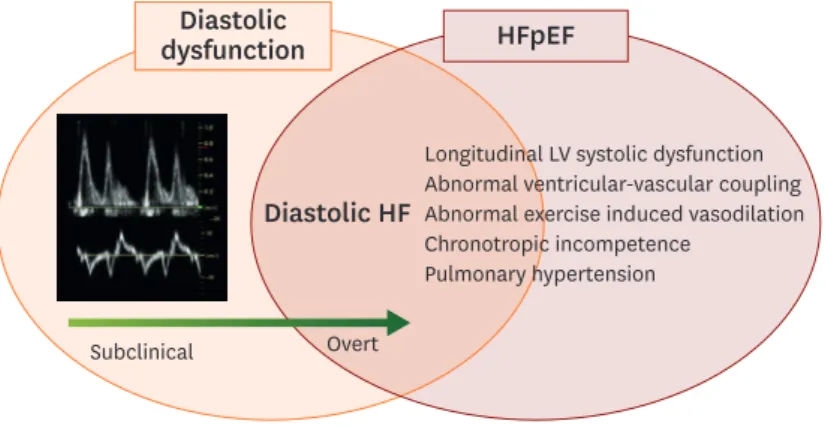
HFpEF was initially termed diastolic HF.7) HFpEF and diastolic HF have been used interchangeably and confusedly for the last 20 years. In principle, HFpEF and diastolic HF are not the same diagnostic terms, and the former should be understood to a larger range of diagnosis since several alternative and complementary mechanisms exist either presence or absence of left ventricular (LV) diastolic dysfunction (Figure 1).8)9) There has been a recent expansion of the ejection fraction derived classification of patients with HF. Accordingly, HF with midrange ejection fraction and HF with improved ejection fraction appeared recently.10)11) Therefore, the term “diastolic HF” tends not to be used although diastolic HF is also an important term in terms of mechanism. Each of these two terms has its pros and cons. For example, if a patient presented with HF but a meaningful diastolic dysfunction or elevation of LV filling pressure are not proven in diagnostic tests, the patients can be diagnosed with HFpEF, but not enough to be diagnosed with diastolic HF. In the case of HFpEF which the diastolic dysfunction is not proven, there is a fundamental limitation in developing a therapeutic agent because of lack of any indicators to monitor. In order to overcome these shortcomings of HFpEF, there is a trend to classify several key phenotypes in this heterogeneous disease, and construct a treatment strategy according to the main pathophysiology.
PATHOPHYSIOLOGY AND PHENOTYPES OF HEART
FAILURE WITH PRESERVED EJECTION FRACTION
There have been a number of efforts to defining phenotypes of patients with HFpEF according to their main features and pathophysiology after a few clinical trials, but repetitively failed to prove benefits of potential pharmacologic treatments in this heterogeneous syndrome.12-15) Different treatment strategies according to the phenotype have been presented very convincingly, however, it is often difficult to specify this as only one phenotype.12-15)
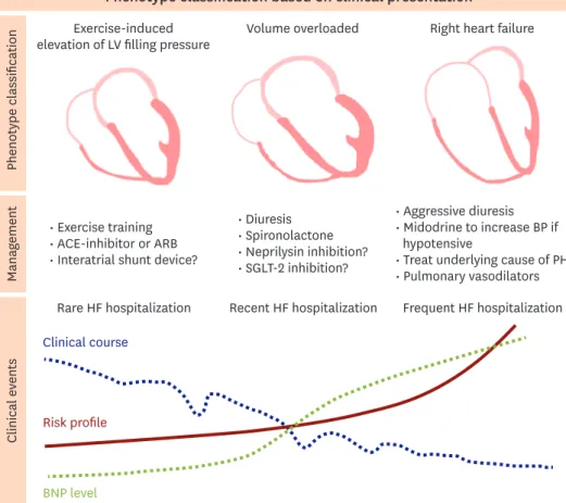
Representative phenotypic classifications of HFpEF are presented in Figures 2 and 3, which might help physicians to understand the main clinical presentation and pathophysiology in managing patients with HFpEF.12)15) As shown in Figure 2, patients with HFpEF can be
Diastolic HF
Subclinical Overt
HFpEF Diastolic
dysfunction
Longitudinal LV systolic dysfunction Abnormal ventricular-vascular coupling Abnormal exercise induced vasodilation Chronotropic incompetence
Pulmonary hypertension
Figure 1. Basic concepts of diastolic HF and HFpEF.
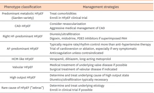
roughly classified into 3 phenotypes: exercise induced elevation of LV filling pressures, volume overload or right HF.12) The first phenotype of “exercise-induced elevation of LV filling pressures”, expressing the lowest risk type, but also the most difficult to diagnose as HFpEF.12) The predominant symptom in these patients is exertional dyspnea and there are no definite clues in echocardiographic features or brain natriuretic peptide (BNP) levels.12) The second phenotype of “volume overload” is the common one that physicians recognize.12) The third phenotype of “right HF” is the highest risk clinical subclass with poor prognosis.12) An another attractive phenotype classification of HFpEF that provides clinicians with a specific and effective therapeutic strategies to individual patients is demonstrated in Figure 3.15) There are 8 phenotypes, which include predominant metabolic syndrome (often referred to as “garden variety” associated with hypertension, obesity, DM, metabolic syndrome, and/ or chronic kidney disease), coronary artery disease, predominant right HF, AF, hypertrophic cardiomyopathy like phenotype, valvular heart disease, high-output HF, rare causes such as restrictive cardiomyopathy or constrictive pericarditis.15) This phenotype classification based on etiology and pathophysiology is quite helpful in guiding initial treatment, but not mutually exclusive, and therefore sometimes difficult to place patients into a single category.15) However, even in patients classified as a garden variety phenotype after exclusion of certain diseases that require specific treatment, the mechanism of HFpEF varies with a more predominant contributing components among aortic stiffness (increased afterload), cardio-metabolic (systemic inflammation), and cardio-renal (increased preload) aspects. Recently,
Phenotype classification based on clinical presentation
Phenotype classification Manag ement Clinical e vents Exercise-induced
elevation of LV filling pressure Volume overloaded Right heart failure
• Exercise training • ACE-inhibitor or ARB • Interatrial shunt device?
• Diuresis • Spironolactone • Neprilysin inhibition? • SGLT-2 inhibition? • Aggressive diuresis • Midodrine to increase BP if hypotensive
• Treat underlying cause of PH • Pulmonary vasodilators Rare HF hospitalization Recent HF hospitalization Frequent HF hospitalization Clinical course
Risk profile
BNP level
Figure 2. Phenotype classification of HF with preserved ejection fraction based on clinical presentations. ACE = angiotensin-converting enzyme; ARB = angiotensin II receptor blocker; BNP = brain natriuretic peptide; BP = blood pressure; HF = heart failure; LV = left ventricular; PH = pulmonary hypertension; SGLT-2 = sodium-glucose cotransporter-2.
a few novel “phenomapping” techniques by machine learning to define clusters of patients based on dense phenotypic data have been suggested to provide an unbiased way to classify heterogeneous clinical syndromes of HFpEF.16)17)
DIAGNOSTIC CHALLENGES IN HEART FAILURE WITH
PRESERVED EJECTION FRACTION
The diagnosis of HFpEF is still challenging, because symptoms are nonspecific and can be explained by several alternative non-cardiac conditions, such as chronic lung disease, anemia, and chronic kidney disease.8) In patients with phenotypes of volume overload or right HF based on clinical presentation,12) it is not difficult to document typical signs and symptoms of HF, such as pulmonary congestion, elevated BNP, and advanced diastolic dysfunction on Doppler echocardiography. However, in patients with the phenotype of exercise-induced elevation of LV filling pressure especially in those with significant LV structural abnormalities, we often encounter a few problems of echocardiogram only assessed at rest for HFpEF diagnosis.18-20) First, diastolic dysfunction as assessed by
echocardiogram is highly prevalent in the elderly population.19)20) Second, echocardiographic Phenotype classification based on etiology/pathophysiology
Phenotype classification Management strategies
Predominant metabolic HFpEF
(Garden variety) Treat comorbiditiesEnroll in HFpEF clinical trial
CAD-HFpEF Consider revascularizationAggressive medical management of CAD
Right HF-predominant HFpEF Diuresis/ultrafiltrationDigoxin, midodrine, PDE5 inhibitors if superimposed PAH
AF-predominant HFpEF Typically require rate/rhythm control more than anti-hypertensive therapyTrial of cardioversion or ablation, especially if very symptomatic Anticoagulation unless contraindicated
HCM-like HFpEF Verapamil, diltiazem, long-acting metoprolol
Valvular HFpEF Medical treatment of underlying valve disease if possibleSurgical treatment of valvular disease if indicated
High output HFpEF Determine and treat underlying cause of high output stateDiuretics/ultrafiltration typically necessary
Rare cause of HFpEF (“zebras”) Determine and treat underlying etiologyEnroll in clinical trial if possible
Heterogeneity of individual pathophysiology within the “garden variety” phenotype
Aortic
stiffness metabolic
Cardio-renal
Figure 3. Phenotype classification of HFpEF based on etiology and pathophysiology.
AF = atrial fibrillation; CAD = coronary artery disease; HCM = hypertrophic cardiomyopathy; HF = heart failure; HFpEF = heart failure with preserved ejection fraction; PAH = pulmonary arterial hypertension; PDE5 = phosphodiesterase type 5.
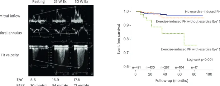
variables indicating diastolic dysfunction may be absent in a significant proportion of patients diagnosed with HFpEF.19) The existing tools to assess LV systolic performance are far from perfect.19) Last, viewing HFpEF as results of ventricular stiffening and diastolic dysfunction only is an oversimplification although there are complementary and complex mechanisms.19) Additionally, exertional dyspnea is a common clinical problem. It can be complex and multiple etiologies such as respiratory causes, cardiac causes, metabolic causes and psychological causes.20) The differential diagnosis of HFpEF is challenging in stable elderly patients with equivocal symptoms. Therefore, we need to perform an exercise stress test because we cannot explain the patients' symptom completely with a resting echocardiography.20)21) A noninvasive diagnostic test for diastolic dysfunction using supine bicycle exercise Doppler echocardiography has been set up about 15 years ago.20) Because this echocardiographic method has advantages for evaluation of diastolic function during exercise. It is possible to get continuous imaging at each stages of exercise.20) Moreover, since the supine position increases myocardial wall tension and oxygen demand, it is suitable for diastolic stress.20) Currently, diastolic stress test is recommended for evaluation of exercise-induced elevation of LV filling pressure if the resting echocardiographic evaluation is inconclusive for diagnosis or exclusion of HFpEF.18)21) In a simultaneous invasive-echo study in patients with HFpEF and non-cardiac dyspnea, much improved diagnostic performance of adding exercise E/e′ on the European Society of Cardiology criteria were proven by demonstrating a 30% increase of sensitivity from 60% to 90%.18) In addition to the improvement of diagnostic performance and risk stratification of HFpEF,21-25) diastolic stress test can provide a prognostic information.26-28) Holland et al. showed the incremental prognostic implications of LV filling pressure with exercise in patients with or without inducible myocardial ischemia.26) Our previous work also demonstrated the prognostic values of exercise-induced elevation of LV filling pressure and consequent exercise-induced pulmonary hypertension in subjects with preserved LV ejection fraction (Figure 4).27) The types and methods of diastolic stress test vary from invasive to noninvasive techniques.21) Therefore, it is needed to apply an appropriate stress test after considering the patient's clinical situation and the established facility in each institution.
0.8 1.0 0.9 0.7 0.6 0 Follow-up (months) L0g-rank p<0.001 Ev ent fr ee survival 20 40 60 80 100 Mitral inflow Mitral annulus TR velocity Resting 25 W Ex 50 W Ex E/e′ PASP 8.6 30 mmHg 16.9 54 mmHg 17.8 71 mmHg n=481 n=430 n=267 n=104 n=17
Exercise-induced PH with exercise E/e′ ↑ Exercise-induced PH without exercise E/e′ ↑ No exercise-induced PH
Figure 4. Exercise-induced elevation of left ventricular filling pressure and exercise-induced PH detected by diastolic stress echocardiography. PASP = pulmonary artery systolic pressure; PH = pulmonary hypertension; TR = tricuspid regurgitation.
COMMON COMORBIDITIES IN HEART FAILURE WITH
PRESERVED EJECTION FRACTION: HYPERTENSION AND
ARTERIAL STIFFNESS
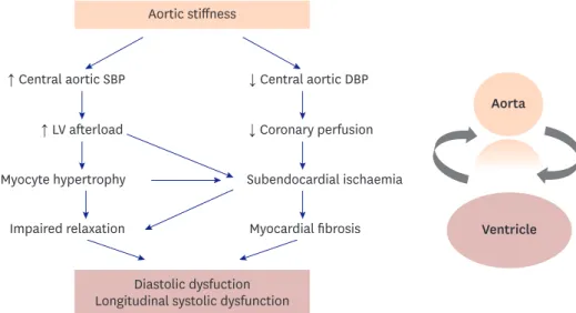
Arterial stiffness is a cardinal feature of vascular aging and hypertension.29) The aorta deliveries blood from the LV to the capillaries, acting as a highly efficient conduit and cushion. As the aorta stiffens, blood travels faster, returns earlier, and boosts pressure in late systole. As a result, arterial stiffness results in elevation of augmentation pressure and augmentation index, and widening of the arterial pulse pressure.30)31)Figure 5 shows the possible relationship between arterial stiffness and LV diastolic/longitudinal systolic dysfunction which was originally proposed by Mottram et al.32) Increased central systolic blood pressure (BP) results in increasing LV afterload and makes myocyte hypertrophy and impaired relaxation. Simultaneously, central aortic diastolic BP decreases.32) It reduces coronary perfusion and induces subendocardial ischemia and fibrosis.32) These adverse effects finally lead to LV diastolic dysfunction and longitudinal systolic dysfunction. Recent studies have demonstrated that noninvasively assessed central BP and variables reflecting arterial mechanical function at rest and with exercise are well correlated with LV diastolic function and LV systolic functional impairments such as LV global longitudinal strain and apical rotation.33-38) In terms of sex difference in HFpEF, the indices reflecting central arterial stiffness in radial arterial tonometry were higher in women than in men.38) Additionally, we found that the relationship between variables of arterial stiffness and LV diastolic function was stronger in women.38) In another study, the dynamic changes of arterial elastance during exercise in patients with hypertension were investigated.39) When age-matched men and women with hypertension were compared, there was a steeper rise of arterial elastance index during exercise in hypertensive women.39) Moreover, the arterial elastance index during exercise was an independent determinant of exercise duration.39) To summarize, arterial stiffness and ventricular-vascular interaction at rest and dynamic changes during stress are considered a principal pathophysiology in patients with HFpEF and should be a therapeutic target for preventing HFpEF.40-43)
Aortic stiffness
↑ Central aortic SBP ↓ Central aortic DBP ↓ Coronary perfusion Subendocardial ischaemia Myocardial fibrosis ↑ LV afterload Myocyte hypertrophy Impaired relaxation Diastolic dysfuction Longitudinal systolic dysfunction
Aorta
Ventricle
Figure 5. Pathophysiologic relationship between arterial stiffness and LV dysfunction. DBP = diastolic blood pressure; LV = left ventricular; SBP = systolic blood pressure.
DIABETES MELLITUS
DM is a common comorbidity in HF and has an adverse impact on prognosis.44)45) Classically, many studies have been conducted on the mechanisms and treatment strategies of diabetic cardiomyopathy, in which myocardial dysfunction occurs in absence of significant coronary artery stenosis in patients with DM.46)47) Although definite pathophysiological mechanisms of DM in HFpEF have yet to be elucidated, several common pathological mechanisms have been suggested including microvascular dysfunction, metabolic derangements and systemic inflammation, sodium retention, and impaired skeletal muscle function.45) Insulin resistance in DM leads to increase free fatty acid utilization by cardiac myocytes, which may lead to mitochondrial dysfunction, production of toxic lipid intermediates, and increased reactive oxygen species.45) Hyperglycemia-induced advanced glycation end products result in microvascular dysfunction and decline nitric oxide availability.48) In addition, hyperglycemia causes up-regulation of the sodium-glucose cotransporter-2 (SGLT-2) leading to increased proximal renal sodium absorption, volume expansion, and decreased of diuretics response.49) As a result, many previous studies have demonstrated LV diastolic dysfunction and impaired LV longitudinal functional reserve in patients with DM.50)51) Furthermore, impaired LV longitudinal functional reserve in patients with DM without overt heart disease provided incremental prognostic information for composite of death and HF hospitalization.28) Recently, as expectations for the effect of SGLT-2 inhibitors on improving HF outcome have increased, interests in DM as a risk factor of HFpEF have been amplified. SGLT-2 inhibitors have demonstrated convincing reductions in the incidence and risks of HF in patients with DM.52-55) Now, studies are underway to prove the beneficial effects of SGLT-2 inhibitors in overt HF patients with or without DM.
OBESITY AND OBSTRUCTIVE SLEEP APNEA
Obesity is a major risk factor of HF and also one of the most common comorbidities in patients with HFpEF.56) Obesity has several harmful effects in aspects of HF, mediated by changes in volume status, cardiac loading, energy substrate utilization, tissue metabolism and systemic inflammation that are believed to promote disease progression.57)58) Typically, obese patients with HFpEF have increased plasma volume with greater biventricular filling pressure, typical myocardial remodeling characterized by increased LV dimensions and mass.57)58) Interestingly, obesity is more important risk factor of occurrence of HFpEF in women than in men.59) Our previous study also demonstrated overweight women had exercise intolerance than lean women through displaying impaired arterial compliance during exercise.37) Because obesity is possible to be treated by weight reduction and physical activity, many studies have demonstrated significant improvement in cardiac structure and function after bariatric surgery, dietary or exercise intervention.60) In obese patients, obstructive sleep apnea (OSA) is frequently combined and OSA might deteriorate LV diastolic dysfunction and longitudinal systolic function by exposing to episodic hypoxemia, nocturnal sympathetic nervous system activation, elevated BP, oxidative stress and inflammation.61)62) In our recent randomized, sham controlled clinical trial, continuous positive airway pressure treatment for 3 months in patients with severe OSA improved LV diastolic function more than sham treatment, and was accompanied by improvements in arterial stiffness and ventricular-vascular coupling.63) Moreover, we additionally proved that LV mechanical function assessed by speckle-tracking echocardiography and right ventricular fractional area change were significantly improved by continuous positive airway pressure.64) Hence, obesity and OSA are
modifiable risk factors, so it is necessary to actively evaluate and treat them before myocardial structural and functional changes are too advanced.
CONCLUSION
HFpEF is a heterogeneous clinical syndrome and the single greatest unmet need in
cardiovascular medicine. We hope that HFpEF will become a solved disease entity with lots of evidence in the future through more scientific phenotype classification in the era of precision medicine, improved diagnostic imaging even in early stage of disease, applying appropriate drugs and intervention focusing each pathophysiologic mechanism.
REFERENCES
1. Owan TE, Hodge DO, Herges RM, Jacobsen SJ, Roger VL, Redfield MM. Trends in prevalence and outcome of heart failure with preserved ejection fraction. N Engl J Med 2006;355:251-9.
PUBMED | CROSSREF
2. Bhatia RS, Tu JV, Lee DS, et al. Outcome of heart failure with preserved ejection fraction in a population-based study. N Engl J Med 2006;355:260-9.
PUBMED | CROSSREF
3. Dunlay SM, Roger VL, Redfield MM. Epidemiology of heart failure with preserved ejection fraction. Nat Rev Cardiol 2017;14:591-602.
PUBMED | CROSSREF
4. Lee JH, Kim MS, Kim EJ, et al. KSHF guidelines for the management of acute heart failure: part I. definition, epidemiology and diagnosis of acute heart failure. Korean Circ J 2019;49:1-21.
PUBMED | CROSSREF
5. Lee JH, Kim MS, Yoo BS, et al. KSHF guidelines for the management of acute heart failure: part II. treatment of acute heart failure. Korean Circ J 2019;49:22-45.
PUBMED | CROSSREF
6. Kim MS, Lee JH, Cho HJ, et al. KSHF guidelines for the management of acute heart failure: part III. specific management of acute heart failure according to the etiology and co-morbidity. Korean Circ J
2019;49:46-68. PUBMED | CROSSREF
7. Zile MR, Baicu CF, Gaasch WH. Diastolic heart failure--abnormalities in active relaxation and passive stiffness of the left ventricle. N Engl J Med 2004;350:1953-9.
PUBMED | CROSSREF
8. Oktay AA, Shah SJ. Diagnosis and management of heart failure with preserved ejection fraction: 10 key lessons. Curr Cardiol Rev 2015;11:42-52.
PUBMED | CROSSREF
9. Pfeffer MA, Shah AM, Borlaug BA. Heart failure with preserved ejection fraction in perspective. Circ Res
2019;124:1598-617. PUBMED | CROSSREF
10. Yancy CW, Jessup M, Bozkurt B, et al. 2013 ACCF/AHA guideline for the management of heart failure: executive summary: a report of the American College of Cardiology Foundation/American Heart Association task force on practice guidelines. Circulation 2013;128:1810-52.
PUBMED | CROSSREF
11. Ponikowski P, Voors AA, Anker SD, et al. 2016 ESC guidelines for the diagnosis and treatment of acute and chronic heart failure: the task force for the diagnosis and treatment of acute and chronic heart failure of the European Society of Cardiology (ESC). Developed with the special contribution of the Heart Failure Association (HFA) of the ESC. Eur J Heart Fail 2016;18:891-975.
PUBMED | CROSSREF
12. Shah SJ, Katz DH, Deo RC. Phenotypic spectrum of heart failure with preserved ejection fraction. Heart Fail Clin 2014;10:407-18.
13. Shah SJ, Kitzman DW, Borlaug BA, et al. Phenotype-specific treatment of heart failure with preserved ejection fraction: a multiorgan roadmap. Circulation 2016;134:73-90.
PUBMED | CROSSREF
14. Lewis GA, Schelbert EB, Williams SG, et al. Biological phenotypes of heart failure with preserved ejection fraction. J Am Coll Cardiol 2017;70:2186-200.
PUBMED | CROSSREF
15. Silverman DN, Shah SJ. Treatment of heart failure with preserved ejection fraction (HFpEF): the phenotype-guided approach. Curr Treat Options Cardiovasc Med 2019;21:20.
PUBMED | CROSSREF
16. Shah SJ, Katz DH, Selvaraj S, et al. Phenomapping for novel classification of heart failure with preserved ejection fraction. Circulation 2015;131:269-79.
PUBMED | CROSSREF
17. Shah SJ. 20th annual Feigenbaum lecture: echocardiography for precision medicine-digital biopsy to deconstruct biology. J Am Soc Echocardiogr 2019;32:1379-1395.e2.
PUBMED | CROSSREF
18. Obokata M, Kane GC, Reddy YNV, Olson TP, Melenovsky V, Borlaug BA. Role of diastolic stress testing in the evaluation for heart failure with preserved ejection fraction: a simultaneous invasive-echocardiographic study. Circulation 2017;135:825-38.
PUBMED | CROSSREF
19. Henning RJ. Diagnosis and treatment of heart failure with preserved left ventricular ejection fraction.
World J Cardiol 2020;12:7-25.
PUBMED | CROSSREF
20. Ha JW, Oh JK, Pellikka PA, et al. Diastolic stress echocardiography: a novel noninvasive diagnostic test for diastolic dysfunction using supine bicycle exercise Doppler echocardiography. J Am Soc Echocardiogr
2005;18:63-8. PUBMED | CROSSREF
21. Ha JW, Andersen OS, Smiseth OA. Diastolic stress test: invasive and noninvasive testing. JACC Cardiovasc Imaging 2020;13:272-82.
PUBMED | CROSSREF
22. Ha JW, Choi D, Park S, et al. Left ventricular diastolic functional reserve during exercise in patients with impaired myocardial relaxation at rest. Heart 2009;95:399-404.
PUBMED | CROSSREF
23. Choi EY, Shim CY, Kim SA, et al. Passive leg-raise is helpful to identify impaired diastolic functional reserve during exercise in patients with abnormal myocardial relaxation. J Am Soc Echocardiogr 2010;23:523-30.
PUBMED | CROSSREF
24. Choi EY, Ha JW, Rim SJ, et al. Incremental value of left ventricular diastolic function reserve index for predicting exercise capacity in patients with hypertrophic cardiomyopathy. J Am Soc Echocardiogr
2008;21:487-92. PUBMED | CROSSREF
25. Moon J, Hong YJ, Kim YJ, et al. Extent of late gadolinium enhancement on cardiovascular magnetic resonance imaging and its relation to left ventricular longitudinal functional reserve during exercise in patients with hypertrophic cardiomyopathy. Circ J 2013;77:1742-9.
PUBMED | CROSSREF
26. Holland DJ, Prasad SB, Marwick TH. Prognostic implications of left ventricular filling pressure with exercise. Circ Cardiovasc Imaging 2010;3:149-56.
PUBMED | CROSSREF
27. Shim CY, Kim SA, Choi D, et al. Clinical outcomes of exercise-induced pulmonary hypertension in subjects with preserved left ventricular ejection fraction: implication of an increase in left ventricular filling pressure during exercise. Heart 2011;97:1417-24.
PUBMED | CROSSREF
28. Kim SA, Shim CY, Kim JM, et al. Impact of left ventricular longitudinal diastolic functional reserve on clinical outcome in patients with type 2 diabetes mellitus. Heart 2011;97:1233-8.
PUBMED | CROSSREF
29. Mitchell GF. Arterial stiffness and hypertension: chicken or egg? Hypertension 2014;64:210-4.
PUBMED | CROSSREF
30. Shim CY. Arterial-cardiac interaction: the concept and implications. J Cardiovasc Ultrasound 2011;19:62-6.
PUBMED | CROSSREF
31. Shim CY, Hong GR, Ha JW. Ventricular stiffness and ventricular-arterial coupling in heart failure: what is it, how to assess, and why? Heart Fail Clin 2019;15:267-74.
32. Mottram PM, Haluska BA, Leano R, Carlier S, Case C, Marwick TH. Relation of arterial stiffness to diastolic dysfunction in hypertensive heart disease. Heart 2005;91:1551-6.
PUBMED | CROSSREF
33. Kim D, Shim CY, Hong GR, et al. Differences in left ventricular functional adaptation to arterial stiffness and neurohormonal activation in patients with hypertension: a study with two-dimensional layer-specific speckle tracking echocardiography. Clin Hypertens 2017;23:21.
PUBMED | CROSSREF
34. Shim CY, Park S, Choi EY, et al. The relationship between ventricular-vascular uncoupling during exercise and impaired left ventricular longitudinal functional reserve in hypertensive patients. J Am Soc Hypertens
2013;7:198-205. PUBMED | CROSSREF
35. Shim CY, Hong GR, Park S, et al. Impact of central haemodynamics on left ventricular function in individuals with an exaggerated blood pressure response to exercise. J Hypertens 2015;33:612-20.
PUBMED | CROSSREF
36. Lee JS, Shim CY, Wi J, et al. Left ventricular diastolic function is closely associated with mechanical function of the left atrium in patients with paroxysmal atrial fibrillation. Circ J 2013;77:697-704.
PUBMED | CROSSREF
37. Shim CY, Yang WI, Park S, et al. Overweight and its association with aortic pressure wave reflection after exercise. Am J Hypertens 2011;24:1136-42.
PUBMED | CROSSREF
38. Shim CY, Park S, Choi D, et al. Sex differences in central hemodynamics and their relationship to left ventricular diastolic function. J Am Coll Cardiol 2011;57:1226-33.
PUBMED | CROSSREF
39. Park S, Ha JW, Shim CY, et al. Gender-related difference in arterial elastance during exercise in patients with hypertension. Hypertension 2008;51:1163-9.
PUBMED | CROSSREF
40. Kass DA. Ventricular arterial stiffening: integrating the pathophysiology. Hypertension 2005;46:185-93.
PUBMED | CROSSREF
41. Chirinos JA, Segers P, Hughes T, Townsend R. Large-artery stiffness in health and disease: JACC state-of-the-art review. J Am Coll Cardiol 2019;74:1237-63.
PUBMED | CROSSREF
42. Weber T, Chirinos JA. Pulsatile arterial haemodynamics in heart failure. Eur Heart J 2018;39:3847-54.
PUBMED | CROSSREF
43. Tan YT, Wenzelburger F, Lee E, et al. The pathophysiology of heart failure with normal ejection fraction: exercise echocardiography reveals complex abnormalities of both systolic and diastolic ventricular function involving torsion, untwist, and longitudinal motion. J Am Coll Cardiol 2009;54:36-46.
PUBMED | CROSSREF
44. De Groote P, Lamblin N, Mouquet F, et al. Impact of diabetes mellitus on long-term survival in patients with congestive heart failure. Eur Heart J 2004;25:656-62.
PUBMED | CROSSREF
45. McHugh K, DeVore AD, Wu J, et al. Heart failure with preserved ejection fraction and diabetes: JACC state-of-the-art review. J Am Coll Cardiol 2019;73:602-11.
PUBMED | CROSSREF
46. Shim CY, Park S, Choi EY, et al. Is albuminuria an indicator of myocardial dysfunction in diabetic patients without overt heart disease? A study with Doppler strain and strain rate imaging. Metabolism 2008;57:448-52.
PUBMED | CROSSREF
47. Shim CY, Song BW, Cha MJ, et al. Combination of a peroxisome proliferator-activated receptor-gamma agonist and an angiotensin II receptor blocker attenuates myocardial fibrosis and dysfunction in type 2 diabetic rats. J Diabetes Investig 2014;5:362-71.
PUBMED | CROSSREF
48. Dei Cas A, Khan SS, Butler J, et al. Impact of diabetes on epidemiology, treatment, and outcomes of patients with heart failure. JACC Heart Fail 2015;3:136-45.
PUBMED | CROSSREF
49. Heerspink HJ, Perkins BA, Fitchett DH, Husain M, Cherney DZ. Sodium glucose cotransporter 2 inhibitors in the treatment of diabetes mellitus: cardiovascular and kidney effects, potential mechanisms, and clinical applications. Circulation 2016;134:752-72.
PUBMED | CROSSREF
50. Ha JW, Lee HC, Park S, et al. Gender-related difference in left ventricular diastolic elastance during exercise in patients with diabetes mellitus. Circ J 2008;72:1443-8.
51. Ha JW, Lee HC, Kang ES, et al. Abnormal left ventricular longitudinal functional reserve in patients with diabetes mellitus: implication for detecting subclinical myocardial dysfunction using exercise tissue Doppler echocardiography. Heart 2007;93:1571-6.
PUBMED | CROSSREF
52. Zinman B, Wanner C, Lachin JM, et al. Empagliflozin, cardiovascular outcomes, and mortality in type 2 diabetes. N Engl J Med 2015;373:2117-28.
PUBMED | CROSSREF
53. Wiviott SD, Raz I, Bonaca MP, et al. Dapagliflozin and cardiovascular outcomes in type 2 diabetes. N Engl J Med 2019;380:347-57.
PUBMED | CROSSREF
54. Oh CM, Cho S, Jang JY, et al. Cardioprotective potential of an SGLT2 inhibitor against doxorubicin-induced heart failure. Korean Circ J 2019;49:1183-95.
PUBMED | CROSSREF
55. Lee SG, Lee SJ, Lee JJ, et al. Anti-inflammatory effect for atherosclerosis progression by sodium-glucose cotransporter 2 (SGLT-2) inhibitor in a normoglycemic rabbit model. Korean Circ J 2020;50:443-57.
PUBMED | CROSSREF
56. Kenchaiah S, Evans JC, Levy D, et al. Obesity and the risk of heart failure. N Engl J Med 2002;347:305-13.
PUBMED | CROSSREF
57. Lauer MS, Anderson KM, Kannel WB, Levy D. The impact of obesity on left ventricular mass and geometry. The Framingham Heart Study. JAMA 1991;266:231-6.
PUBMED | CROSSREF
58. Obokata M, Reddy YN, Pislaru SV, Melenovsky V, Borlaug BA. Evidence supporting the existence of a distinct obese phenotype of heart failure with preserved ejection fraction. Circulation 2017;136:6-19.
PUBMED | CROSSREF
59. Tadic M, Cuspidi C. Obesity and heart failure with preserved ejection fraction: a paradox or something else? Heart Fail Rev 2019;24:379-85.
PUBMED | CROSSREF
60. Wolfe BM, Kvach E, Eckel RH. Treatment of obesity: weight loss and bariatric surgery. Circ Res
2016;118:1844-55. PUBMED | CROSSREF
61. Fung JW, Li TS, Choy DK, et al. Severe obstructive sleep apnea is associated with left ventricular diastolic dysfunction. Chest 2002;121:422-9.
PUBMED | CROSSREF
62. McNicholas WT, Bonsigore MRManagement Committee of EU COST ACTION B26. Sleep apnoea as an independent risk factor for cardiovascular disease: current evidence, basic mechanisms and research priorities. Eur Respir J 2007;29:156-78.
PUBMED | CROSSREF
63. Shim CY, Kim D, Park S, et al. Effects of continuous positive airway pressure therapy on left ventricular diastolic function: a randomised, sham-controlled clinical trial. Eur Respir J 2018;51:1701774.
PUBMED | CROSSREF
64. Kim D, Shim CY, Cho YJ, et al. Continuous positive airway pressure therapy restores cardiac mechanical function in patients with severe obstructive sleep apnea: a randomized, sham-controlled study. J Am Soc Echocardiogr 2019;32:826-35.