355
Influence of replacement with Ringer’s lactate for preoperative
NPO deficits on blood glucose concentration in children
undergoing strabismus surgery
Department of Anesthesiology and Pain Medicine, and Anesthesia and Pain Research Institute, Yonsei University Health System, Seoul, *Department of Anesthesiology and Pain Medicine, Kwandong University College of Medicine, Gangneung, Korea
Na Hyung Lee, M.D., Woo Kyung Lee, M.D.*, Seung-Ho Choi, M.D., and Ki-Young Lee, M.D.
Received: July 7, 2009.
Revised: 1st, July 14, 2009; 2nd, July 28, 2009. Accepted: September 18, 2009.
Corresponding author: Ki-Young Lee, M.D., Department of Anesthesiology and Pain Medicine, and Anesthesia and Pain Research Institute, Yonsei University Health System, Sinchon-dong, Seodaemun-gu, Seoul 120-752, Korea. Tel: 82-2-2228-2421, Fax: 82-2-2227-7897, E-mail: [email protected] Background: We investigated the effect that replacement with Ringer’s lactate (RL) for preoperative NPO deficits might have on blood glucose concentration in children undergoing strabismus surgery.
Methods: Sixty children scheduled for strabismus surgery were enrolled in this study and RL was administered to all subjects for replacement of preoperative NPO deficits. Patients were randomly assigned to three groups according to the types of maintenance fluid employed during anesthesia. RL, 5% dextrose in one-fourth strength normal saline (D51/4NS), and an equal volume of D51/4NS and RL each were used as maintenance fluids for Group 1, 2, and 3, respectively. After glycopyrrolate 0.004 mg/kg IV, anesthesia was induced with propofol 3 mg/kg and rocuronium 0.6 mg/kg. After tracheal intubation, anesthesia was maintained with 2−3 vol% sevoflurane in 50% air with oxygen. Blood glucose concentrations were checked from blood samples through a 22 gauge catheter inserted into a saphenous vein at the time of induction, 30 and 60 min after induction of anesthesia.
Results: There were no significant differences in baseline blood glucose levels at the time of induction of anesthesia among three groups. And the mean blood glucose concentrations remained unchanged throughout the study period in all groups. None of the patients were found to be hypoglycemic or hyperglycemic throu-ghout the study period.
Conclusions: This study shows that the replacement of preo-perative NPO deficits with RL maintains the blood glucose con-centration within physiological range throughout the operation and anesthetic recovery phase, regardless of the types of maintenance fluid. (Anesth Pain Med 2009; 4: 355∼359)
Key Words: Blood glucose concentration, Children, Hyperglyce-mia, HypoglyceHyperglyce-mia, Replacement fluid.
INTRODUCTION
Intraoperative glucose administration has been advocated to prevent hypoglycemia and provide sufficient energy during starvation of the perioperative period. However, the validity of intravenous glucose administration during surgery still remains controversial in pediatric patients.
Infants and children who fast in the preoperative period are at risk for perioperative hypoglycemia. And the increased risk of unrecognized hypoglycemia in pediatric patients has tradi-tionally dictated the use of glucose containing solutions during surgery [1-3]. On the other hand, pediatric patients who receive large quantities of glucose containing solutions during surgery are at risk for perioperative hyperglycemia [2,4].
Generally, it is recommended that a balanced salt solution (e.g. Ringer’s lactate solution, RL) should be used for all deficits and third-space losses, and 5% dextrose in 0.45% normal saline should be administered at maintenance rates for long surgical procedures or for patients thought to be at risk for hypoglycemia [5]. This procedure is thought to be able to minimize the chance of a bolus administration of glucose and satisfy the concern for unrecognized hypoglycemia or accidental hyperglycemia [5]. Also, it has been known that a balanced salt solution or a glucose containing solution can be used as a maintenance fluid in children undergoing brief surgeries.
Meanwhile, replacing the preoperative NPO deficits with RL is needed to prevent dehydration due to less fluid intake and postoperative nausea and vomiting (PONV) in pediatric patients undergoing eye surgery because they have a high incidence of PONV. However, it is still not known about the effect that a large amount of RL for replacing the preoperative NPO deficits
would have on blood glucose concentration in children. Also, currently available recommendations for intraoperative fluid regi-mens are not clear with regard to the amount of intraoperative glucose administered to pediatric patients.
In this randomized study, we investigated the effect that preoperative NPO deficit replacement with RL during anesthesia would have on blood glucose concentration in children under-going strabismus surgery.
MATERIALS AND METHODS
After obtaining the approval of the institutional review board and written informed consent from the parents of all children, sixty otherwise healthy children scheduled for strabismus surgery as inpatients, ranging from 3 to 9 years of age, were enrolled in this study. Patients with diabetes, hepatic or renal disease were excluded from the study.
They were divided into three groups of 20 patients each randomly using the sealed envelope method according to the types of maintenance fluid administered during anesthesia. RL, 5% dextrose in one-fourth strength normal saline (D51/4NS),
and an equal volume of D51/4NS and RL each were used as
maintenance fluid for Group 1, 2, and 3, respectively. RL was administered to all subjects for replacement of preoperative NPO deficits.
During anesthesia, fluids for replacement of preoperative deficits and maintenance were given to all patients in amounts sufficient to cover the estimated preoperative fluid deficit and maintenance requirements. The amount of maintenance fluid administered hourly was calculated from the equation: the amount of maintenance fluid administered hourly (ml) = 4 × (the first 10 kg of B.W.) + 2 × (the next 10 kg of B.W.) + 1 × (B.W. over than 20 kg). Body weight was abbreviated to B.W. Also, the total amount of replacement fluid was calculated from the equation: the total amount of replacement fluid (ml) = the fasting time (8 h) × the amount of maintenance fluid administered hourly (ml/hr). The half volume of the calculated total amount of replacement fluid was administered to the patients for the initial first hour, and the remaining half was administered at the equally divided infusion rate for the next two hours. The administration of replacement fluid was discontinued after the last blood sampling for checking for blood glucose concentration.
Following the overnight fasting for 8 h without IV fluid administration, an IV cannula of 24 G was secured at an upper limb of the patient after application of local anesthetic
cream (EMLAⓇ 5% cream, AstraZeneca, Sweden) for 1 h in ward. All patients were monitored with electrocardiography, noninvasive blood pressure and pulse oximetry when they arrived in the operating room. The anesthetic techniques were standardized. After premedication with glycopyrrolate 0.004 mg/kg IV in operation room, anesthesia was induced with propofol 3 mg/kg with 100% oxygen via face mask. Propofol solution was made of mixing 10 ml of 1% propofol with 2 ml of 1% lidocaine to reduce pain on injection. Rocuronium 0.6 mg/kg was given intravenously for neuromuscular blockade. After tracheal intubation, anesthesia was maintained with 2−3 vol% sevoflurane in 50% air with oxygen. Additional fluids, such as blood or colloids were not administrated during the study period. At the end of surgery, neuromuscular block was reversed with neostigmine 0.03 mg/kg and atropine 0.01 mg/kg IV. After adequate spontaneous respiration, the endotracheal tube was extubated and the patient was transferred to recovery room.
To obtain blood samples to determine blood glucose concen-tration, a 22 gauge catheter was inserted into a saphenous vein immediately after the disappearance of the eyelash reflex. The IV catheterization site was prepared with local anesthetic cream (EMLAⓇ 5% cream, AstraZeneca, Sweden) for at least 1 hr to reduce pain. The first blood sample was taken during induction of anesthesia just before the beginning of the infusion of RL for replacement, and subsequent blood samples were taken at 30 and 60 min after induction of anesthesia. Blood glucose concentrations were estimated by reflectance meter techniques, Glucometer (AccuChekⓇ, Roche, Germany). Hypoglycemia was defined as a blood glucose concentration of less than 60 mg/dl and hyperglycemia, greater than 200 mg/dl. Maintenance fluid was administered through a previously prepared catheter in an upper limb by a microinfusion set (FMC 5905 Control-A-Flo Set, Baxter Ltd., Malta) and replacement fluid through a saphenous route by a syringe pump (TE-331, Terumo, Japan). A power analysis based on preliminary observations had indicated that a sample size of 20 patients per group would provide 90% power and α = 0.05 to detect a difference of at least 20% in the blood glucose concentration among the groups.
All data were expressed as mean ± standard deviation. The Chi-square test was used to compare gender ratios among the groups. Clinical data were compared using analysis of variance or Kruskal-Wallis test as appropriate. The differences of blood glucose concentration were evaluated by analysis of variance for repeated measures with Bonferroni correction. A P value
Fig. 1. Changes in blood glucose concentration. There were no significant differences in the baseline blood glucose levels at anesthetic induction among three groups, and the mean blood glucose concentrations remained unchanged throughout the study period in all groups. Group 1, 2 and 3 received Ringer’s lactate solution (RL), 5% dextrose in one-fourth strength normal saline (D51/4NS), and equal volume of D51/4NS and RL each, respectively, for maintenance fluid therapy. BST1: during induction of anesthesia; BST2: 30 min after induction; BST3: 60 min after induction.
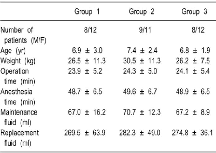
Table 1. Demographic Data of the Patients and Administered Fluid Volumes
Group 1 Group 2 Group 3
Number of 8/12 9/11 8/12 patients (M/F) Age (yr) 6.9 ± 3.0 7.4 ± 2.4 6.8 ± 1.9 Weight (kg) 26.5 ± 11.3 30.5 ± 11.3 26.2 ± 7.5 Operation 23.9 ± 5.2 24.3 ± 5.0 24.1 ± 5.4 time (min) Anesthesia 48.7 ± 6.5 49.6 ± 6.7 48.9 ± 6.5 time (min) Maintenance 67.0 ± 16.2 70.7 ± 12.3 67.2 ± 8.9 fluid (ml) Replacement 269.5 ± 63.9 282.3 ± 49.0 274.8 ± 36.1 fluid (ml)
Values are mean ± SD and number of cases. Group 1, 2 and 3 received Ringer’s lactate solution (RL), 5% dextrose in one-fourth strength normal saline (D51/4NS), and equal volume of D51/4NS and RL each, respectively, for maintenance fluid therapy. Maintenance fluid and Replacement fluid refer to the total amounts of fluid administered to the patients till 60 min after anesthetic induction as maintenance and replacement fluids, respectively.
< 0.05 was considered as statistically significant.
RESULTS
The demographic characteristics of the patients and the infused amounts of maintenance and replacement fluids were similar among three groups (Table 1).
There were no significant differences in the baseline blood glucose levels at anesthetic induction among three groups (Fig. 1). The mean blood glucose concentrations remained unchanged throughout the study period in all groups (Fig. 1). No patients were found to be hypoglycemic or hyperglycemic throughout the study period.
DISCUSSION
In the present study, the replacement of preoperative NPO deficits with RL maintained the blood glucose concentration within physiological range throughout the operation and anesthetic recovery phase, regardless of the types of maintenance fluid. No patients were found to be hypoglycemic or hyperglycemic throughout the study period.
Pediatric anesthesiologists were greatly concerned about the risk of hyperglycemia and hypoglycemia during the perioperative period. Compared with normoglycemia, hyperglycemia associated
with cerebral hypoxia results in accumulation of lactate in the brain [6,7]. This has been described as an important risk factor for development of neurological damage. So it is recommended that unnecessary glucose administration leading to intraoperative hyperglycemia should be avoided, since hyperglycemia can worsen the neurological outcome [8]. Furthermore, hyperglycemia can induce osmotic diuresis and consequently dehydration and electrolyte imbalance, especially in infants [9]. Thus, it has been suggested that glucose-free solutions should be used during surgery [10].
Adverse respiratory effects from hyperglycemia include in-creased carbon dioxide production, tidal volume, minute venti-lation and minute oxygen consumption [6]. Intraoperative glucose administration increased respiratory quotient during anesthesia [11,12]. These respiratory effects from hyperglycemia may have contribution to the prolonged extubation time after surgery [13]. In general, surgery has been shown to increase blood glucose concentrations despite the use of glucose-free solutions during surgery, independent of the anesthetic technique [14]. Stress hormone such as cortisol has been shown to inhibit the release of insulin and trigger a cascade of catabolic changes, which result in the hyperglycemia during and after surgery. The increase in plasma cortisol concentrations during surgery failed to inhibit insulin release and to increase plasma glucose concentrations, and cortisol and catecholamines released by noxious stimuli change the conformation of the insulin receptor, resulting in insulin-resistance in peripheral tissue [15]. Some
investigators demonstrated that most children also respond to surgery with an increase in blood glucose [10,15].
Perioperative fasting and the low carbohydrate reserves in infants and children were considered as major risk factors for hypoglycemia, especially in neonates and patients receiving total parenteral nutrition [16]. Moreover, general anesthesia conceals the symptoms of hypoglycemia. In the current study, children administered RL solution for replacement fluid therapy did not become hypoglycemic perioperatively as other study [17]. These findings suggested that glycogen stores are likely to be sufficient to prevent hypoglycemia in otherwise healthy children during short duration of surgery. Despite of low incidence of hypoglycemia, dextrose containing solutions may be necessary for certain specific conditions such as premature babies or neonates, poor general condition, and according to the type and duration of surgery.
Whether preoperative fasting causes hypoglycemia during pediatric anesthesia is controversial. Bevan and Burn observed that to shorten the preoperative fasting time could decrease the prevalence of hypoglycemia in children [18]. Other study did not confirm that the duration of starvation influenced the preoperative blood glucose concentration [19]. Our results are based on the overnight fasting for 8 hours in inpatients, and the use of different NPO guidelines and outpatients undergoing ambulatory surgery of shorter duration may have led to different results.
Recently, RL solution with low concentration dextrose has been recommended for routine fluid therapy during surgery in pediatric patients [5]. Its dextrose concentration is a compro-mise to avoid hypoglycemia and hyperglycemia and it also reduces the risk of severe hyponatremia. In our study, no patients were hypoglycemic or hyperglycemic. This finding shows that infusion of dextrose less than 5% concentration is sufficient to maintain blood glucose concentration within the physiological range during the perioperative period.
In conclusion, the current study shows that the replacement of preoperative NPO deficits with RL maintains the blood glucose concentration within physiological range throughout the operation and anesthetic recovery phase, regardless of the types of maintenance fluid. Although we inserted an additional catheter to the patients for infusion of replacement fluid and blood sampling, it is thought to be more desirable to secure an IV catheter of large diameter enough to infuse maintenance and replacement fluid in clinical practice.
REFERENCES
1. Payne K, Ireland P: Plasma glucose levels in the perioperative period in children. Anaesthesia 1984; 39: 868-72.
2. Wellborn LG, McGill WA, Hannallah RS, Nisselson CL, Ruttimann UE, Hicks JM: Perioperative blood glucose concentrations in pediatric outpatients. Anesthesiology 1986; 65: 543-7. 3. Wellborn LG, Hannallah RS, McGill WA, Ruttimann UE, Hicks
JM: Glucose concentrations for routine intravenous infusion in pediatric outpatient surgery. Anesthesiology 1987; 67: 427-30. 4. Nishina K, Mikawa K, Maekawa N, Asano M, Obara H: Effects
of exogenous intravenous glucose on plasma glucose and lipid homeostasis in anesthetized infants. Anesthesiology 1995; 83: 258-63.
5. Coté CJ: Pediatric anesthesia. In: Miller RD, eds. Anesthesia. 6th ed. New York: Churchill Livingstone, 2005: 2388-9.
6. Sieber FE, Smith DS, Traystman RJ, Wollman H: Glucose: A reevaluation of its intraoperative use. Anesthesiology 1987; 67: 72-81.
7. Li PA, Siesjö BK: Role of hyperglycaemia-related acidosis in ischaemic brain damage. Acta Physiol Scand 1997; 161: 567-80. 8. Steward DJ: Hyperglycaemia, something to worry about! Paediatr
Anaesth 1992; 2: 81-3.
9. Wright PD, Henderson K, Johnston IDA: Glucose utilization and insulin secretion during surgery in man. Br J Surg 1974; 61: 5-8. 10. Sandström K, Larsson LE, Nilsson K, Stenqvist O: Intraoperative
glucose administration influences respiratory quotient during paediatric anaesthesia. Acta Anaesthesiol Scand 1999; 43: 302-7. 11. Hagerdal M, Caldwell CB, Gross TB: Intraoperative fluid mana-gement influences carbon dioxide production and respiratory quotient. Anesthesiology 1983; 59: 48-50.
12. Bell C, Hughes CW, Oh TH, Donielson DW, O’Connor T: The effect of intravenous dextrose infusion on postbypass hyperg-lycemia in pediatric patients undergoing cardiac operations. J Clin Anesth 1993; 5: 381-5.
13. Schricker T, Lattermann R, Fiset P, Wykes L, Carli F: Integrated analysis of protein and glucose metabolism during surgery: effects of anesthesia. J Appl Physiol 2001; 91: 2523-30.
14. Nishina K, Mikawa K, Maekawa N, Asano M, Obara H: Effects of exogenous intravenous glucose on plasma glucose and lipid homeostasis in anesthetized infants. Anesthesiology 1995; 83: 258-63.
15. Ayers J, Graves SA: Perioperative management of total parenteral nutrition, glucose containing solutions, and intraoperative glucose monitoring in paediatric patients: a survey of clinical practice. Paediatr Anaesth 2001; 11: 41-4.
16. Chambrier C, Aouifi A, Bon C, Saudin F, Paturel B, Bouletreau P: Effects of intraoperative glucose administration on circulating metabolites and nitrogen balance during prolonged surgery. J Clin Anesth 1999; 11: 646-51.
17. Bevan JC, Burn MC: Acid-base and blood glucose levels of paediatric cases at induction of anaesthesia: the effects of
preoperative starvation and feeding in paediatric surgical patients. Br J Anaesth 1973; 45: 415-22.
18. Graham IF: Preoperative starvation and plasma glucose concen-trations in children undergoing outpatient anaesthesia. Br J Anaesth 1979; 51: 161-4.
19. Berleur MP, Dahan A, Murat I, Hazebroucq G: Perioperative infusion in paediatric patients: rationale for using Ringer-lactate solution with low dextrose concentration. J Clin Pharm Ther 2003; 28: 31-40.